

TO THE EDITOR:
Chronic infection with hepatitis delta virus (HDV) often induces severe liver inflammation and fibrosis.( 1 ) Infection with HDV is dependent on coinfection with hepatitis B virus (HBV) as HDV requires HBV surface antigen (HBsAg) in its envelope in order to infect hepatocytes. Patients with chronic HBV infection harbor hepatocytes with integrated HBV DNA,( 2 ) but the importance of integrated HBV DNA for HDV remains largely unknown. We addressed this by analyzing multiple pieces of explant liver tissue from 2 patients with HDV‐induced cirrhosis.
Thirty‐five liver pieces were analyzed by droplet digital polymerase chain reaction assays to quantify viral nucleic acids (details in the Supporting Information). We detected core‐gene RNA (core‐RNA), which reflects ongoing replication of HBV, in a minor proportion of liver pieces and covalently closed circular DNA (cccDNA) in no piece; however, we observed expression of S‐gene RNA (S‐RNA) and HDV RNA in almost all pieces (Fig. 1A‐C). This is consistent with expression of HBsAg from integrated HBV DNA in which the S gene but not the core gene has a preserved upstream promoter. The high levels of HDV RNA in the absence of HBV core‐RNA and presence of S‐RNA imply that HDV replication does not require ongoing HBV replication in the same cell and support that HDV production is supplied by HBsAg from integrations. This is suggested by experimental data( 3 ) and by our previous findings of abundant RNA with junctions between HBV and human sequences in tissue from the 2 patients.( 4 )
FIG. 1.
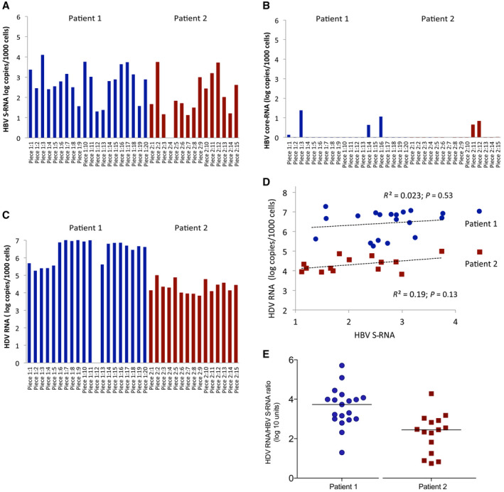
HDV RNA production, HBV infection, and HBV integration show different tissue distribution in the liver. Levels of (A) HBV S‐RNA, (B) HBV core‐RNA, and (C) HDV RNA in liver pieces from 2 patients by ddPCR quantification. (D) HDV RNA and HBV S‐RNA levels did not correlate. (E) The ratio between HDV RNA and S‐RNA levels in different liver pieces varied up to 1,000‐fold (the horizontal lines represent median values). Abbreviation: ddPCR, droplet digital polymerase chain reaction.
Interestingly, we observed a lack of correlation between levels of HDV RNA and S‐RNA (Fig. 1D), with HDV RNA:S‐RNA ratios ranging from 10:1 to >10,000:1 (Fig. 1E). Such high variation indicates that HDV RNA probably is present in a large number of hepatocytes that lack expression of S‐RNA and that cells not containing HBV infection (cccDNA) or integrated HBV DNA may contain and produce HDV RNA. This would be in line with experimental data showing persistence of HDV in the absence of HBV.( 5 ) Such spread of HDV to hepatocytes that do not replicate HBV and lack integrated HBV DNA might be abundant. Synthesis of HDV antigen in those cells should lead to exposure of HDV epitopes on the cell surface, potentially triggering cytotoxic T‐cell activation. This might help explain the severe necroinflammation that is often observed during HDV infection.( 1 )
In summary, our data suggest that integrated HBV DNA may be a significant source of HBsAg in patients coinfected with HBV/HDV, at least in those with cirrhosis, and that HDV RNA may be expressed in cells lacking both cccDNA and integrated HBV DNA.
Supporting information
Table S1‐S2
Potential conflict of interest: Nothing to report.
References
- 1. Grabowski J, Wedemeyer H. Hepatitis delta: immunopathogenesis and clinical challenges. Dig Dis 2010;28:133‐138. [DOI] [PubMed] [Google Scholar]
- 2. Lindh M, Rydell GE, Larsson SB. Impact of integrated viral DNA on the goal to clear hepatitis B surface antigen with different therapeutic strategies. Curr Opin Virol 2018;30:24‐31. [DOI] [PubMed] [Google Scholar]
- 3. Freitas N, Cunha C, Menne S, Gudima SO. Envelope proteins derived from naturally integrated hepatitis B virus DNA support assembly and release of infectious hepatitis delta virus particles. J Virol 2014;88:5742‐5754. [DOI] [PMC free article] [PubMed] [Google Scholar]
- 4. Ringlander J, Skoglund C, Prakash K, Andersson ME, Larsson SB, Tang K‐W, et al. Deep sequencing of liver explant transcriptomes reveals extensive expression from integrated hepatitis B virus DNA. J Viral Hepat 2020;11:1162‐1170. [DOI] [PubMed] [Google Scholar]
- 5. Giersch K, Bhadra OD, Volz T, Allweiss L, Riecken K, Fehse B, et al. Hepatitis delta virus persists during liver regeneration and is amplified through cell division both in vitro and in vivo. Gut 2019;68:150‐157. [DOI] [PubMed] [Google Scholar]
Associated Data
This section collects any data citations, data availability statements, or supplementary materials included in this article.
Supplementary Materials
Table S1‐S2