ABSTRACT
COVID-19 patients produce circulating and mucosal antibodies. In adults, specific saliva antibodies have been detected. Nonetheless, seroprevalence is routinely investigated, while little attention has been paid to mucosal antibodies. We therefore assessed SARS-CoV-2-specific antibody prevalence in serum and saliva in children in the Netherlands. We assessed SARS-CoV-2 antibody prevalence in serum and saliva of 517 children attending medical services in the Netherlands (irrespective of COVID-19 exposure) from April to October 2020. The prevalence of SARS-CoV-2 spike (S), receptor binding domain (RBD), and nucleocapsid (N)-specific IgG and IgA were evaluated with an exploratory Luminex assay in serum and saliva and with the Wantai SARS-CoV-2 RBD total antibody enzyme-linked immunosorbent assay in serum. Using the Wantai assay, the RBD-specific antibody prevalence in serum was 3.3% (95% confidence interval [CI]. 1.9 to 5.3%). With the Luminex assay, we detected heterogeneity between antibodies for S, RBD, and N antigens, as IgG and IgA prevalence ranged between 3.6 and 4.6% in serum and between 0 and 4.4% in saliva. The Luminex assay also revealed differences between serum and saliva, with SARS-CoV-2-specific IgG present in saliva but not in serum for 1.5 to 2.7% of all children. Using multiple antigen assays, the IgG prevalence for at least two out of three antigens (S, RBD, or N) in serum or saliva can be calculated as 3.8% (95% CI, 2.3 to 5.6%). Our study displays the heterogeneity of the SARS-CoV-2 antibody response in children and emphasizes the additional value of saliva antibody detection and the combined use of different antigens.
IMPORTANCE Comprehending humoral immunity to SARS-CoV-2, including in children, is crucial for future public health and vaccine strategies. Others have suggested that mucosal antibody measurement could be an important and more convenient tool to evaluate humoral immunity compared to circulating antibodies. Nonetheless, seroprevalence is routinely investigated, while little attention has been paid to mucosal antibodies. We show the heterogeneity of SARS-CoV-2 antibodies, in terms of both antigen specificity and differences between circulating and mucosal antibodies, emphasizing the additional value of saliva antibody detection next to detection of antibodies in serum.
KEYWORDS: SARS-CoV-2, antibodies, children, humoral immunity, prevalence, saliva
INTRODUCTION
Severe acute respiratory syndrome coronavirus 2 (SARS-CoV-2) is a positive-sense single-stranded RNA virus from the Coronaviridae family causing coronavirus disease 2019 (COVID-19). In 2020, this novel virus emerged as the cause of a pandemic. In the Netherlands, the first COVID-19 patient was confirmed on 27 February 2020. After the first peak in hospital admission rates during March and April 2020, the Netherlands endured a second peak during the second half of 2020. Epidemiological data on immunity are essential to understand disease pathology and to guide national prevention measures and possibilities for vaccine development (1).
Generally, humoral immunity is measured as the presence of pathogen-specific antibodies in serum. The prevalence of SARS-CoV-2 antibodies in serum has been described in several countries, including the Netherlands (2–4), with a potential durability of over 8 months (5, 6). Although the mucosa of the upper respiratory tract is the primary entry site of SARS-CoV-2, little attention has been paid to the presence of mucosal antibodies as part of the humoral immune response (7). For other viruses such as hepatitis B virus, norovirus, and human immunodeficiency virus type 1, studies have shown high similarity in circulating and mucosal antibody profiles, advocating for the use of saliva samples to measure humoral immunity (8–10). In respiratory syncytial virus (RSV), mucosal anti-RSV IgA and IgG combined proved at least as reliable as serum to detect infection. These saliva antibodies could help to distinguish current RSV (re-)infection from a false-positive result due to preexisting maternal antibodies (11). In adult COVID-19 patients, promising results on SARS-CoV-2-specific saliva antibodies with neutralizing capacities and a durability similar to serum have been reported (12–14). Interestingly, in asymptomatic or mild COVID-19, mucosal antibodies were detected, even in some seronegative patients (15). Mucosal antibody measurement could be an important and more convenient tool to evaluate humoral immunity in children, since they often have asymptomatic or mild disease (16, 17).
Among children, circulating antibody prevalence ranges from 0.9% in the United States to 7.3% in Switzerland (16, 18). The use of saliva antibody assays has yet to be explored in asymptomatic cases and in a pediatric population. Thus, the primary aim of this study was to evaluate the SARS-CoV-2-specific antibody prevalence in saliva compared to serum in a pediatric population during the COVID-19 outbreak in 2020 in the Netherlands. Since the Luminex assay provides a very sensitive method which is easily adjustable to different antigens, antibody isotypes, and sample types, we utilized this explorative assay in addition to the validated Wantai assay.
RESULTS
Study population.
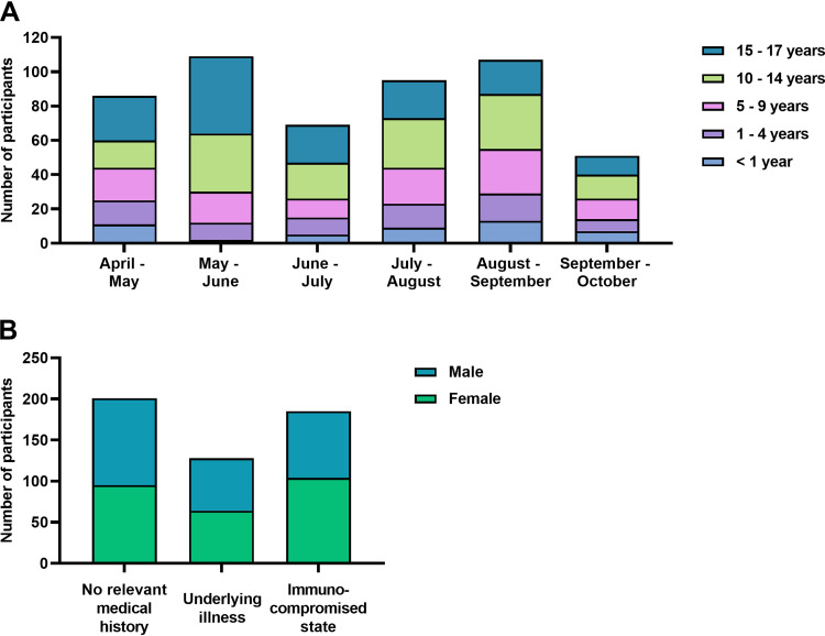
A total of 589 children were approached, 517 of which were included (see Fig. S1 in the supplemental material). The characteristics of the participants are shown in Table S2. Figure 1A shows the age distribution across the inclusion period. The median age was 11 years (interquartile range [IQR], 5 to 15 years). An immunocompromised state and an underlying illness were described in 35.8% and 24.8% of children, respectively, while 38.9% did not have a relevant medical history. Sex was equally distributed among comorbidity groups (Fig. 1B). SARS-CoV-2 PCR on nasopharyngeal swabs had been performed previously by the treating physician in 107 children, either due to clinical suspicion of COVID-19 or as preprocedural testing. Paired serum and saliva samples of sufficient volume for all assays were available for 413/517 (82%) children, with a median age of 12 years (IQR, 7 to 15 years).
FIG 1.
Age distribution, comorbidity, and sex. (A) The distribution of age across the inclusion period of 24 weeks is depicted for the total number of cases (n = 517), with each bar representing 4 weeks. (B) Comorbidity and male/female ratio of the study sample is calculated for the nonmissing values (n = 514, excluding 3 missing values for comorbidity).
Antibody prevalence.
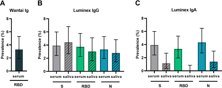
Figure 2 shows the estimated prevalence determined by the Wantai receptor binding domain (RBD) total antibody assay (Fig. 2A) and the Luminex assay for spike (S), RBD, and nucleocapsid (N)-specific IgG (Fig. 2B) and IgA (Fig. 2C) in serum and saliva. Titers from the Luminex assay are shown in Fig. S2, and the assay cutoff and assay performance parameters are in Table S1. The prevalence of RBD-specific antibodies in serum was 16/487 (3.3%; 95% confidence interval [CI], 1.9 to 5.3%) with the Wantai assay. With the Luminex assay, the prevalence of SARS-CoV-2-specific antibodies in serum ranged between 3.3% (95% CI, 1.9 to 5.2%) and 4.3% (95% CI, 2.7 to 6.4%) depending on the antigen and isotype measured. The prevalence of SARS-CoV-2-specific antibodies in saliva ranged between 0.0% (95% CI, 0.0 to 0.7%) and 4.4% (95% CI, 2.7 to 6.8%).
FIG 2.
SARS-CoV-2 antibody prevalence estimates in serum and saliva. (A) Prevalence estimate of SARS-CoV-2 RBD total antibodies in the Wantai assay for serum. (B and C) Prevalence estimates of SARS-CoV-2 S- (gray bars), RBD- (green bars), and N-specific (blue bars) IgG (B) and IgA (C) in the Luminex assays for serum (solid bars) and saliva (hatched bars). Prevalence was the calculated proportion with a value above the determined cutoff out of nonmissing values. Estimates are shown with 95% confidence intervals. S, trimeric SARS-CoV-2 spike protein; RBD, the monomeric receptor binding domain of the SARS-CoV-2 spike protein; N, SARS-CoV-2 nucleocapsid protein.
The prevalence of SARS-CoV-2-specific antibodies did not increase over calendar months in the Luminex assays or the Wantai assay (Fig. S3). The seroprevalence was 4/200 (2.0%) in children aged 0 to 10 years and 12/287 (4.2%) in children aged 10 to 17 years in the Wantai assay (Table S2). The seroprevalence was 3.3% in children with an immunocompromised state, 5.0% in children with underlying illness, and 2.2% in children with no relevant medical history. Most of the children that were positive in the Wantai assay (13/16, 81%) had COVID-19 symptoms at the time of inclusion or in the previous 4 weeks or reported a COVID-19-positive household member.
Comparison of serum and saliva SARS-CoV-2 antibody prevalence.
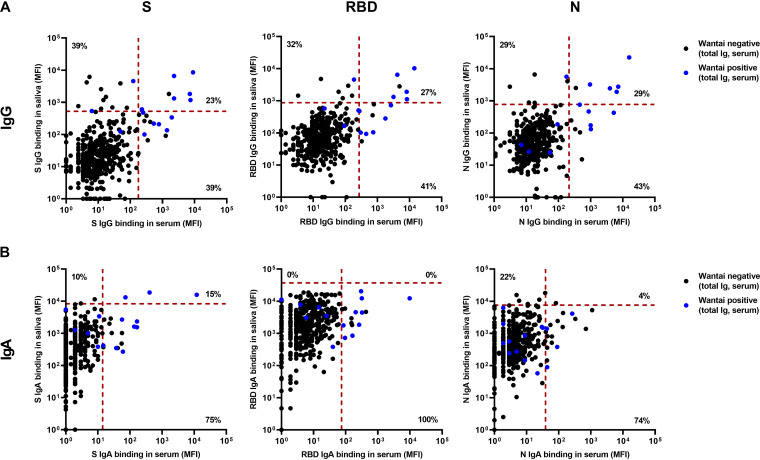
In the Luminex assay, 31/422 (7.4%) children had detectable S-specific IgG, while 22/422 (5.2%) had detectable RBD-specific IgG, and 21/422 (5.0%) had detectable N-specific IgG in serum and/or saliva (Fig. 2B). Figure 3A shows the correspondence between serum and saliva IgG for all paired samples in the Luminex and Wantai assays. Between 7/31 and 6/21 (23 to 29%) of these children showed corresponding positive titers in both serum and saliva, depending on the antigen used. However, 12/31 to 9/21 (38 to 43%) children had positive SARS-CoV-2-specifc IgG titers in serum but not in saliva, and 6/21 to 12/31 (29 to 39%) had positive titers in saliva but not in serum.
FIG 3.
Correspondence of Luminex assays for serum and saliva. (A and B) SARS-CoV-2 S-, RBD-, and N-specific IgG (A) and IgA (B), measured in paired serum and saliva samples (n = 413) by Luminex assay, expressed as MFI. Only samples also measured in the Wantai assay are shown, with the positive Wantai results indicated in blue. Serum and saliva are plotted against each other to reveal the differences between each compartment. The red dotted lines are the cutoff values to discriminate positive and negative measurements in the Luminex assays. Percentages represent data points which are positive for both compartments or for a single compartment as the percentage of total positives in each graph. S, trimeric SARS-CoV-2 spike protein; RBD, protein of only the monomeric receptor binding domain of the SARS-CoV-2 spike protein; N, SARS-CoV-2 nucleocapsid protein; MFI, median fluorescence intensity.
Comparable with these findings, between 6/15 and 8/15 (40 to 53%) Wantai positive children also had positive saliva IgG titers in the Luminex assay, but between 6/12 and 11/19 (50 to 58%) children with SARS-CoV-2-specific saliva IgG in the Luminex assay were negative in the Wantai assay. This corresponds to 6 to 11/413 (1.5 to 2.7%) of the total paired sample, depending on the antigen used.
We compared IgA responses in serum and saliva (Fig. 3B), but there was low agreement between the two compartments (0/15 to 3/20; 0 to 15% with detectable IgA in both serum and saliva). The Luminex IgA assays were positive in saliva while negative in serum in 0/15 to 5/23 (0 to 22%) of children with IgA antibodies.
Combined Luminex assay.
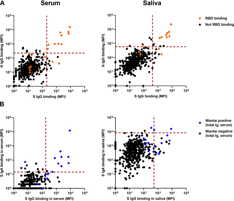
Figure 4A shows the correspondence between the three SARS-CoV-2-specific antigens. All children with S- and N-specific IgG in serum also had positive titers of RBD-specific IgG. Comparing IgG and IgA antibodies (Fig. 4B) in serum, a few children showed S-specific IgA but not IgG antibodies (10/509; 2.0%). All but one child positive for both serum IgG and IgA in the Luminex assay were also positive in the Wantai total antibody assay (9/487; 1.8%). There was low correspondence between IgG and IgA in saliva.
FIG 4.
Correspondence of Luminex assays for different antigens and isotypes. (A) S-, RBD-, and N protein-specific IgG, measured in serum (n = 509, left) and saliva (n = 430, right) in the Luminex assay, expressed as MFI. S and N are plotted against each other, and RBD-positive samples are shown in orange. The red dotted lines are the cutoff values to discriminate positive and negative measurements for S and N. (B) SARS-CoV-2 S-specific IgG and IgA in serum (n = 487, left) and saliva (n = 413, right) measured in the Luminex assay, expressed as MFI. IgG and IgA are plotted against each other to reveal the correspondence between the two isotypes. Samples that were also positive in the Wantai RBD total antibody assay are shown in blue. S, trimeric SARS-CoV-2 spike protein; RBD, monomeric receptor binding domain of the SARS-CoV-2 spike; N, SARS-CoV-2 nucleocapsid protein; MFI, median fluorescence intensity.
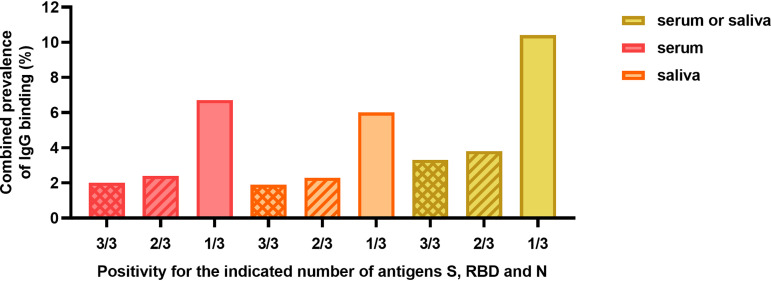
We explored combining multiantigen assays to calculate the SARS-CoV-2 IgG prevalence in several ways (Fig. 5). The total prevalence of IgG binding to any of the antigens is 34/509 (6.7%) in serum and 26/430 (6.0%) in saliva. To increase the specificity, the IgG prevalence for at least two out of three antigens (S, RBD, or N protein) can be calculated as 2.4% for serum, 2.3% for saliva, and 3.8% (95% CI, 2.3 to 5.6%) when both serum and saliva are measured. Considering only children with positive Luminex titers for two out of three antigens in saliva, 4/413 children had saliva antibodies, while they were negative in the Wantai assay, corresponding to 1.0% (95% CI, 0.3 to 2.2%) of the total sample.
FIG 5.
Combined SARS-CoV-2 IgG antibody prevalence. Combined prevalence of the explorative Luminex assay of IgG in serum (pink bars), in saliva (orange bars), or in serum and saliva combined (yellow bars). The combined prevalence was calculated for children positive for 3/3 SARS-CoV-2 antigens (crosshatched bars) at least 2/3 antigens (hatched bars), and for children positive for at least 1/3 antigens (solid bars). S, trimeric SARS-CoV-2 spike protein; RBD, monomeric receptor binding domain of the SARS-CoV-2 spike; N, SARS-CoV-2 nucleocapsid protein.
DISCUSSION
This is the first study evaluating concurrent mucosal and circulating SARS-CoV-2-specific antibodies in a large pediatric cohort. We found children with detectable SARS-CoV-2-specific saliva IgG, while circulating antibody levels (measured with the Wantai assay) were undetectable. Similarly, not all seropositive children had detectable saliva IgG. Additionally, we detected heterogeneity in the presence of antibodies to different SARS-CoV-2-specific antigens.
We found an antibody seroprevalence comparable to that of the S-specific IgG seroprevalence among 1,000 Dutch children during June and July 2020 in a population-based study (19, 20). Most children in our population had an underlying illness or immunocompromised state. Therefore, the similarities to national seroprevalence studies are particularly interesting and suggest similar COVID-19 incidence in our hospital-based cohort compared to the general community. Similar to the Netherlands, other European countries report lower seroprevalence in children (range, 0.8 to 7.3%) compared to adults (range, 1 to 20%) (18, 21).
Although many reports are appearing on SARS-CoV-2 seroprevalence, the initial confrontation of SARS-CoV-2 with the adaptive immune system is located at the mucosal surface of the respiratory tract. To our knowledge, only a few adult cohorts report on the associations between circulating and mucosal SARS-CoV-2-specific antibodies. For example, a sensitivity of 84.2% and a specificity of 100% for saliva S-specific IgG to detect PCR- and seropositive patients in a symptomatic population was reported (12). The durability appears similar to that of circulating antibodies, as salivary antibodies were shown to be measurable for up to 9 months after infection in convalescent mild COVID-19 patients (22). The findings from three cohorts of SARS-CoV-2-infected adults suggest that mucosal IgG antibodies can be used as a surrogate for circulating IgG due to the high similarity (12–14).
However, a proportion of seronegative children in our sample did have saliva antibodies (mostly IgG). Corroborating these findings, 15 to 20% of seronegative health care workers had mucosal SARS-CoV-2 S-specific antibodies with, in some cases, comparable in vitro neutralizing capacity to serum (15, 23). A possible explanation for the discordance between circulating and mucosal antibodies could be the disease severity. Cervia et al. hypothesize that the mucosal antibody response is more prominent in mild infection and in younger individuals, although their youngest participant was 30 years old (15). Similarly, a preprint validation study of a saliva IgA assay observed asymptomatic individuals with saliva SARS-CoV-2-specific antibodies despite a negative PCR and/or serum antibody tests (24). Hence, reporting seroprevalence alone may result in an underestimation of humoral immunity, particularly in younger and mildly infected patients.
We still lack full understanding of how IgA provides additional value for evaluating humoral immunity (25). IgA can appear earlier in blood than IgG following SARS-CoV-2 infection (26). Although IgA is the key immunoglobulin for mucosal immunity, evidence of SARS-CoV-2-specific saliva IgA is inconsistent. Compared to IgG, saliva IgA is less correlated with serum IgA (3). Saliva IgG is mostly derived from circulatory IgG through transudation, whereas saliva IgA can be produced locally (27). This is also observed in vaccination response studies, showing saliva S-specific IgG in all fully vaccinated participants, while only 60% showed saliva S-specific IgA (28). Consistent with our findings, MacMullan et al. and Pisanic et al. emphasize the lower sensitivity for IgA to detect PCR-positive patients compared to IgG (12, 13). The high variation in mucosal IgA titers complicates detection of SARS-CoV-2-specific saliva IgA, possibly caused by polyreactive IgA known to function as a nonspecific mucosal immune barrier (24).
There is growing evidence on the use of SARS-CoV-2 multiantigen assays in epidemiological surveys. In line with other studies, we observed that positive individuals often do not show equally elevated titers across all three SARS-CoV-2-specific antigens. In an epidemiological survey of 1,225 blood donors, seroprevalence with single-antigen assays also varied widely between 0.8% and 7.5% depending on the antigen type (29). A potential association between specific antigens and disease severity has been proposed. Outpatients with mild or asymptomatic infection showed higher ratios of S- and RBD-specific antibodies than of N-specific antibodies, whereas all three antigens were effective for detecting responses in hospitalized patients (23, 30). Considering their different kinetics (31) and functions in the humoral immune response and the broad clinical spectrum of COVID-19, combining multiple SARS-CoV-2 antigens seems an appropriate method when investigating population prevalence (32).
Targeting multiple antigens in multiple compartments to evaluate the humoral response can increase sensitivity but may consequently increase false positives. A golden standard is unfortunately still missing to determine true rates, as both PCR and serology can have false-negative results (33, 34). To increase the certainty of SARS-CoV-2 exposure and minimize the possibility of a false-positive result (particularly in a presumed low-prevalence context) (35), we could define a sample as positive only when the IgG level is above the cutoff for at least two antigen assays as proposed in other studies (13, 31, 32, 36). In adult cohorts of (symptomatic) PCR-proven COVID-19 patients and pre-COVID-19 era controls, combining multiple N and S/RBD antibody assays resulted in higher diagnostic accuracy (13, 32, 36, 37). Although Luminex assays in serum are known to have high accuracy to detect previous SARS-CoV-2 infection (31, 32, 36, 37) and the validated Wantai assay provides the context for the results, follow-up research should focus on validation of established assays on materials other than serum as well as the combined use of different antigens in high- and low-prevalence settings.
Our study encountered several other limitations. We did not evaluate the potential moment of infection; thus, we could have missed SARS-CoV-2-exposed children, as IgG can be detected after 10 days and for several months post-symptom onset, while IgM and IgA wane more quickly (3). We minimized this effect by detecting multiisotype antibodies. Moreover, cross-reactive antibodies from previous coronavirus types could have resulted in positivity without a history of actual SARS-CoV-2 infection. Antibody cross-reactivity is a known phenomenon for pathogens with shared structural motifs (38, 39). In a cohort sampled in the pre-COVID-19 era, detectable SARS-CoV-2 S-specific antibodies were found and at a higher frequency in children than adults, peaking to 62% between 6 and 16 years of age (39). Although these antibody titers were lower than those in COVID-19 patients, their sera exhibited neutralizing activity against SARS-CoV-2 (39). Thus, even if some of the antibody titers we found are the result of cross-reactivity, they could still be functional. Since we did not perform in vitro neutralization testing, the functionality of the (mucosal) antibodies in our cohort against SARS-CoV-2 remains unknown.
Conclusion and implications.
Comprehending humoral immunity to SARS-CoV-2, including in children, is crucial for future public health and vaccine strategies. We therefore detected SARS-CoV-2 antibody prevalence in serum and saliva of children. Our study displays the heterogeneity of the SARS-CoV-2 antibody response in children and emphasizes the additional value of saliva assays for antibody detection as well as the combined use of different antigens. Validation of multiantigen saliva antibody assays is recommended for further research.
MATERIALS AND METHODS
Study design and participants.
This prospective cross-sectional study included simultaneous convenience blood and saliva sampling of children attending medical services at one of seven participating secondary- and tertiary-care hospitals in the Northwest region of the Netherlands during 24 consecutive weeks (12 April to 2 October 2020). Inclusion criteria were children aged 0 to 18 years residing in the Netherlands who required blood testing or intravenous cannulation for any reason. Eligibility was irrespective of (suspected) acute or prior COVID-19 infection. Children were excluded if sample collection of neither serum nor saliva was sufficient.
We recorded age, sex, COVID-19-related symptoms, and proven or suspected COVID-19 in household members. COVID-19-related symptoms were defined as fever (>37.5°C), sore throat, cough, shortness of breath, tachypnea, headache, abdominal cramps, and diarrhea. Electronic patient files were used to extract previous SARS-CoV-2 PCR assay results and medical history that could influence the humoral immune response or infection severity. Children with immunodeficiency, autoimmune disease, hematological malignancies, and/or use of immunomodulating drugs were defined as having an “immunocompromised state.” Immunomodulating drugs included azathioprine, methotrexate, monoclonal antibodies, immunoglobulins, and corticosteroids. We defined children with an “underlying illness” as children with obesity (body mass index [BMI], ≥30); respiratory, cardiovascular, endocrine, metabolic, hematologic, or kidney diseases; solid malignancies; or psychomotor retardation. Previously healthy children were categorized as having “no relevant medical history.” The study protocol was approved by the ethics committee of the Amsterdam University Medical Centers (NL73556.018.20) and conducted in accordance with good clinical practice standards. Written informed consent was obtained from both parents/guardians and/or from children above the legal age of consent.
Serum and saliva sampling.
Saliva was obtained using a sterile container or a buccal swab (ORACOL saliva collection device, product code S10; Malvern Medical Developments Ltd.). Saliva samples were either stored at −70°C until centrifuging or directly centrifuged (at 1,000 rpm for 10 min). Supernatant and pellets were stored in aliquots at −80°C. During venipuncture, a blood sample of 1 to 5 ml was collected and centrifuged, and was serum stored at −20°C.
Serum assays.
Seroprevalence was assessed with the FDA-approved Wantai SARS-CoV-2 RBD total antibody enzyme-linked immunosorbent assay following the manufacturer’s instructions, with a sensitivity of 96.7% (95% CI, 83.3 to 99.4%) and specificity of 97.5% (95% CI, 91.3 to 99.3%) (40), and confirmed with an in-house-developed SARS-CoV-2 RBD total antibody bridge assay as described previously (sensitivity, 98.1%; specificity, 99.5%) (41).
Protein coupling to Luminex beads.
An explorative Luminex assay was developed to investigate antigen-specific IgG and IgA in serum and saliva. A recombinant prefusion ectodomain trimer of SARS-CoV-2 S protein, the monomeric RBD of the S protein, and N protein were designed, produced, and purified as previously described (42, 43). The proteins were covalently coupled to Magplex beads (Luminex) using a two-step carbodiimide reaction at a ratio of 75 μg protein to 12.5 million beads for S, at an equimolar concentration for N, and at 3× the equimolar concentration for RBD. Beads were washed with 100 mM monobasic sodium phosphate, pH 6.2, activated with sulfo-N-hydroxysulfosuccinimide and 1-ethyl-3-(3-dimethylaminopropyl) carbodiimide (Thermo Fisher Scientific), and incubated for 30 min on a rotator at room temperature (RT). Activated beads were washed 3 times with 50 mM MES (morpholineethanesulfonic acid), pH 5.0, proteins were added, and beads were incubated for 3 h on a rotator at RT. The beads were washed with phosphate-buffered saline (PBS) and blocked with PBS containing 2% bovine serum albumin (BSA), 3% fetal calf serum, and 0.02% Tween 20 at pH 7.0 (PBS-blocking) for 30 min on a rotator at RT. Finally, the beads were washed, stored in PBS containing 0.05% sodium azide at 4°C, and used within 6 weeks.
Luminex assays.
A total of 50 μl of PBS-blocking containing 20 of each bead per μl was incubated with 50 μl of 1:10,000 diluted serum or 1:10 diluted saliva supernatant in PBS-blocking overnight on a plate shaker at 4°C. Subsequently, plates were washed with Tris-buffered saline (TBS) containing 0.05% Tween 20 (TBST) using a magnetic separator. Beads were resuspended in 50 μl of goat anti-human IgG-PE (Southern Biotech) or goat anti-human IgA-PE (Southern Biotech) in PBS-blocking and incubated on a plate shaker at RT for 2 h. Afterward, beads were washed with TBST and resuspended in 70 μl MAGPIX drive fluid (Luminex). Read-out was performed on a MAGPIX system (Luminex). The resulting median fluorescence intensity (MFI) values per bead were background-corrected by subtraction of the MFI values of buffer only. A titration of serum and saliva of an adult convalescent COVID-19 patient was used to normalize data between plates. The cutoff was determined at 2 (geometric) standard deviations above the geometric mean of the total sample (n = 509 for serum, n = 430 for saliva), separately for each combination of antigen, sample type and secondary antibody (Table S1). Beads with no antigen were included to confirm the absence of antibodies binding to beads or blocking components for each individual sample. Prepandemic serum pools (n = 3) or healthy donor saliva (early in the pandemic with negative PCR and/or absence of symptoms; n = 4) were included on each assay plate as negative controls. The replicability was calculated on samples measured twice, and the assay precision was calculated using positive-control sera (n = 6) or saliva (n = 5) included on each plate (Table S1).
Statistical analyses.
We estimated the prevalence of circulating and mucosal antibodies as the proportion of children with a result above the cutoff value. Confidence intervals were calculated with the Clopper-Pearson method in IBM SPSS Statistics version 26 predictive analytics software. In accordance with the World Health Organization’s Investigation Protocol for COVID-19 seroepidemiological research, we stratified participants into predefined age groups as follows: <1 year, 1 to 4 years, 5 to 9 years, 10 to 14 years, and 15 to 17 years (1). We calculated a minimum sample size of 139 children per age group (0 to 1 year, 1 to 5 years, and 5 to 18 years) to detect a seroprevalence of 10% (95% CI, 5 to 15%).
Supplementary Material
ACKNOWLEDGMENTS
This study would not have been possible without the instrumental and passionate support of Stichting Steun Emma Kinderziekenhuis, of the physicians, laboratory, and nursing staff of all participating care centers, the AUMC microbiology staff, the student team for inclusion of patients and the student team for data entry, the donors of control samples, all participants and their caregivers, and the creative contribution of Eli Vlessing.
This work was supported by the Contribute Foundation.
Footnotes
Supplemental material is available online only.
Contributor Information
Maya W. Keuning, Email: m.w.keuning@amsterdamumc.nl.
Jennifer Dien Bard, Children's Hospital Los Angeles, University of Southern California.
REFERENCES
- 1.World Health Organization. 2020. Population-based age-stratified seroepidemiological investigation protocol for coronavirus 2019 (COVID-19) infection, 26 May 2020. World Health Organization, Geneva, Switzerland. [Google Scholar]
- 2.Pollán M, Pérez-Gómez B, Pastor-Barriuso R, Oteo J, Hernán MA, Pérez-Olmeda M, Sanmartín JL, Fernández-García A, Cruz I, Fernández de Larrea N, Molina M, Rodríguez-Cabrera F, Martín M, Merino-Amador P, León Paniagua J, Muñoz-Montalvo JF, Blanco F, Yotti R, Group E-CS. 2020. Prevalence of SARS-CoV-2 in Spain (ENE-COVID): a nationwide, population-based seroepidemiological study. Lancet 396:535–544. doi: 10.1016/S0140-6736(20)31483-5:S0140-6736(20)31483-5. [DOI] [PMC free article] [PubMed] [Google Scholar]
- 3.Gudbjartsson DF, Norddahl GL, Melsted P, Gunnarsdottir K, Holm H, Eythorsson E, Arnthorsson AO, Helgason D, Bjarnadottir K, Ingvarsson RF, Thorsteinsdottir B, Kristjansdottir S, Birgisdottir K, Kristinsdottir AM, Sigurdsson MI, Arnadottir GA, Ivarsdottir EV, Andresdottir M, Jonsson F, Agustsdottir AB, Berglund J, Eiriksdottir B, Fridriksdottir R, Gardarsdottir EE, Gottfredsson M, Gretarsdottir OS, Gudmundsdottir S, Gudmundsson KR, Gunnarsdottir TR, Gylfason A, Helgason A, Jensson BO, Jonasdottir A, Jonsson H, Kristjansson T, Kristinsson KG, Magnusdottir DN, Magnusson OT, Olafsdottir LB, Rognvaldsson S, Le Roux L, Sigmundsdottir G, Sigurdsson A, Sveinbjornsson G, Sveinsdottir KE, Sveinsdottir M, Thorarensen EA, Thorbjornsson B, Thordardottir M, Saemundsdottir J, et al. 2020. Humoral immune response to SARS-CoV-2 in Iceland. N Engl J Med 383:1724–1734. doi: 10.1056/NEJMoa2026116. [DOI] [PMC free article] [PubMed] [Google Scholar]
- 4.Slot E, Hogema BM, Reusken CBEM, Reimerink JH, Molier M, Karregat JHM, Ijlst J, Novotný VMJ, van Lier RAW, Zaaijer HL. 2020. Low SARS-CoV-2 seroprevalence in blood donors in the early COVID-19 epidemic in the Netherlands. Nat Commun 11:5744. doi: 10.1038/s41467-020-19481-7. [DOI] [PMC free article] [PubMed] [Google Scholar]
- 5.Hartley GE, Edwards ESJ, Aui PM, Varese N, Stojanovic S, McMahon J, Peleg AY, Boo I, Drummer HE, Hogarth PM, O’Hehir RE, van Zelm MC. 2020. Rapid generation of durable B cell memory to SARS-CoV-2 spike and nucleocapsid proteins in COVID-19 and convalescence. Sci Immunol 5:eabf8891. doi: 10.1126/sciimmunol.abf8891. [DOI] [PMC free article] [PubMed] [Google Scholar]
- 6.Choe PG, Kim K-H, Kang CK, Suh HJ, Kang E, Lee SY, Kim NJ, Yi J, Park WB, Oh M-D. 2021. Antibody responses 8 months after asymptomatic or mild SARS-CoV-2 infection. Emerg Infect Dis 27:928–931. doi: 10.3201/eid2703.204543. [DOI] [PMC free article] [PubMed] [Google Scholar]
- 7.Russell MW, Moldoveanu Z, Ogra PL, Mestecky J. 2020. Mucosal immunity in COVID-19: a neglected but critical aspect of SARS-CoV-2 infection. Front Immunol 11:611337. doi: 10.3389/fimmu.2020.611337. [DOI] [PMC free article] [PubMed] [Google Scholar]
- 8.Hettegger P, Huber J, Paßecker K, Soldo R, Kegler U, Nöhammer C, Weinhäusel A. 2019. High similarity of IgG antibody profiles in blood and saliva opens opportunities for saliva based serology. PLoS One 14:e0218456. doi: 10.1371/journal.pone.0218456. [DOI] [PMC free article] [PubMed] [Google Scholar]
- 9.Chohan BH, Lavreys L, Mandaliya KN, Kreiss JK, Bwayo JJ, Ndinya-Achola JO, Martin HL Jr.. 2001. Validation of a modified commercial enzyme-linked immunoassay for detection of human immunodeficiency virus type 1 immunoglobulin G antibodies in saliva. Clin Diagn Lab Immunol 8:346–348. doi: 10.1128/CDLI.8.2.346-348.2001. [DOI] [PMC free article] [PubMed] [Google Scholar]
- 10.Tamminen K, Malm M, Vesikari T, Blazevic V. 2018. Norovirus-specific mucosal antibodies correlate to systemic antibodies and block norovirus virus-like particles binding to histo-blood group antigens. Clin Immunol 197:110–117. doi: 10.1016/j.clim.2018.09.009. [DOI] [PubMed] [Google Scholar]
- 11.Okiro E, Sande C, Mutunga M, Medley G, Cane P, Nokes D. 2008. Identifying infections with respiratory syncytial virus by using specific immunoglobulin G (IgG) and IgA enzyme-linked immunosorbent assays with oral-fluid samples. J Clin Microbiol 46:1659–1662. doi: 10.1128/JCM.02190-07. [DOI] [PMC free article] [PubMed] [Google Scholar]
- 12.MacMullan MA, Ibrayeva A, Trettner K, Deming L, Das S, Tran F, Moreno JR, Casian JG, Chellamuthu P, Kraft J, Kozak K, Turner FE, Slepnev VI, Le Page LM. 2020. ELISA detection of SARS-CoV-2 antibodies in saliva. Sci Rep 10:20818. doi: 10.1038/s41598-020-77555-4. [DOI] [PMC free article] [PubMed] [Google Scholar]
- 13.Pisanic N, Randad PR, Kruczynski K, Manabe YC, Thomas DL, Pekosz A, Klein SL, Betenbaugh MJ, Clarke WA, Laeyendecker O, Caturegli PP, Larman HB, Detrick B, Fairley JK, Sherman AC, Rouphael N, Edupuganti S, Granger DA, Granger SW, Collins MH, Heaney CD. 2020. COVID-19 serology at population scale: SARS-CoV-2-specific antibody responses in saliva. J Clin Microbiol 59:e02204-20. doi: 10.1128/JCM.02204-20. [DOI] [PMC free article] [PubMed] [Google Scholar]
- 14.Isho B, Abe KT, Zuo M, Jamal AJ, Rathod B, Wang JH, Li Z, Chao G, Rojas OL, Bang YM, Pu A, Christie-Holmes N, Gervais C, Ceccarelli D, Samavarchi-Tehrani P, Guvenc F, Budylowski P, Li A, Paterson A, Yue FY, Marin LM, Caldwell L, Wrana JL, Colwill K, Sicheri F, Mubareka S, Gray-Owen SD, Drews SJ, Siqueira WL, Barrios-Rodiles M, Ostrowski M, Rini JM, Durocher Y, McGeer AJ, Gommerman JL, Gingras A-C. 2020. Persistence of serum and saliva antibody responses to SARS-CoV-2 spike antigens in patients with COVID-19. Science Immunology 5:eabe5511. doi: 10.1126/sciimmunol.abe5511. [DOI] [PMC free article] [PubMed] [Google Scholar]
- 15.Cervia C, Nilsson J, Zurbuchen Y, Valaperti A, Schreiner J, Wolfensberger A, Raeber ME, Adamo S, Weigang S, Emmenegger M, Hasler S, Bosshard PP, De Cecco E, Bächli E, Rudiger A, Stüssi-Helbling M, Huber LC, Zinkernagel AS, Schaer DJ, Aguzzi A, Kochs G, Held U, Probst-Müller E, Rampini SK, Boyman O. 2020. Systemic and mucosal antibody responses specific to SARS-CoV-2 during mild versus severe COVID-19. J Allergy and Clin Immunol 147:545–557. doi: 10.1016/j.jaci.2020.10.040. [DOI] [PMC free article] [PubMed] [Google Scholar]
- 16.Dingens AS, Crawford KHD, Adler A, Steele SL, Lacombe K, Eguia R, Amanat F, Walls AC, Wolf CR, Murphy M, Pettie D, Carter L, Qin X, King NP, Veesler D, Krammer F, Dickerson JA, Chu HY, Englund JA, Bloom JD. 2020. Serological identification of SARS-CoV-2 infections among children visiting a hospital during the initial Seattle outbreak. Nat Commun 11:4378. doi: 10.1038/s41467-020-18178-1. [DOI] [PMC free article] [PubMed] [Google Scholar]
- 17.Statista. 2020. Distribution of hospitalizations due to the coronavirus (COVID-19) in the Netherlands as of September 29, 2020, by age group. https://www.statista.com/statistics/1129037/coronavirus-hospitalization-by-age-in-netherlands/. Accessed 2 October 2020.
- 18.Stringhini S, Wisniak A, Piumatti G, Azman AS, Lauer SA, Baysson H, De Ridder D, Petrovic D, Schrempft S, Marcus K, Yerly S, Arm Vernez I, Keiser O, Hurst S, Posfay-Barbe KM, Trono D, Pittet D, Gétaz L, Chappuis F, Eckerle I, Vuilleumier N, Meyer B, Flahault A, Kaiser L, Guessous I. 2020. Seroprevalence of anti-SARS-CoV-2 IgG antibodies in Geneva, Switzerland (SEROCoV-POP): a population-based study. Lancet 396:313–319. doi: 10.1016/S0140-6736(20)31304-0. [DOI] [PMC free article] [PubMed] [Google Scholar]
- 19.Vos ERA, den Hartog G, Schepp RM, Kaaijk P, van Vliet J, Helm K, Smits G, Wijmenga-Monsuur A, Verberk JDM, van Boven M, van Binnendijk RS, de Melker HE, Mollema L, van der Klis FRM. 2021. Nationwide seroprevalence of SARS-CoV-2 and identification of risk factors in the general population of the Netherlands during the first epidemic wave. J Epidemiol Community Health 75:489–495. doi: 10.1136/jech-2020-215678. [DOI] [PMC free article] [PubMed] [Google Scholar]
- 20.National Institute for Public Health and the Environment Ministry of Health, Welfare and Sport. n.d. PIENTER Corona Study. https://www.rivm.nl/en/pienter-corona-study/results.
- 21.Lai C-C, Wang J-H, Hsueh P-R. 2020. Population-based seroprevalence surveys of anti-SARS-CoV-2 antibody: an up-to-date review. Int J Infect Dis 101:314–322. doi: 10.1016/j.ijid.2020.10.011. [DOI] [PMC free article] [PubMed] [Google Scholar]
- 22.Alkharaan H, Bayati S, Hellström C, Aleman S, Olsson A, Lindahl K, Bogdanovic G, Healy K, Tsilingaridis G, De Palma P, Hober S, Månberg A, Nilsson P, Pin E, Sällberg Chen M. 2021. Persisting salivary IgG against SARS-CoV-2 at 9 months after mild COVID-19: a complementary approach to population surveys. J Infect Dis 224:407–414. doi: 10.1093/infdis/jiab256. [DOI] [PMC free article] [PubMed] [Google Scholar]
- 23.Faustini SE, Jossi SE, Perez-Toledo M, Shields A, Allen JD, Watanabe Y, Newby ML, Cook A, Willcox CR, Salim M, Goodall M, Heaney JL, Marcial-Juarez E, Morley GL, Torlinska B, Wraith DC, Veenith T, Harding S, Jolles S, Mark PJ, Plant T, Huissoon A, O'Shea MK, Willcox BE, Drayson MT, Crispin M, Cunningham AF, Richter AG. 2020. Detection of antibodies to the SARS-CoV-2 spike glycoprotein in both serum and saliva enhances detection of infection. medRxiv. doi: 10.1101/2020.06.16.20133025. [DOI] [PMC free article] [PubMed] [Google Scholar]
- 24.Varadhachary A, Chatterjee D, Garza J, Garr RP, Foley C, Letkeman AF, Dean J, Haug D, Breeze J, Traylor R, Malek A, Nath R, Linbeck L. 2020. Salivary anti-SARS-CoV-2 IgA as an accessible biomarker of mucosal immunity against COVID-19. medRxiv. doi: 10.1101/2020.08.07.20170258. [DOI] [Google Scholar]
- 25.Horton RE, Vidarsson G. 2013. Antibodies and their receptors: different potential roles in mucosal defense. Front Immunol 4:200. doi: 10.3389/fimmu.2013.00200. [DOI] [PMC free article] [PubMed] [Google Scholar]
- 26.Guo L, Ren L, Yang S, Xiao M, Chang D, Yang F, Dela Cruz CS, Wang Y, Wu C, Xiao Y, Zhang L, Han L, Dang S, Xu Y, Yang Q, Xu S, Zhu H, Xu Y, Jin Q, Sharma L, Wang L, Wang J. 2020. Profiling early humoral response to diagnose novel coronavirus disease (COVID-19). Clin Infect Dis 71:778–785. doi: 10.1093/cid/ciaa310. [DOI] [PMC free article] [PubMed] [Google Scholar]
- 27.Brandtzaeg P. 2013. Secretory immunity with special reference to the oral cavity. J Oral Microbiol 5:20401. doi: 10.3402/jom.v5i0.20401. [DOI] [PMC free article] [PubMed] [Google Scholar]
- 28.Ketas TJ, Chaturbhuj D, Portillo VMC, Francomano E, Golden E, Chandrasekhar S, Debnath G, Díaz-Tapia R, Yasmeen A, Kramer KD, Munawar T, Leconet W, Zhao Z, Brouwer PJM, Cushing MM, Sanders RW, Cupo A, Klasse PJ, Formenti SC, Moore JP. 2021. Antibody responses to SARS-CoV-2 mRNA vaccines are detectable in saliva. Pathog Immun 6:116–134. doi: 10.20411/pai.v6i1.441. [DOI] [PMC free article] [PubMed] [Google Scholar]
- 29.Fotis C, Meimetis N, Tsolakos N, Politou M, Akinosoglou K, Pliaka V, Minia A, Terpos E, Trougakos IP, Mentis A, Marangos M, Panayiotakopoulos G, Dimopoulos MA, Gogos C, Spyridonidis A, Alexopoulos LG. 2021. Accurate SARS-CoV-2 seroprevalence surveys require robust multi-antigen assays. Sci Rep 11:6614. doi: 10.1038/s41598-021-86035-2. [DOI] [PMC free article] [PubMed] [Google Scholar]
- 30.Röltgen K, Powell AE, Wirz OF, Stevens BA, Hogan CA, Najeeb J, Hunter M, Wang H, Sahoo MK, Huang C, Yamamoto F, Manohar M, Manalac J, Otrelo-Cardoso AR, Pham TD, Rustagi A, Rogers AJ, Shah NH, Blish CA, Cochran JR, Jardetzky TS, Zehnder JL, Wang TT, Narasimhan B, Gombar S, Tibshirani R, Nadeau KC, Kim PS, Pinsky BA, Boyd SD. 2020. Defining the features and duration of antibody responses to SARS-CoV-2 infection associated with disease severity and outcome. Science Immunology 5:eabe0240. doi: 10.1126/sciimmunol.abe0240. [DOI] [PMC free article] [PubMed] [Google Scholar]
- 31.Rosado J, Pelleau S, Cockram C, Hélène Merkling S, Nekkab N, Demeret C, Meola A, Kerneis S, Terrier B, Fafi-Kremer S, de Seze J, Dejardin F, Petres S, Longley R, Backovic M, Mueller I, White MT. 2020. Serological signatures of SARS-CoV-2 infection: implications for antibody-based diagnostics. medRxiv. doi: 10.1101/2020.05.07.20093963:2020.05.07.20093963. [DOI] [PMC free article] [PubMed] [Google Scholar]
- 32.Dobaño C, Vidal M, Santano R, Jiménez A, Chi J, Barrios D, Ruiz-Olalla G, Rodrigo Melero N, Carolis C, Parras D, Serra P, Martínez de Aguirre P, Carmona-Torre F, Reina G, Santamaria P, Mayor A, García-Basteiro AL, Izquierdo L, Aguilar R, Moncunill G. 2021. Highly sensitive and specific multiplex antibody assays to quantify immunoglobulins M, A, and G against SARS-CoV-2 antigens. J Clin Microbiol 59:e01731-20. doi: 10.1128/JCM.01731-20. [DOI] [PMC free article] [PubMed] [Google Scholar]
- 33.Woloshin S, Patel N, Kesselheim AS. 2020. False negative tests for SARS-CoV-2 infection: challenges and implications. N Engl J Med 383:e38. doi: 10.1056/NEJMp2015897. [DOI] [PubMed] [Google Scholar]
- 34.Vashisht R, Patel A, Crews BO, Garner OB, Dahm L, Wilson C, Butte AJ. 2021. Age- and sex-associated variations in the sensitivity of serological tests among individuals infected with SARS-CoV-2. JAMA Netw Open 4:e210337. doi: 10.1001/jamanetworkopen.2021.0337. [DOI] [PMC free article] [PubMed] [Google Scholar]
- 35.Bryant JE, Azman AS, Ferrari MJ, Arnold BF, Boni MF, Boum Y, Hayford K, Luquero FJ, Mina MJ, Rodriguez-Barraquer I, Wu JT, Wade D, Vernet G, Leung DT. 2020. Serology for SARS-CoV-2: apprehensions, opportunities, and the path forward. Science Immunology 5:eabc6347. doi: 10.1126/sciimmunol.abc6347. [DOI] [PubMed] [Google Scholar]
- 36.Ayouba A, Thaurignac G, Morquin D, Tuaillon E, Raulino R, Nkuba A, Lacroix A, Vidal N, Foulongne V, Le Moing V, Reynes J, Delaporte E, Peeters M. 2020. Multiplex detection and dynamics of IgG antibodies to SARS-CoV2 and the highly pathogenic human coronaviruses SARS-CoV and MERS-CoV. J Clin Virol 129:104521. doi: 10.1016/j.jcv.2020.104521. [DOI] [PMC free article] [PubMed] [Google Scholar]
- 37.Mariën J, Ceulemans A, Michiels J, Heyndrickx L, Kerkhof K, Foque N, Widdowson M-A, Mortgat L, Duysburgh E, Desombere I, Jansens H, Van Esbroeck M, Ariën KK. 2021. Evaluating SARS-CoV-2 spike and nucleocapsid proteins as targets for antibody detection in severe and mild COVID-19 cases using a Luminex bead-based assay. J Virol Methods 288:114025. doi: 10.1016/j.jviromet.2020.114025. [DOI] [PMC free article] [PubMed] [Google Scholar]
- 38.Anderson EM, Goodwin EC, Verma A, Arevalo CP, Bolton MJ, Weirick ME, Gouma S, McAllister CM, Christensen SR, Weaver J, Hicks P, Manzoni TB, Oniyide O, Ramage H, Mathew D, Baxter AE, Oldridge DA, Greenplate AR, Wu JE, Alanio C, D’Andrea K, Kuthuru O, Dougherty J, Pattekar A, Kim J, Han N, Apostolidis SA, Huang AC, Vella LA, Kuri-Cervantes L, Pampena MB, Betts MR, Wherry EJ, Meyer NJ, Cherry S, Bates P, Rader DJ, Hensley SE, UPenn COVID Processing Unit. 2021. Seasonal human coronavirus antibodies are boosted upon SARS-CoV-2 infection but not associated with protection. Cell 184:1858–1864.e10. doi: 10.1016/j.cell.2021.02.010. [DOI] [PMC free article] [PubMed] [Google Scholar]
- 39.Ng KW, Faulkner N, Cornish GH, Rosa A, Harvey R, Hussain S, Ulferts R, Earl C, Wrobel AG, Benton DJ, Roustan C, Bolland W, Thompson R, Agua-Doce A, Hobson P, Heaney J, Rickman H, Paraskevopoulou S, Houlihan CF, Thomson K, Sanchez E, Shin GY, Spyer MJ, Joshi D, O'Reilly N, Walker PA, Kjaer S, Riddell A, Moore C, Jebson BR, Wilkinson M, Marshall LR, Rosser EC, Radziszewska A, Peckham H, Ciurtin C, Wedderburn LR, Beale R, Swanton C, Gandhi S, Stockinger B, McCauley J, Gamblin SJ, McCoy LE, Cherepanov P, Nastouli E, Kassiotis G. 2020. Preexisting and de novo humoral immunity to SARS-CoV-2 in humans. Science 370:1339–1343. doi: 10.1126/science.abe1107. [DOI] [PMC free article] [PubMed] [Google Scholar]
- 40.GeurtsvanKessel CH, Okba NMA, Igloi Z, Bogers S, Embregts CWE, Laksono BM, Leijten L, Rokx C, Rijnders B, Rahamat-Langendoen J, van den Akker JPC, van Kampen JJA, van der Eijk AA, van Binnendijk RS, Haagmans B, Koopmans M. 2020. An evaluation of COVID-19 serological assays informs future diagnostics and exposure assessment. Nat Commun 11:3436. doi: 10.1038/s41467-020-17317-y. [DOI] [PMC free article] [PubMed] [Google Scholar]
- 41.Vogelzang EH, Loeff FC, Derksen NIL, Kruithof S, Ooijevaar-de Heer P, van Mierlo G, Linty F, Mok JY, van Esch W, de Bruin S, Vlaar APJ, Seppen B, Leeuw M, van Oudheusden AJG, Buiting AGM, Jim KK, Vrielink H, Swaneveld F, Vidarsson G, van der Schoot CE, Wever PC, Li W, van Kuppeveld F, Murk J-L, Bosch BJ, Wolbink G-J, Rispens T, Amsterdam University Medical Center COVID-19 Biobank Study Group. 2020. Development of a SARS-CoV-2 total antibody assay and the dynamics of antibody response over time in hospitalized and nonhospitalized patients with COVID-19. J Immunol 205:3491–3499. doi: 10.4049/jimmunol.2000767. [DOI] [PubMed] [Google Scholar]
- 42.Brouwer PJM, Caniels TG, van der Straten K, Snitselaar JL, Aldon Y, Bangaru S, Torres JL, Okba NMA, Claireaux M, Kerster G, Bentlage AEH, van Haaren MM, Guerra D, Burger JA, Schermer EE, Verheul KD, van der Velde N, van der Kooi A, van Schooten J, van Breemen MJ, Bijl TPL, Sliepen K, Aartse A, Derking R, Bontjer I, Kootstra NA, Wiersinga WJ, Vidarsson G, Haagmans BL, Ward AB, de Bree GJ, Sanders RW, van Gils MJ. 2020. Potent neutralizing antibodies from COVID-19 patients define multiple targets of vulnerability. Science 369:643–650. doi: 10.1126/science.abc5902. [DOI] [PMC free article] [PubMed] [Google Scholar]
- 43.Larsen MD, de Graaf EL, Sonneveld ME, Plomp HR, Nouta J, Hoepel W, Chen HJ, Linty F, Visser R, Brinkhaus M, Šuštić T, de Taeye SW, Bentlage AEH, Toivonen S, Koeleman CAM, Sainio S, Kootstra NA, Brouwer PJM, Geyer CE, Derksen NIL, Wolbink G, de Winther M, Sanders RW, van Gils MJ, de Bruin S, Vlaar APJ, Rispens T, den Dunnen J, Zaaijer HL, Wuhrer M, Ellen van der Schoot C, Vidarsson G, Biobank Study Group. 2021. Afucosylated IgG characterizes enveloped viral responses and correlates with COVID-19 severity. Science 371:eabc8378. doi: 10.1126/science.abc8378. [DOI] [PMC free article] [PubMed] [Google Scholar]
Associated Data
This section collects any data citations, data availability statements, or supplementary materials included in this article.
Supplementary Materials
Supplemental material. Download SPECTRUM00731-21_Supp_1_seq9.pdf, PDF file, 0.6 MB (598.7KB, pdf)


