Abstract
Serpiginous choroiditis (SC) is a rare, chronic, recurrent, progressive disease of unknown origin. The inflammatory process of SC can disrupt Bruch’s membrane, allowing occasional choroidal vascular growth, leading to significant visual loss even in the healed stages of the disease. Optical coherence tomography angiography (OCTA) can help in the detection of choroidal neovascular membrane (CNV), leading to a definitive diagnosis and thereby guide the initiation of intravitreal anti-vascular endothelial growth factor (anti-VEGF) treatment. We report herein two cases of SC complicated with a CNV detected with OCTA and treated with a series of anti-VEGF injections.
Keywords: Serpiginous choroiditis, neovascular membrane, optical coherence tomography angiography, anti-VEGF
Introduction
Serpiginous choroiditis (SC), is a rare clinical entity characterized by irreversible damage primarily to the choriocapillaries and secondarily to the retinal pigment epithelium, the rest of the choroid, and the outer retina.1,2 Lesions typically appear in the peripapillary area, but may extend to the macula as well.2 The most common complication of SC is the development of choroidal neovascular membrane (CNV), which occurs in 10-35% of all cases.3,4
Optical coherence tomography angiography (OCTA) is a novel non-invasive imaging modality that detects flow changes in the choroid, enabling stratified vascular analyses. Compared to fluorescein angiography (FA), OCTA provides high-resolution digital images of the different vascular layers, including the choriocapillaries and choroidal vessels. As a result, OCTA contributes significantly to the identification and monitoring of chorioretinopathies and their related complications such as CNV.5,6,7,8,9,10
Within this context, we would like to present two cases of SC complicated with CNV, confirmed with OCTA, and treated with ranibizumab (Lucentis, Novartis, Greece).
Case Reports
Case 1
A 60-year-old woman was referred to the outpatient service of our department due to reduced visual acuity in her left eye. Her systemic history included systemic hypertension, dyslipidemia, and type 2 diabetes mellitus. A full ophthalmological examination was performed. Her best-corrected visual acuity (BCVA) was 20/25 and 20/40 in her right (OD) and left eye (OS), respectively. On dilated fundoscopy, no signs of diabetic retinopathy could be observed; however, the presence of peripapillary scarring in both eyes was detected (Figure 1A, B). OCT demonstrated macular pseudohole in OD and stage 2 macular hole in OS (Figure 2A, B). FA was performed and the diagnosis of SC was established (Figure 3A, B). The patient underwent a full systematic check-up, including Quantiferon Tb Gold test, which resulted negative. As there were no signs of active inflammation, no treatment was suggested at that time. Twenty-three months after initial presentation, the patient urgently visited our department due to bilateral blurred vision. Her BCVA had decreased to 20/50 and 20/200 in OD and OS, respectively. In OD a small subretinal hemorrhage with edema at the edge of the scar adjacent to the macula could be visualized. OCT showed the presence of hyperreflective lesion with intraretinal fluid (IRF) in the macular area of OD, while the macular hole in OS progressed to stage 4 (Figure 4A, B). OCTA revealed a CNV in the area corresponding to the hyperreflective lesion detected by OCT in the OD. To address the worsening clinical picture in OD, the patient was administered intravitreal ranibizumab. After three injections at 2-month intervals, BCVA improved to 20/25 with OCTA showing complete regression of the neovascular membrane (Figure 5). During the follow-up period of 16 months, the patient’s BCVA remained stable and no further intravitreal injection was required.
Figure 1.
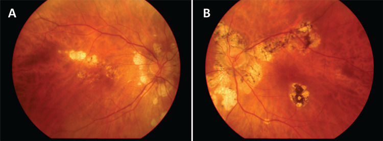
Color fundus photographs of patient 1 showing the typical peripapillary scarring with satellite lesions in the right eye (A) and left eye (B)
Figure 2.
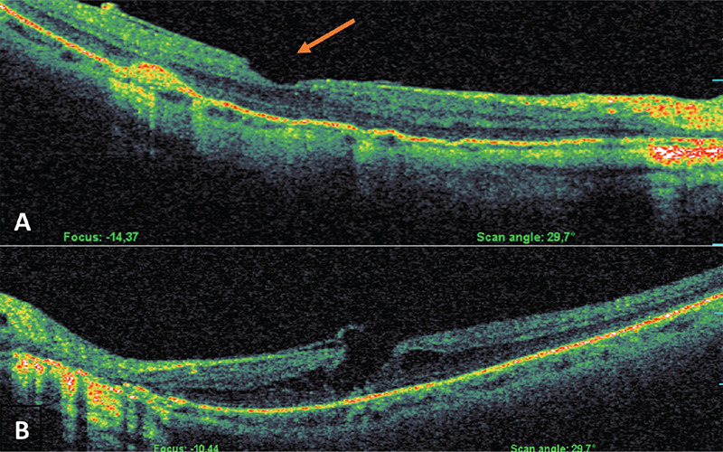
Optical coherence tomography images of patient 1 showing macular pseudohole in the right eye (A, red arrow) and stage 2 macular hole in the left eye (B)
Figure 3.
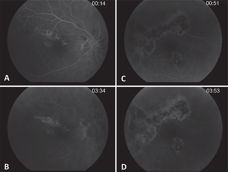
Fluorescein angiography images of patient 1 showing early hypofluorescence and late hyperfluorescence of the peripapillary and satellite lesions in the right eye (A, B) and at the lesion margins in the left eye (C, D)
Figure 4.
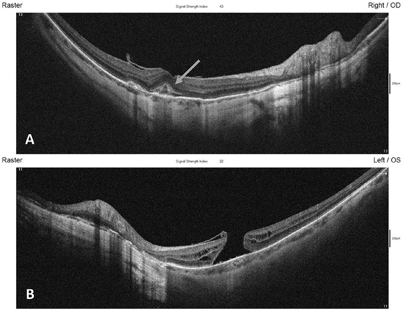
Optical coherence tomography images of patient 1 showing a subfoveal hyperreflective lesion and intraretinal fluid in the right eye (A, arrow) and a stage 4 macular hole in the left eye (B)
Figure 5.
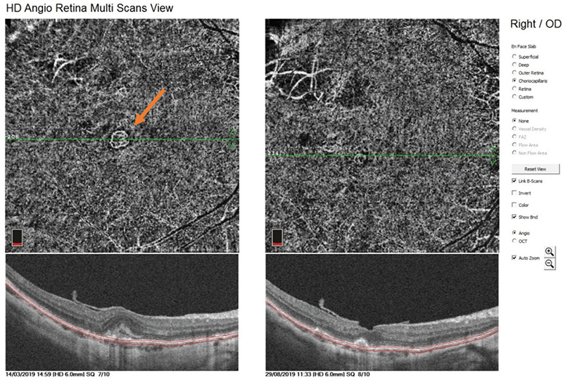
Optical coherence tomography angiography image of the right eye of patient 1 showing a fine anastomotic network of vessels indicative of type 2 choroidal neovascular membrane (left, red arrow) and the same eye after three intravitreal injections of ranibizumab (right)
Case 2
A 55-year-old woman first presented to our outpatient service in March 2010 due to blurred vision in OS. Her systemic and ophthalmic history was uneventful. On presentation, BCVA was 20/20 in OD and counting fingers with positive Amsler test in OS. Dilated fundus examination revealed retinal edema and hemorrhage within the macular region in OS, while OD was normal (Figure 6). OCT detected the presence of subretinal fluid (SRF) under the fovea in OS (Figure 7). Subsequently, FA was performed and a neovascular membrane into the macular area was detected (Figure 8). Since there were no signs of any underlying disease at that time, the CNV was characterized as idiopathic and the patient was treated with 5 injections of intravitreal ranibizumab during the time period of 10 months. Her BCVA improved to 20/200 in OS, while the OCT showed complete regression of the SRF (Figure 9). In July 2012, her follow-up examination revealed the presence of peripapillary chorioretinal atrophy in OD (Figure 10A) and chorioretinal atrophy extending from the optic disc to the macula and the periphery in OS (Figure 10B), therefore the patient was diagnosed with SC. She underwent a full systemic check-up, including Quantiferon Tb Gold test, which resulted negative, and a regular follow-up program was suggested. Oral methotrexate (10 mg per week) was attempted; however, it was discontinued due to elevation of liver enzymes. As an alternative, short courses of oral methylprednisolone (Medrol, Pfizer, Greece) 16 mg per day was administered. Methylprednisolone contributed to the improvement of the SC-related retinal clinical picture of her OS which remained steady for a follow-up period of 6 years.
Figure 6.
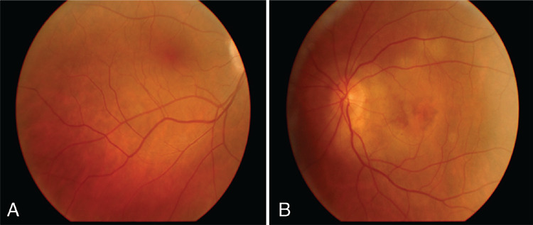
Color fundus photographs of patient 2. The right eye looks normal (A), while the left eye presents retinal edema and hemorrhage within the macular area (B)
Figure 7.

Optical coherence tomography of the left eye of patient 2 showing subretinal fluid under the fovea
Figure 8.
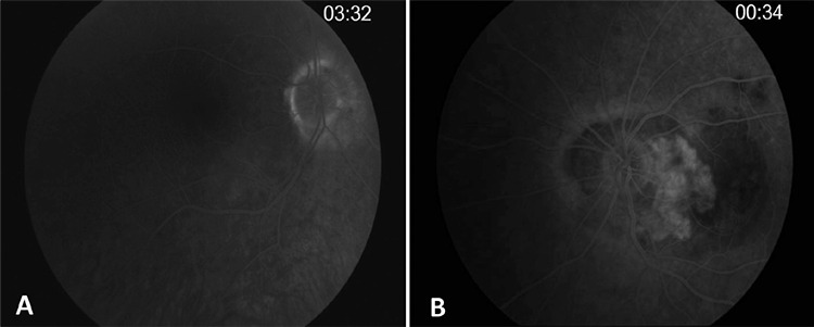
Fluorescein angiography in patient 2. No abnormal findings were observed in the right eye (A), while in the left eye a classic choroidal neovascular membrane with leakage into the macular area was detected (B)
Figure 9.

Optical coherence tomography of the left eye of patient 2 after five intravitreal injections of ranibizumab. Complete regression of the subretinal fluid was observed
Figure 10.
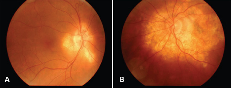
Color fundus photographs of patient 2 two years after initial presentation showing the peripapillary chorioretinal atrophy typical of SC in the right eye (A) and left eye (B)
In June 2018, the patient visited our outpatient service again due to blurred vision in OD. Dilated fundoscopy and OCT imaging indicated no change in OS, however, BCVA in OD dropped to 20/50 with IRF and SRF (Figure 11). Due to deterioration of the clinical picture in OD, a course of oral methylprednisolone (32 mg per day) was administered, but there was no favorable response in BCVA or the clinical picture. Further increase in methylprednisolone was ruled out due to the patient’s development of Cushing-like systemic symptomatology. OCTA demonstrated a fine anastomotic network of vessels, suggestive of type 2 CNV secondary to SC. We concluded that the deterioration of the clinical picture was due to the CNV that was misdiagnosed as recurrence of the disease. Subsequently, the patient received three intravitreal injections of ranibizumab. Her BCVA stabilized at 20/25 with complete regression of both macular edema and CNV (Figure12).
Figure 11.
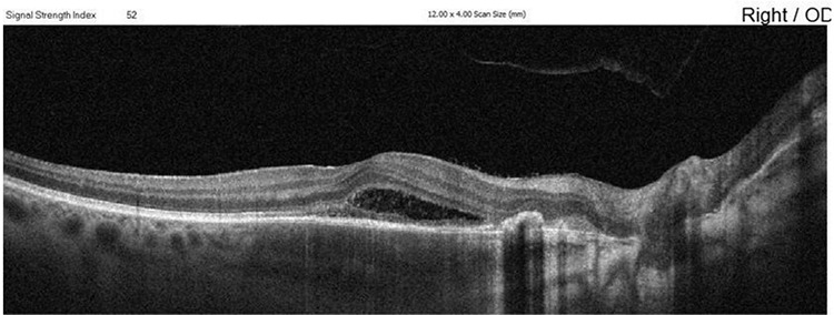
Optical coherence tomography of the right eye of patient 2 eight years after initial presentation shows the presence of subretinal and intraretinal fluid
Figure 12.
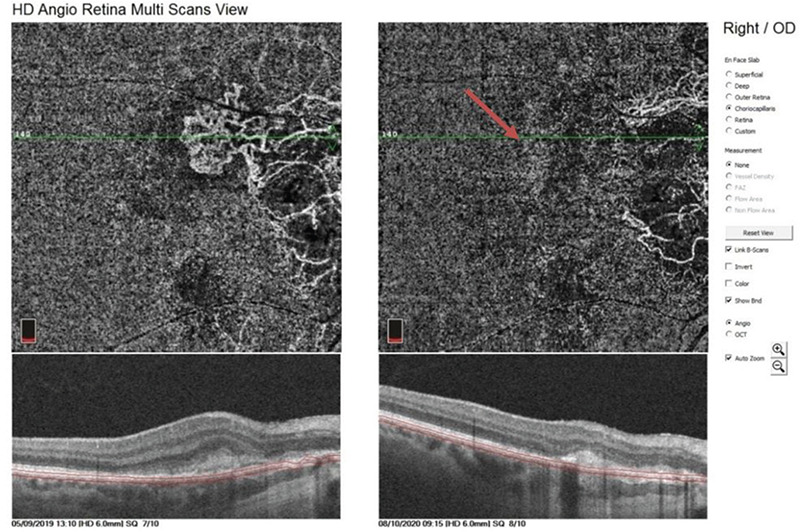
Optical coherence tomography angiography of the right eye of patient 2 before (left) and after (right) three intravitreal injections of ranibizumab. Thirteen months later, complete regression of choroidal neovascularization (red arrow) was observed
Discussion
SC, first described by Hutchinson3 in 1900, is a rare, vision-threatening disease with a prevalence of between 0.2% and 5% of all uveitis cases. It affects individuals from 30 to 70 years old and presents with painless, blurred vision, metamorphopsias, and paracentral scotomas.4,11 Initially it is unilateral, with the majority of cases demonstrating fellow eye involvement within 5 years. Confirmed unilateral cases are most commonly reported from tuberculosis-endemic countries.11 Dilated fundus examination reveals asymmetric bilateral grayish-yellow lesions and mild inflammation of the vitreous body.4 Recurrences, usually symptomatic, occur at the edges of the scars.11 Interestingly, the interval between recurrences varies from weeks to years.4 When no macular scarring exists, significant improvement of visual acuity is expected, similar to patient 1 and OD of patient 2 in our report. When CNV progresses to macular scarring, the visual outcome is less fortunate (patient 2, OS). The literature suggests that inflammation is the primary cause of CNV following age-related macular degeneration and myopia. Both infectious and non-infectious pathomechanisms can lead to the development of CNV. Although the overall incidence of CNV in non-infectious uveitides is only 2%, its prevalence is significantly higher in multifocal choroiditis, punctate inner choroiditis, and SC, which present with choroidal neovascularization in 32-46%, 17-40%, and 10-25% of all cases, respectively.12,13 Especially in SC, CNV constitutes the most common complication. It typically originates from the border of the choroidal lesions inducing ischemia in the outer retina and inner choroidal layers.14 Taking into account that SC affects patients at their productive age, prompt diagnosis and treatment of SC-related CNV is of paramount importance to preserve visual capacity.7
It is known that among the prevalent theories regarding the pathogenesis of CNV is inflammatory-induced angiogenesis mediated by vascular endothelial growth factor (VEGF).6,12,15 It is no surprise then, that the anti-VEGF medications exert a beneficial impact on CNV. Furthermore, damage to the RPE and Bruch’s membrane complex caused by chronic inflammation enables these new capillaries to pass from the choroid to the sub-RPE and subretinal space.7,12,13 It should be mentioned that in patient 2, CNV in OD manifested prior to the characteristic clinical lesions of SC. To our knowledge, this is the first report of this peculiar incidence.
Contemporary imaging technologies contribute significantly to the diagnosis of SC and its related manifestations. OCTA is a non-invasive imaging modality that facilitates the diagnosis and management of retinal and choroidal diseases.3,18 Recently, El Ameen and Herbort9 reported a case of SC diagnosed by OCTA and concluded that OCTA could even substitute for indocyanine green angiography.
OCTA seems to be useful in the diagnosis of SC-related complications as well, since its associated lesions present diagnostic difficulties with FA.7 OCTA has excellent reproducibility, is non-invasive, and is easily performed in few minutes.14 Although the use of OCTA in the diagnosis of type 1 CNV in age-related macular degeneration has been widely studied, published reports referring to the detection of the same complication in uveitis are limited.7
Campos Polo et al.6 used OCTA for the detection of CNV and its response to anti-VEGF injections in SC. Aggarwal et al.10 reported that OCTA is a helpful diagnostic tool for the detection of type 1 CNV in patients with tuberculous serpiginious-like choroiditis TB-SLC when conventional multimodal imaging methods fail to assist in the diagnosis. Astroz et al.16 indicated the diagnostic superiority of OCTA over FA in the detection of inflammatory CNV. Furthermore, Karti and Saatci13 reported that OCTA is useful not only in the differentiation of CNV from active inflammatory lesions, but it is also able to picture the characteristic lesions of white dot syndromes.
Until recently, the treatment options for SC-related CNV were photocoagulation, photodynamic therapy, and surgical excision.15 However, today intravitreal anti-VEGF injections are considered to be the first line of treatment.7 In our reported cases, we used ranibizumab, which contributed to BCVA improvement and regression of the inflammatory CNV. Our outcomes suggest that this therapeutic intervention is promising. Further cohorts of patients with SC are needed in order to draw safer conclusions in this direction.
Footnotes
Ethics
Informed Consent: Obtained.
Peer-review: Externally and internally peer reviewed.
Authorship Contributions
Surgical and Medical Practices: D.D., Concept: A.P., S.T., M.G., Design: A.P., S.T., M.G., G.L., Data Collection or Processing: A.P., D.K., E-K.P., Analysis or Interpretation: D.D., I.P., G.L., Literature Search: A.P., E-K.P., Writing: A.P., D.K., G.L.
Conflict of Interest: No conflict of interest was declared by the authors.
Financial Disclosure: The authors declared that this study received no financial support.
References
- 1.Borrego-Sanz L, Gómez-Gómez A, Gurrea-Almela M, Esteban-Ortega M, Pato E, Díaz-Valle D, Díaz-Valle T, Muñoz-Fernández S, Rodriguez-Rodriguez L; Madrid Uveitis Study Group. Visual acuity loss and development of ocular complications in white dot syndromes: a longitudinal analysis of 3 centers. Graefes Arch Clin Exp Ophthalmol. 2019;257:2505–2516. doi: 10.1007/s00417-019-04429-5. [DOI] [PubMed] [Google Scholar]
- 2.Neri P, Lettieri M, Fortuna C, Manoni M, Giovannini A. Inflammatory choroidal neovascularization. Middle East Afr J Ophthalmol. 2009;16:245–251. doi: 10.4103/0974-9233.58422. [DOI] [PMC free article] [PubMed] [Google Scholar]
- 3.Majumder PD, Biswas J, Gupta A. Enigma of serpiginous choroiditis. Indian J Ophthalmol. 2019;67:325–333. doi: 10.4103/ijo.IJO_822_18. [DOI] [PMC free article] [PubMed] [Google Scholar]
- 4.Parodia MB, Iaconob P, Verbraakc FD, Bandelloa F. Antivascular Endothelial Growth Factors for Inflammatory Chorioretinal Disorders. Ophthalmol. Basel, Karger. 2010:84–95. doi: 10.1159/000320011. [DOI] [PubMed] [Google Scholar]
- 5.Pakzad-Vaezi K, Khaksari K, Chu Z, Van Gelder RN, Wang RK, Pepple KL. Swept-Source OCT Angiography of Serpiginous Choroiditis. Ophthalmol Retina. 2018;2:712–719. doi: 10.1016/j.oret.2017.11.001. [DOI] [PMC free article] [PubMed] [Google Scholar]
- 6.Campos Polo R, Rubio Sánchez C, Sánchez Trancón A. Choroidal neovascularisation secondary to serpiginous choroiditis: The value of OCT-angiography in diagnosis and response to therapy with aflibercept. Arch Soc Esp Oftalmol. 2019;94:460–464. doi: 10.1016/j.oftal.2018.12.013. [DOI] [PubMed] [Google Scholar]
- 7.Agarwal A, Invernizzi A, Singh RB, Foulsham W, Aggarwal K, Handa S, Agrawal R, Pavesio C, Gupta V. An update on inflammatory choroidal neovascularization: epidemiology, multimodal imaging, and management. J Ophthalmic Inflamm Infect. 2018;8:13. doi: 10.1186/s12348-018-0155-6. [DOI] [PMC free article] [PubMed] [Google Scholar]
- 8.Montorio D, Giuffrè C, Miserocchi E, Modorati G, Sacconi R, Mercuri S, Querques L, Querques G, Bandello F. Swept-source optical coherence tomography angiography in serpiginous choroiditis. Br J Ophthalmol. 2018;102:991–995. doi: 10.1136/bjophthalmol-2017-310989. [DOI] [PubMed] [Google Scholar]
- 9.El Ameen A, Herbort CP Jr. Serpiginous choroiditis imaged by optical coherence tomography angiography. Retin Cases Brief Rep. 2018;12:279–285. doi: 10.1097/ICB.0000000000000512. [DOI] [PubMed] [Google Scholar]
- 10.Aggarwal K, Agarwal A, Sharma A, Sharma K, Gupta V; OCTA Study Group. Detection of type 1 choroidal neovascular membranes using optical coherence tomography angiography in tubercular posterior uveitis. Retina. 2019;39:1595–1606. doi: 10.1097/IAE.0000000000002176. [DOI] [PubMed] [Google Scholar]
- 11.Nazari Khanamiri H, Rao NA. Serpiginous choroiditis and infectious multifocal serpiginoid choroiditis. Surv Ophthalmol. 2013;58:203–232. doi: 10.1016/j.survophthal.2012.08.008. [DOI] [PMC free article] [PubMed] [Google Scholar]
- 12.Karti O, Ipek SC, Ates Y, Saatci AO. Inflammatory Choroidal Neovascular Membranes in Patients With Noninfectious Uveitis: The Place of Intravitreal Anti-VEGF Therapy. Med Hypothesis Discov Innov Ophthalmol. 2020;9:118–126. [PMC free article] [PubMed] [Google Scholar]
- 13.Karti O, Saatci AO. Optical Coherence Tomography Angiography in Eyes with Non-infectious Posterior Uveitis; Some Practical Aspects. Med Hypothesis Discov Innov Ophthalmol. 2019;8:312–322. [PMC free article] [PubMed] [Google Scholar]
- 14.Parodi MB, Iacono P, La Spina C, Knutsson KA, Mansour A, Arevalo JF, Bandello F. Intravitreal bevacizumab for choroidal neovascularisation in serpiginous choroiditis. Br J Ophthalmol. 2014;98:519–522. doi: 10.1136/bjophthalmol-2013-304237. [DOI] [PubMed] [Google Scholar]
- 15.Saatci AO, Ayhan Z, Engin Durmaz C, Takes O. Simultaneous Single Dexamethasone Implant and Ranibizumab Injection in a Case with Active Serpiginous Choroiditis and Choroidal Neovascular Membrane. Case Rep Ophthalmol. 2015;6:408–414. doi: 10.1159/000442346. [DOI] [PMC free article] [PubMed] [Google Scholar]
- 16.Astroz P, Miere A, Mrejen S, Sekfali R, Souied EH, Jung C, Nghiem-Buffet S, Cohen SY. Optical Coherence Tomography Angiography to Distinguish Choroidal Neovascularization from Macular Inflammatory Lesions in Multifocal Choroiditis. Retina. 2018;38:299–309. doi: 10.1097/IAE.0000000000001617. [DOI] [PubMed] [Google Scholar]


