Abstract
The Kinematic Alignment (KA) technique for total knee arthroplasty (TKA) is an alternative surgical technique aiming to resurface knee articular surfaces.
The restricted KA (rKA) technique for TKA applies boundaries to the KA technique in order to avoid reproducing extreme constitutional limb/knee anatomies.
The vast majority of TKA cases are straightforward and can be performed with KA in a standard (unrestricted) fashion.
There are some specific situations where performing KA TKA may be more challenging (complex KA TKA cases) and surgical technique adaptations should be included.
To secure good clinical outcomes, complex KA TKA cases must be preoperatively recognized, and planned accordingly.
The proposed classification system describes six specific issues that must be considered when aiming for a KA TKA implantation.
Specific recommendations for each situation type should improve the reliability of the prosthetic implantation to the benefit of the patient.
The proposed classification system could contribute to the adoption of a common language within our orthopaedic community that would ease inter-surgeon communication and could benefit the teaching of the KA technique. This proposed classification system is not exhaustive and will certainly be improved over time.
Cite this article: EFORT Open Rev 2021;6:881-891. DOI: 10.1302/2058-5241.6.210042
Keywords: complex cases, Kinematic Alignment, total knee replacement
Introduction
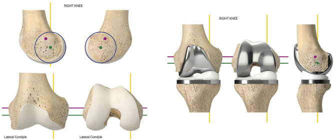
The Kinematic Alignment (KA) technique for total knee arthroplasty (TKA) is an alternative surgical technique aiming to resurface knee articular surfaces.1,2 Ultimately, the goal of KA is to alter the knee physiological biomechanics as little as possible by restoring native (pre-arthritic) knee joint line alignment and ligaments laxities (Fig. 1, Supplementary Video (121.5MB, mp4) ).1,2 Setting up the orientation and height of the KA bone cuts is done by referencing the articular surfaces, by compensating for cartilage and/or bone loss, and by considering the thickness of the implants. By doing so, components are aligned on knee kinematic axes. This allows for native articular surface height and orientation to be restored to its pre-disease position (Fig. 1, Supplementary Video (121.5MB, mp4) ).1,2
Fig. 1.
The kinematic alignment technique for total knee arthroplasty (TKA) has the aim of restoring the pre-arthritic tri-dimensional knee anatomy and to align components on the knee’s kinematic axes. (Reproduced from Fig 16.2, Chap 16, Riviere C, Vendittoli P-A eds. Personalized Hip and Knee Joint Replacement. Cham, Springer, 2020. Used with permission)
Since its first development by Stephen Howell in 2007, the KA technique has generated much debate in the orthopaedic community. One particular debate is its applicability to the spectrum of arthritic phenotypes that a surgeon commonly treats.1,3,4 Other debates are represented by situations where the disease process leading to a TKA indication has created soft tissue stretching/contracture, and patients with acquired extra-articular deformity affecting native knee kinematics and loads. It leads us to question whether there are indications where performing KA TKA is more challenging. Complex cases for performing KA TKA have to be preoperatively recognized, and reproducible adequate implantation requires appropriate planning. In addition, there are rare cases where KA TKA should not be recommended at all. This instructional review aims to describe, through a classification system, the most frequent situations where KA TKA is challenging, and to provide planning recommendations for each of them.
Classification system
Performing KA TKA may be challenging when the surgeon faces at least one of the six situations that are illustrated in Table 1. There may be a combination of these situations, which further complicates the KA TKA implantation.
Table 1.
PAS (Personalized Arthroplasty Society) classification of most commonly occurring challenging situations that a surgeon may face when aiming for ‘physiological’ TKA implantation
| PAS classification of most commonly occurring challenging situations that a surgeon may face when aiming for ‘physiological’ TKA implantation | |||||||
|---|---|---|---|---|---|---|---|
| Knee types | 1 | 2 | 3 | 4 | 5 | 6 | Exceptional cases where KA/rKA is not recommended |
| Description of the osteoarthritic knee/limb | Severe constitutional varus limb | Severe constitutional valgus limb | Extreme constitutional joint line orientation (severe frontal joint line obliquity, high tibial posterior slope) | Patella maltracking | Difficulty in estimating native knee anatomy (mainly articular bone loss) | Acquired lower limb malalignment from previous fracture malunion, osteotomy or metabolic bone disease for example | Cases with severe soft tissue modifications - global instability (recurvatum) - severe contractures (knee arthrodesis) Cases with important anatomy destruction preventing a KA reconstruction Cases requiring diaphyseal implant fixation |
| Treatment recommendations | option 1: pure (unrestricted) KA-TKA option 2: restricted KA-TKA option 3: KA-TKA + realignment osteotomy |
Regarding high JLO:
option 1: pure KA-TKA option 2: restricted KA-TKA Regarding high tibial slope: - slightly reduce the large native tibial slope when using postero-stabilized TKA designs. The increased flexion gap resulting from the resection of the posterior cruciate ligament would prevent tightness in flexion - restore native slope when using cruciate-retaining TKA design |
Patella resurfacing Lateral facetectomy Lateral arthrotomy with Z-plasty of retinaculm +/-Restricted KA-TKA to reduce severe constitutional valgus deformity, if any. +/- Tibial tuberosity osteotomy +/- MPFL reconstruction |
Surgeon should carefully estimate the quantity and location of bone loss by planning TKA on the contralateral knee, and by intraoperatively assessing femorotibial laxities. The objective is to perform either unrestricted or restricted KA-TKA, depending on whether there is an extreme constitutional limb/knee anatomy or not. |
Options 1: KA-TKA 2: KA-TKA + realignment osteotomy 3: rKA-TKA |
||
Note. TKA, total knee arthroplasty; KA, Kinematic Alignment; rKA, restricted Kinematic Alignment; JLO, joint line obliquity; MPFL, medial patella-femoral ligament.
Type 1
Type 1 defines a lower limb with important constitutional varus alignment (Types 3 and 4 of the Lin et al5 classification of native lower limb alignment). From the authors’ experience, alteration of the physiological ligament length (contracture and stretching of medial and lateral collateral ligaments, respectively) in this type of patient is generally negligible. Some surgeons believe that restoring such patients’ extreme anatomy with KA (unrestricted) could carry the risk of generating potentially suboptimal prosthetic kinetics6 with subsequent complications (accelerated polyethylene wear and/or implant loosening). On the other hand, such fear remains without scientific evidence. By waiting for more evidence regarding the acceptable limb alignment boundaries when performing KA TKA, it is reasonable and understandable that some surgeons aim to maintain limb and knee alignment within acceptable limits by performing restricted KA (rKA) TKA (Fig. 2).7–9 The aim of rKA is to limit the patient’s anatomy restoration to within certain boundaries for cases with extreme anatomies, potentially unfavourable with current implant bearing and fixation methods.7–9 This reduces the extent of soft tissue releases compared to more traditional mechanical alignment surgery.10,11 In a study assessing 4884 lower limb computerized tomography (CT) scans of patients scheduled for TKA (performed with patient-specific instrumentation - PSI), Almaawi et al8 found that around 9% of patients had over 5 degrees of constitutional varus lower limb deformity. This may be an overestimation, given that articular bone loss (often present in severe long-standing knee osteoarthritis) was not accounted for when measuring knee/limb alignment, in addition to the likely presence of selection bias (many surgeons opt for a PSI solution only for complex cases).
Fig. 2.
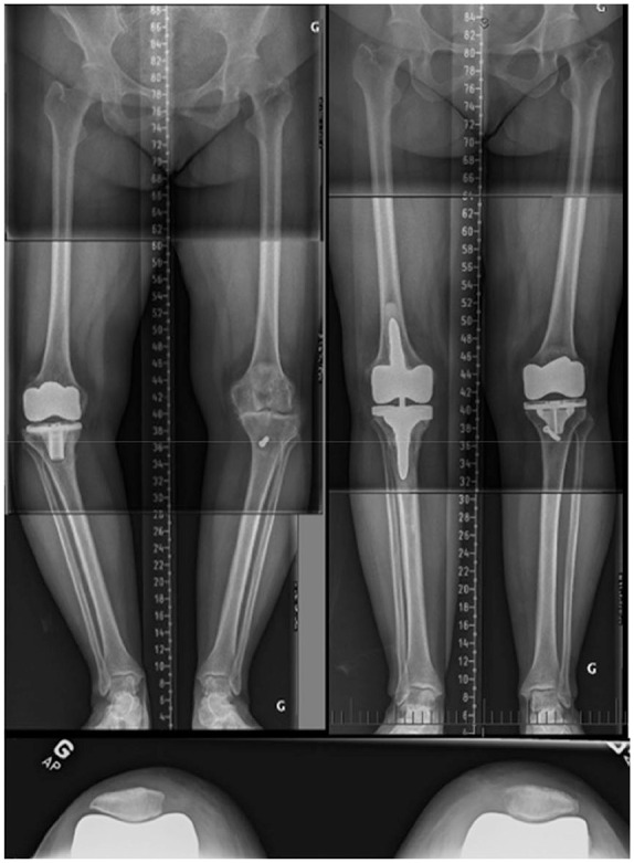
This composite figure illustrates a 65-year-old female patient with constitutional ‘Type 1’ left knee. The patient undertook a right MA TKA that failed after four years. On the left knee, the native lateral distal femoral angle (LDFA) was 2 degrees valgus and the medial proximal tibial angle (MPTA) 9 degrees varus. In the same setup, a left computational assisted rKA TKA and a right TKA revision were performed. rKA boundaries were applied when implanting the left knee: the tibial varus was reduced to 5 degrees with a resulting HKA of 3 degrees varus. Deep MCL had to be released to obtain medio-lateral compartment balance.
Note. MA, mechanical alignment; TKA, total knee arthroplasty; rKA, restricted kinematic alignment; HKA, hip-knee-ankle; MCL, medial collateral ligament.
Type 2
Type 2 defines a lower limb with important constitutional valgus deformity (Type 5 of the Lin et al5 classification of native lower limb alignment). Similarly to Type 1, some surgeons believe that restoring the patient’s extreme anatomy with KA (unrestricted) could carry the risk of generating potentially suboptimal prosthetic kinetics, suboptimal patella tracking and early implant failure. Almaawi et al8 found that around 10% of patients had over 5 degrees of constitutional valgus lower limb deformity (likely overestimated proportion). By waiting for more evidence regarding the acceptable limb alignment boundaries, a reasonable option is to reduce the valgus deformity by performing rKA TKA with limited soft tissue release (Fig. 3)7,8 or by combining KA TKA with an extra-articular realignment osteotomy.
Fig. 3.
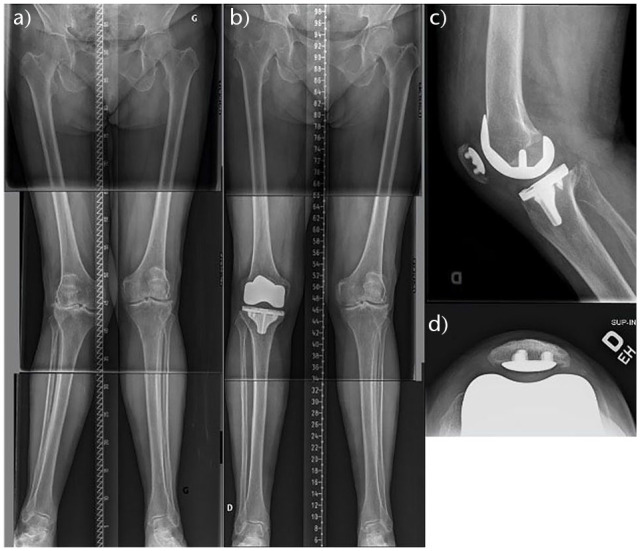
This composite figure illustrates an 82-year-old female patient with a constitutional ‘Type 2’ knee who was treated with a rKA TKA. Her right LDFA was measured at 9 degrees of valgus and tibial MPTA at 2 degrees of varus. Respecting rKA boundaries, the LDFA was reduced to 5 degrees. Reproducing the tibial MPTA and the neutral femoral rotation, the surgeon obtained a final HKA of 3 degrees valgus and a congruent patella-femoral joint. To obtain medio-lateral soft tissue balance, postero-lateral capsule pie crusting was performed.
Notes. rKA, restricted Kinematic Alignment; TKA, total knee arthroplasty; LDFA, lateral distal femoral angle; MPTA, medial proximal tibial angle; HKA, hip-knee-ankle.
The chronic degenerative process of severe valgus cases may have altered the native ligament laxities. The medial collateral ligament is often functional but slightly stretched (by a few millimetres). To maintain medial compartment stability and avoid using thicker polyethylene, we suggest performing a conservative tibial resection on the medial plateau (reducing medial tibial plateau cut by one or a few millimetres compared to the tibial implant thickness). We do not recommend to under-resect the distal medial femoral condyle as this would increase tension on the medial retinaculum when flexing the knee.
These severe valgus cases may be associated with severe patella-femoral joint degeneration from poor patella tracking (lateral tilt and subluxation, lateral retinaculum contracture), with even, sometimes, notable history of patella instability. Reproducing the native knee anatomy with KA TKA should optimize patella-femoral joint kinematics but may not be sufficient to generate adequate patella tracking. Howell et al12 observed three (1.5%) re-operations out of 198 KA TKAs followed-up for four years; the indications for re-operation were anterior knee pain or patellofemoral instability, and these occurred in patients with the more valgus phenotypes. Please refer to the ‘Type 4’ section of this article to learn about the surgical options to improve patella tracking in the setting of a physiological TKA implantation.
Type 3
Type 3 defines a lower limb with severe constitutional joint line orientation either in frontal (severe joint line obliquity –Type 2 of the Lin et al5 classification of native lower limb alignment) or sagittal (high posterior tibial slope that was shown to be up to 15 degrees13) planes.
Regarding patients with severe joint line obliquity, performing unrestricted KA TKA involves positioning the tibial component in substantial varus, which is feared to carry risk of generating suboptimal prosthetic shear stresses6 leading to early tibial implant loosening. As far as we are aware, no published study has ever assessed the isolated influence of joint line obliquity on implant lifespan. Nevertheless, Howell et al14 reported excellent 10-year clinical outcomes (efficacy and safety) out of 213 consecutive unselected unrestricted KA TKA cases, which suggest that the severity of the pre-arthritic standing limb and knee alignment and joint line obliquity has negligible effect on KATKA performances. By waiting for more evidence regarding the acceptable joint line obliquity boundaries when performing a KA TKA, surgeons may wish to exercise caution and slightly reduce its obliquity by performing rKA TKA (Fig. 4).7–9
Fig. 4.
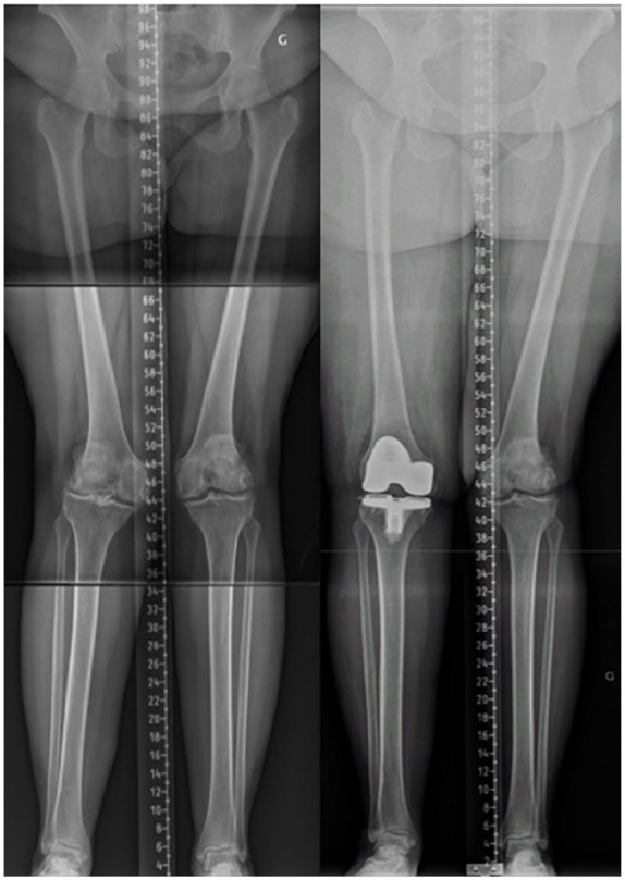
This figure illustrates long-leg radiographs of a patient with constitutional bilateral valgus limbs. Regarding the right knee, LDFA is 11° valgus and MPTA 6° varus. Resultant HKA is not extreme, but the joint line orientation has a pronounced obliquity (‘Type 3’ knee). In this case, the surgeon felt uncomfortable to reproduce the patient’s anatomy and reduced the joint line obliquity.
Note. LDFA, lateral distal femoral angle; MPTA, medial proximal tibial angle; HKA, hip-knee-ankle.
Regarding tibial slope, reproducing extreme posterior slope with KA is feared to carry increased risk of shear stress on the bone–implant fixation interface and on the polyethylene, with potential impairment of TKA lifespan. Nevertheless, there is no scientific evidence supporting those fears. In addition, current posterior-stabilized TKA implants may not be designed to accommodate such high posterior slope and anterior impingement may occur (post against notch). Until we have more evidence regarding the acceptable limit for the tibial slope when performing a KA implantation and more forgiving implants, surgeons should be aware of the mechanical limits of its implant and reduce accordingly the patient’s native tibial slope when exceeding that limit. The increased flexion gap resulting from the resection of the posterior cruciate ligament15 would prevent tightness in flexion. In contrast, it is likely that the surgeon may safely restore the native slope when using a cruciate-retaining TKA design.
Type 4
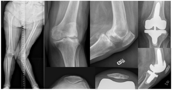
Type 4 defines knees that preoperatively present with laterally tilted, fixed (lateral retinaculum contracture), and severely degenerated patella (Fig. 5). Patients may sometimes have notable history of patella instability. Reproducing the native knee anatomy with pure KA TKA, although often of benefit for optimizing patella femoral joint kinematics (by replicating the normal valgus angle of the femur it prevents the lateral retinacular structures from becoming over tensioned in flexion, as is the case with a mechanical alignment technique – Fig. 6), may in itself not be enough for extreme cases. Anatomic resurfacing may recreate the patient’s native poor patella-femoral joint biomechanics, and lead to disappointing outcomes caused by patella instability, patella implant accelerated wear and/or early loosening. In addition, performing a medial parapatellar approach associated with extended lateral retinaculum release could jeopardize the blood supply of the patella, leading to avascular necrosis and collapse. For optimal KA implantation for Type 4 the surgeon may need to think about additional measures:
Fig. 5.

This figure illustrates a patient with a ‘Type 4’ knee: poor patella tracking (tilted and laterally subluxed patella) causing severe, bone-on-bone, lateral compartment osteoarthritis.
Fig. 6.
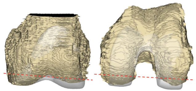
This composite figure illustrates the simulation on a tri-dimensional knee model, in the frontal (left image) and axial (right image) knee planes, of a mechanically aligned femoral component using the measured resection technique. This alignment technique generates a distal (left image) and a posterior (right image) lateral condylar prosthetic overstuffing. The red dotted lines represent the frontal orientation of the distal femoral cut (left image) and the axial orientation of the posterior femoral cut (right image). The prosthetic overstuffing of native lateral condyle articular surfaces would likely cause a lateral retinaculum stretching when flexing the knee. (Reproduced from Fig. 9, Rivière C, Iranpour F, Auvinet E, et al. Mechanical alignment technique for TKA: Are there intrinsic technicallimitations? Orthopaedics & Traumatology: Surgery & Research. 2017;103:1057–1067, with kind permission).
1) Resurfacing the patella with an additional lateral facetectomy of the patella is often sufficient to allow it to track centrally and de-tension the lateral retinacular tissues.
2) For more extreme cases the surgeon may prefer to perform a lateral arthrotomy (with Z-plasty of the retinaculum) in order to release its tension. If the patient presents with severe constitutional valgus limb deformity, a rKA TKA lowering the deformity may be considered.
3) As with standard mechanical alignment (MA) techniques if poor patella tracking still remains it needs addressing, tibial tuberosity osteotomy and/or reconstruction of the medial patellofemoral ligament could present as an option.
Type 5
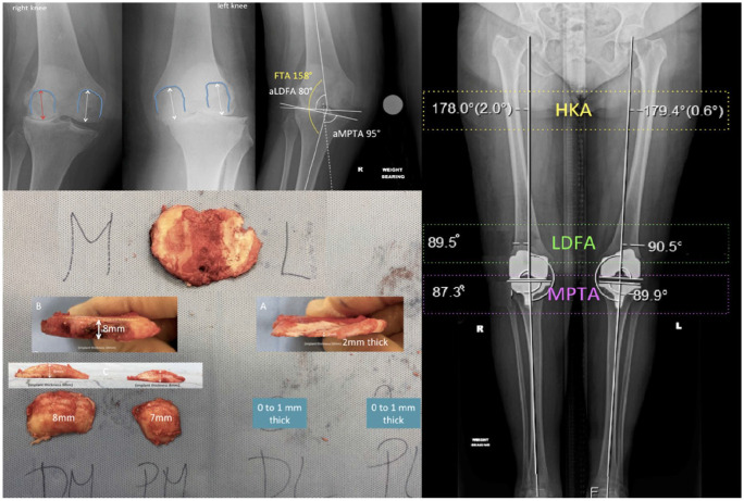
Type 5 defines cases where the pre-arthritic knee anatomy is difficult to estimate because of substantial articular bone loss for valgus (Fig. 7 and Fig. 8) and varus knee (Fig. 9 and Fig. 10).16 Situations like this may be found in cases of severe long-lasting osteoarthritis, collapsed avascular necrotic bone, or sequels of depression-type tibial plateau fracture. The limb deformity generated by those conditions is mainly correctable. Malunion of split-type tibial plateau fractures should not be considered here, as the generated limb deformity is often not correctable.
Fig. 7.
This composite figure illustrates a patient with a ‘Type 5’ right knee. The patient initially suffered bilateral knee osteoarthritis with windswept deformity. The left varus deformed knee was successfully implanted with a mechanically aligned TKA one year prior to the right KA TKA. Regarding the right valgus deformed knee, there was substantial bipolar (femoral and tibial) lateral compartment bone loss and a slight (1–2 mm) MCL stretching (top left images). The patient was treated with a KA TKA with the KA tibial cut being slightly adjusted (millimetric under resection of the medial tibia plateau) to adapt the MCL stretching. Very little bone was resected laterally (bottom left image).
Note. KA, kinematic alignment; TKA, total knee arthroplasty; MCL, medial collateral ligament; FTA, femoro-tibial angle; aLDFA, anatomical lateral distal femoral angle; aMPTA, anatomical medial proximal tibial angle; HKA, hip-knee-ankle; LDFA, lateral distal femoral angle; MPTA, medial proximal tibial angle. (Reproduced from Rivière C, Webb J, Vendittoli PA. Kinematic Alignment Technique for TKA on Degenerative Knees with Severe Bone Loss: A Report of 3 Cases. The Open Orthopaedics Journal, 2021;15:27-34, with permission)
Fig. 8.
This composite figure illustrates a 79-year-old woman with a severe ‘Type 5’ valgus left knee. The patient developed a severe deformity secondary to avascular necrosis of her lateral femoral condyle. There was substantial bipolar (femoral and tibial) lateral compartment bone loss and a stretched MCL. The right knee anatomy was used to plan the left knee TKA. Using computer navigation, the left femoral implant was aligned with 5° of valgus and the tibial component with 2° of varus. A 10 mm lateral femoral augment was used to fill the bone defect and a short-cemented stem for supplementary fixation. A postero-stabilized semi-constrained insert was used to compensate for the stretched MCL.
Note. MCL, medial collateral ligament; TKA, total knee arthroplasty.
Fig. 9.
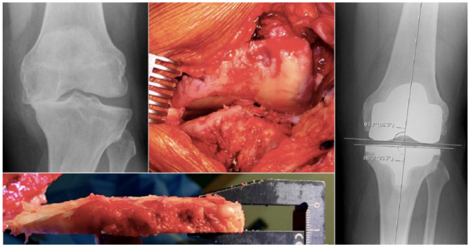
This composite figure illustrates a patient with a ‘Type 5’ varus knee who was treated with ‘calipered unrestricted KA TKA’. There was some medial tibial plateau bone loss, as suggested by knee radiograph (top left image) and intraoperative observation of a large medial gap when stressing the knee in valgus (top middle image). The tibial cut was thin medially in order to account for the medial plateau bone loss (bottom left image). (Reproduced from Fig 16.7, Chap 16, Riviere C, Vendittoli P-A eds. Personalized Hip and Knee Joint Replacement. Cham, Springer, 2020. Used with permission)
Note. KA, Kinematic Alignment; TKA, total knee arthroplasty.
Fig. 10.
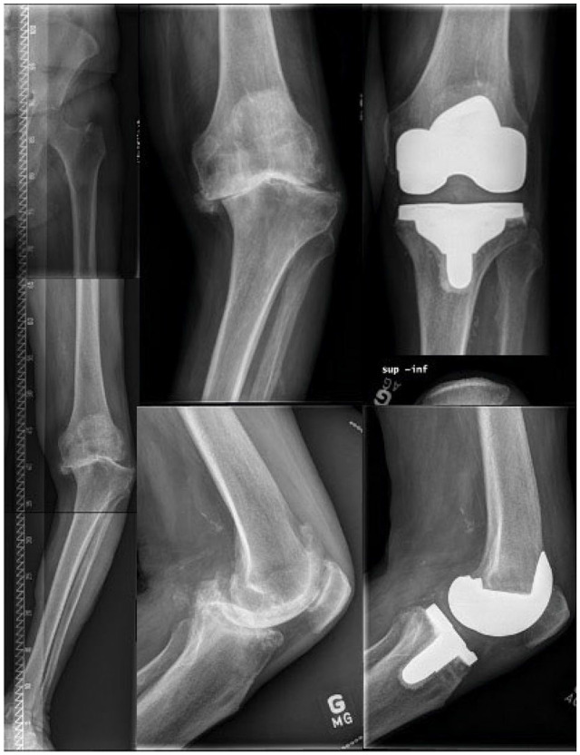
This composite figure illustrates a 71-year-old man with a severe ‘Type 5’ varus left knee. Preoperative HKA was measured at 25°. Important bone loss on the medial compartment (tibial and femoral sides) was present. Using computer navigation, the distal medial femoral condyle cut was performed by compensating for 5 mm thickness articular surface loss (2 mm of cartilage plus 3 mm of bone), and the medial tibial cut was performed by compensating for 8 mm surface loss (2 mm of cartilage plus 6 mm of bone loss). Using spacer blocks and trial implants, we confirmed adequate soft tissue balance. Increased LCL laxity was compensated for by deep MCL release and +2 mm polyethylene. Final LDFA was 2° valgus and MPTA 5° varus. As the PCL was altered, a PS insert was used.
Note. HKA, hip-knee-ankle; LCL, lateral collateral ligament; MCL, medial collateral ligament; LDFA, lateral distal femoral angle; MPTA, medial proximal tibial angle; PCL, posterior cruciate ligament; PS, postero-stabilsed.
In order to restore the pre-arthritic knee anatomy, the surgeon should carefully estimate the quantity and location of bone loss. When performing KA surgery, the key steps used to estimate quantity and location of bone loss are:
1) Preoperative planning on radiographs using the healthy or mildly diseased contralateral knee as a guide for determining the constitutional knee anatomy.
2) Intraoperative assessment, after removal of osteophytes, of the gap of the worn knee compartment by correcting the limb deformity (stress test) before making any cuts. The normal mean ligament laxity is around 1–2 mm medially and 3–4 mm laterally (in flexion),17 and the cartilage thicknesses on the femur and tibia are approximately 2 mm.18,19 With full cartilage loss, gaps would be expected to be 5–6 mm medially (cartilage loss plus physiological ligament laxity). A larger gap suggests the presence of bone loss.
3) Intraoperative joint surface inspection to identify preserved areas of articular surfaces that could serve as references.
4) Assessment of laxity of collateral ligaments with spacer blocks or trial implants to refine bone resections when needed, using the average healthy knee ligament laxity as a target (1–2 mm medially and 3–4 mm laterally).17
An atypical ‘Type 5’ knee can be found in situations of knee osteoarthritis secondary to a malunited articular fracture modifying the anatomical ligament attachment position (Fig. 11). In this case, knee anatomy has been altered and the acquired limb deformity is not correctable with stress test. Restoring the native knee anatomy with KA TKA implies accepting the malunion and releasing soft tissues on the side of the malunion. Extreme cases may require a higher level of implant constraint. The other option is to correct the position of the malunited fragment (osteotomy). The selected treatment option will depend on fragment displacement magnitude and the extent of the soft tissue release that would be required to restore native knee anatomy when performing KA TKA.
Fig. 11.
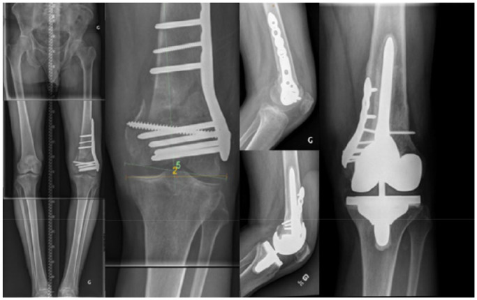
This composite figure illustrates a left KA TKA performed on a post-traumatic knee. The patient sustained a complex articular distal femoral fracture that was treated with ORIF, and developed a medial femoral condyle malunion (substantial proximal migration). The right knee anatomy (LDFA 3.5° valgus and MPTA 5° varus) was used as a template for planning the left KA TKA. To balance the left KA TKA, an osteotomy of the medial femoral condyle had to be performed in order to correct the malunion. Postoperative radiograph shows a left TKA with LDFA of 3° valgus and MPTA of 5° varus. A 100 mm loose fit 12 mm cemented femoral stem was used to supplement femoral fixation and protect the medial condyle fixation.
Note. KA, Kinematic Alignment; TKA, total knee arthroplasty; ORIF, open reduction and internal fixation; LDFA, lateral distal femoral angle; MPTA, medial proximal tibial angle.
Type 6
Type 6 defines acquired lower limb malalignment (in the frontal, sagittal or axial planes) resulting from secondary disease (e.g. rickets, Volkmann syndrome20), previous osteotomy, or the malunion of an extra-articular long bone fracture. In such cases of acquired deformity, the surgeon has the choice of:
1) Accepting the alteration of the patient’s native alignment and perform a KA technique ignoring the extra-articular deformity (Fig. 12 and Fig. 13). By doing so, the knee joint spaces and soft tissue laxities will be preserved. On the other hand, joint load and kinematics may remain altered by the extra-articular pathology.
2) Correcting the acquired deformity with an osteotomy can be performed in a one-stage or two-stage procedure along with a KA TKA (Fig. 14). Thresholds above which modification of extra-articular deformity is required need to be defined, especially for coronal deformity/malrotation which may be difficult to assess.
3) The last option is to carry out a hybrid technique by correcting the extra-articular deformity with intra-articular cut adjustments on the deformed bone while restoring the rest of the knee anatomy (Fig. 15). The decision-making will depend on the severity of the deformity and the alignment boundaries defined by the treating surgeon.
Fig. 12.
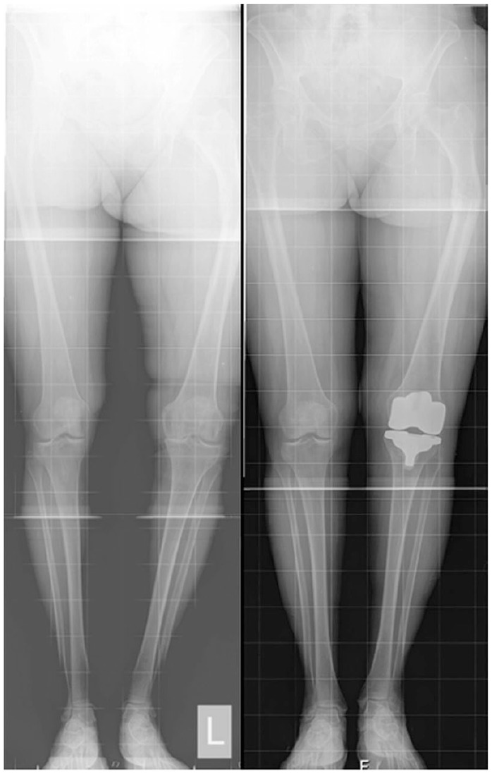
This figure illustrates a patient with a post-traumatic (diaphyseal malunion) ‘Type 6’ knee who was treated with pure (unrestricted) KA TKA, ignoring the extra-articular femoral malunion.
Note. KA, Kinematic Alignment; TKA, total knee arthroplasty.
Fig. 13.
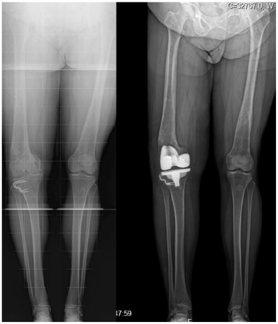
This composite figure illustrates a patient with a history of distal femoral varus malunion followed by high tibial valgus osteotomy, which has left him with a reverse joint line obliquity, ‘Type 6’ knee. This patient was successfully treated with an unrestricted KA TKA that has generated a balanced knee without the need for ligament release.
Note. KA, Kinematic Alignment; TKA, total knee arthroplasty.
Fig. 14.
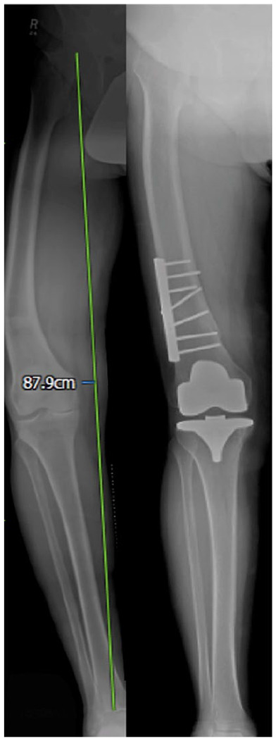
This composite figure illustrates a patient with a ‘Type 6’ knee who was treated with a two-stage surgical procedure: the first surgery consisted of performing a distal femoral osteotomy to correct a severe extra-articular acquired deformity; the second surgery was the implantation of an unrestricted KA TKA.
Note. KA, Kinematic Alignment; TKA, total knee arthroplasty.
Fig. 15.
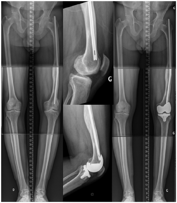
This composite figure presents a patient with a malunited diaphyseal femoral fracture with a bended Kuntscher nail in place, ‘Type 6’ knee. Right and left LDFA were 0.5° and 5° in varus, respectively, suggesting a 4.5° acquired frontal femoral deformity. Right and left MPTA were 9° in varus. The plan was to perform a left KA TKA with the nail in place, and to correct the femoral deformity with intra-articular femoral cuts adjustments. The surgeon reproduced the right-side anatomy. Medial soft tissue release was needed to accommodate the 4° of alignment modification.
Note. LDFA, lateral distal femoral angle; MPTA, medial proximal tibial angle; KA, Kinematic Alignment; TKA, total knee arthroplasty.
Exceptional knees where KA/rKA TKA is not recommended
There are exceptional cases where primary TKA implantation using the KA or rKA techniques should not be recommended. It is the authors’ view not to recommend using the KA technique when a supplementary diaphyseal fixation is required. Current long-stemmed components have a fixed stem–surface angle designed to be mechanically aligned. Revision components with short stems could theoretically be used in KA in situations of favourable knee anatomy, but this requires careful preoperative planning to estimate the risk of impingement between stem and metaphyseal cortex. Therefore, cases with important instability (e.g. incompetent medial collateral ligament or global/recurvatum) are better treated with mechanically aligned higher constrained stemmed implants (Fig. 16 and Fig. 17). In addition, patients with indefinable native knee anatomy (e.g. severe bilateral haemophilic arthropathy – Fig. 18) should be managed with traditional TKA techniques.
Fig. 16.
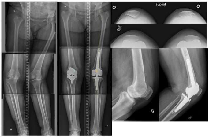
This composite image includes the preoperative long-leg radiograph of a 79-year-old patient with windswept deformity. The left knee, in valgus, had an incompetent medial collateral ligament. Left knee replacement was performed using mechanically aligned (dictated by diaphyseal stem fixation) hinge TKA implants.
Note. TKA, total knee arthroplasty.
Fig. 17.
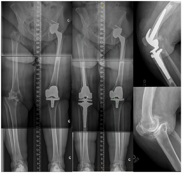
This composite figure illustrates an 88-year-old patient with a severely degenerated, globally unstable left knee. Constrained MA TKA with diaphyseal fixation was performed.
Note. MA, mechanical alignment; TKA, total knee arthroplasty.
Fig. 18.
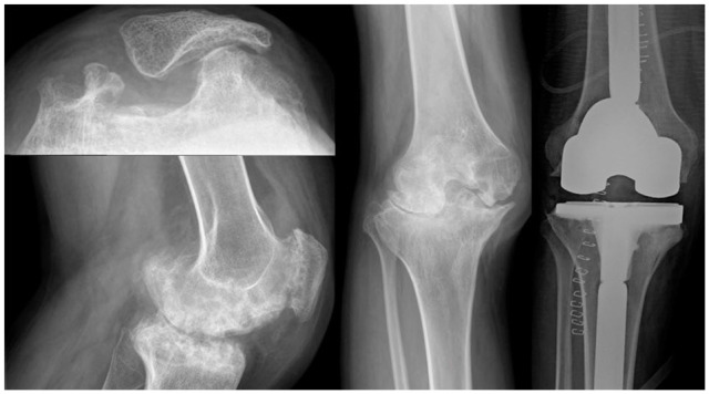
This composite figure illustrates a patient with end-stage bilateral haemophilic knee arthropathy. As the bone loss was dramatic and prevented reliable estimate of the native knee anatomy, the decision was made to perform a mechanically aligned TKA using stemmed implants.
Note. TKA, total knee arthroplasty.
Conclusion
The proposed classification system clarifies the complex situations that arthroplasty surgeons commonly face when aiming for KA TKA. Recognizing complex cases and following planning recommendations would likely improve the reliability of KA implantations for the benefit of the patient. In addition, the classification system eases inter-surgeon communication and benefits the teaching of the KA technique. This classification system is not exhaustive, and will certainly be improved over time.
Footnotes
ICMJE Conflict of interest statement: Charles Rivière and Yaron Barziv declare being consultants for Medacta, William Jackson declares being a consultant for Zimmer-Biomet and Medacta, Loïc Villet and Sivan Sivaloganathan have no conflict of interest to declare. Pascal-André Vendittoli is a consultant for Stryker and Johnson & Johnson and receives royalties from Microport.
OA licence text: This article is distributed under the terms of the Creative Commons Attribution-Non Commercial 4.0 International (CC BY-NC 4.0) licence (https://creativecommons.org/licenses/by-nc/4.0/) which permits non-commercial use, reproduction and distribution of the work without further permission provided the original work is attributed.
Supplemental Material: Supplemental material is available for this paper at https://online.boneandjoint.org.uk/doi/suppl/10.1302/2058-5241.6.210042
Funding statement
No benefits in any form have been received or will be received from a commercial party related directly or indirectly to the subject of this article.
References
- 1. Rivière C, Villet L, Jeremic D, Vendittoli P-A. What you need to know about kinematic alignment for total knee arthroplasty. Orthop Traumatol Surg Res 2021;107:102773. [DOI] [PubMed] [Google Scholar]
- 2. Nisar S, Palan J, Rivière C, Emerton M, Pandit H. Kinematic alignment in total knee arthroplasty. EFORT Open Rev 2020;5:380–390. [DOI] [PMC free article] [PubMed] [Google Scholar]
- 3. Howell SM, Papadopoulos S, Kuznik K, Ghaly LR, Hull ML. Does varus alignment adversely affect implant survival and function six years after kinematically aligned total knee arthroplasty? Int Orthop 2015;39:2117–2124. [DOI] [PubMed] [Google Scholar]
- 4. Howell SM, Shelton TJ, Gill M, Hull ML. A cruciate-retaining implant can treat both knees of most windswept deformities when performed with calipered kinematically aligned TKA. Knee Surg Sports Traumatol Arthrosc 2021;29:437–445. [DOI] [PubMed] [Google Scholar]
- 5. Lin Y-H, Chang F-S, Chen K-H, Huang K-C, Su K-C. Mismatch between femur and tibia coronal alignment in the knee joint: classification of five lower limb types according to femoral and tibial mechanical alignment. BMC Musculoskelet Disord 2018;19:411. [DOI] [PMC free article] [PubMed] [Google Scholar]
- 6. Nakamura S, Tian Y, Tanaka Y, et al. The effects of kinematically aligned total knee arthroplasty on stress at the medial tibia: a case study for varus knee. Bone Joint Res 2017;6:43–51. [DOI] [PMC free article] [PubMed] [Google Scholar]
- 7. Blakeney WG, Vendittoli P-A. Restricted Kinematic Alignment: the ideal compromise? In: Rivière C, Vendittoli P-A, eds. Personalized hip and knee joint replacement. Cham, Switzerland: Springer, 2020:197–206. [PubMed] [Google Scholar]
- 8. Almaawi AM, Hutt JRB, Masse V, Lavigne M, Vendittoli P-A. The impact of mechanical and restricted kinematic alignment on knee anatomy in total knee arthroplasty. J Arthroplasty 2017;32:2133–2140. [DOI] [PubMed] [Google Scholar]
- 9. Laforest G, Kostretzis L, Kiss M-O, Vendittoli P-A. Restricted kinematic alignment leads to uncompromised osseointegration of cementless total knee arthroplasty. Knee Surg Sports Traumatol Arthrosc 2021. 10.1007/s00167-020-06427-1 [Epub ahead of print]. [DOI] [PMC free article] [PubMed]
- 10. Blakeney W, Beaulieu Y, Kiss M-O, Rivière C, Vendittoli P-A. Less gap imbalance with restricted kinematic alignment than with mechanically aligned total knee arthroplasty: simulations on 3-D bone models created from CT-scans. Acta Orthop 2019;90:602–609. [DOI] [PMC free article] [PubMed] [Google Scholar]
- 11. Hutt JRB, LeBlanc M-A, Massé V, Lavigne M, Vendittoli P-A. Kinematic TKA using navigation: surgical technique and initial results. Orthop Traumatol Surg Res 2016;102:99–104. [DOI] [PubMed] [Google Scholar]
- 12. Howell SM, Gill M, Shelton TJ, Nedopil AJ. Reoperations are few and confined to the most valgus phenotypes 4 years after unrestricted calipered kinematically aligned TKA. Knee Surg Sports Traumatol Arthrosc 2021. 10.1007/s00167-021-06473-3 [Epub ahead of print]. [DOI] [PMC free article] [PubMed]
- 13. Meier M, Janssen D, Koeck FX, Thienpont E, Beckmann J, Best R. Variations in medial and lateral slope and medial proximal tibial angle. Knee Surg Sports Traumatol Arthrosc 2021;29:939–946. [DOI] [PubMed] [Google Scholar]
- 14. Howell SM, Shelton TJ, Hull ML. Implant survival and function ten years after kinematically aligned total knee arthroplasty. J Arthroplasty 2018;33:3678–3684. [DOI] [PubMed] [Google Scholar]
- 15. Warth LC, Deckard ER, Meneghini RM. Posterior cruciate ligament resection does not consistently increase the flexion space in contemporary total knee arthroplasty. J Arthroplasty 2021;36:963–969. [DOI] [PubMed] [Google Scholar]
- 16. Rivière C, Webb J, Vendittoli PA. Kinematic alignment technique for TKA on degenerative knees with severe bone loss: a report of 3 cases. Open Orthop J 2021;15:27–34. [Google Scholar]
- 17. Van Damme G, Defoort K, Ducoulombier Y, Van Glabbeek F, Bellemans J, Victor J. What should the surgeon aim for when performing computer-assisted total knee arthroplasty? J Bone Joint Surg [Am] 2005;87-A:52–58. [DOI] [PubMed] [Google Scholar]
- 18. Li G, Park SE, DeFrate LE, et al. The cartilage thickness distribution in the tibiofemoral joint and its correlation with cartilage-to-cartilage contact. Clin Biomech (Bristol, Avon) 2005;20:736–744. [DOI] [PubMed] [Google Scholar]
- 19. Nam D, Lin KM, Howell SM, Hull ML. Femoral bone and cartilage wear is predictable at 0° and 90° in the osteoarthritic knee treated with total knee arthroplasty. Knee Surg Sports Traumatol Arthrosc 2014;22:2975–2981. [DOI] [PubMed] [Google Scholar]
- 20. Colyn W, Agricola R, Arnout N, Verhaar JAN, Bellemans J. How does lower leg alignment differ between soccer players, other athletes, and non-athletic controls? Knee Surg Sports Traumatol Arthrosc 2016;24:3619–3626. [DOI] [PubMed] [Google Scholar]


