Abstract
Purpose
Persistent chronic inflammation of chronic obstructive pulmonary disease (COPD) is associated with poor outcomes and frequently results in acute exacerbation. Predicting the number of exacerbations is important. Because interleukin 6 (IL-6) plays an important role in inducing and maintaining chronic inflammation, we sought to observe whether IL-6 measurement can predict the frequency of acute exacerbation of COPD.
Methods
We reviewed serum IL-6 concentrations of stable COPD patients from January 2016 to December 2017 and statistically analyzed them to determine the optimal threshold value to predict the frequency of COPD acute exacerbations. Outpatients with stable COPD were then recruited between January 2018 and December 2019 and grouped into a low IL-6 group and a high IL-6 group according to this threshold value. We then compared the number of exacerbations of COPD in 1 year between the two groups.
Results
We reviewed data from 95 COPD patients, who had a median of 1.00 exacerbations in preceding year; 35 of these patients had no fewer than two. The median IL-6 concentration was 8.80 pg/mL. IL-6 and hs-CRP were positively correlated with frequency of acute exacerbation in the preceding year, COPD assessment test (CAT) score and British medical research council (mMRC) score, and negatively correlated with forced expiratory volume in one second as percentage of predicted value (FEV1%pred) and FEV1/FVC% (forced vital capacity). IL-6 was the risk factor of COPD patients with two or more exacerbations in 1 year. Finally, we enrolled 65 COPD patients and divided into low IL-6 group and high IL-6 group; the high IL-6 group experienced more frequent exacerbations than did the low IL-6 group.
Conclusion
An IL-6 measurement of 14.030 pg/mL or more is a risk factor for ≥2 acute exacerbations of COPD in the following year.
Keywords: hs-CRP, IL-6, risk factor, prediction, stable COPD, biomarkers
Introduction
Chronic obstructive pulmonary disease (COPD) is a common, preventable, and treatable chronic airway inflammatory disease. COPD was the fifth leading cause of death in China and the fourth leading cause of death globally; it ranked eighth in causes of disease burden as measured by disability-adjusted life years in 2015.1,2
COPD was previously considered to primarily affect the lungs, but now it is increasingly accepted that COPD is a systemic inflammation.3 Persistent systemic inflammation is associated with poor clinical outcomes in COPD and results in frequent acute exacerbation.4 This reduces quality of life, speeds disease progression, and increases the risk of death. Therefore, reducing the number of exacerbations is a primary goal of treatment;5 predicting the number of acute exacerbations in the year following diagnosis is of particular importance.
Interleukin 6 (IL-6) is a soluble inflammatory factor that synthesized in a location in the early stages of inflammation, and then moves to the liver, followed by rapid induction of an extensive range of acute phase proteins such as C-reactive protein (CRP), fibrinogen, and serum amyloid A (SAA).6 IL-6 plays an important role in inducing and maintaining chronic inflammation. It has been shown that IL-6 induces the differentiation of Th17 combined with transforming growth factor (TGF)-β, while inhibits differentiation of regulatory T cells (Treg) induced by TGF-β.7 And up-regulation of the Th17/Treg balance is considered to involve in the development of chronic inflammatory.6 Overproduction of IL-6 has been found in many autoimmune and chronic inflammatory disease including rheumatoid arthritis, systemic lupus erythematosus, Crohn’s disease, asthma, and other inflammatory pulmonary diseases.7,8 IL-6 has also been linked to chronic inflammation-related cancers, such as lung cancer, colorectal cancer, and so on.7 Increasing evidence shows that IL-6 levels are associated with COPD: IL-6 levels are increased in induced sputum of COPD patients, and are inversely correlated with lung function measures such as forced expiratory volume in one second as percentage of predicted value (FEV1%pred).9,10 Two 3-year follow-up studies have shown that elevated IL-6 levels in serum were associated with increased mortality in COPD.11,12 Increased serum IL-6 levels have also been associated with worse six-minute walking distance (6MWD) performance and poor clinical outcomes in COPD patients.4,12 Some studies have suggested that the −572C allele in IL-6 reduced the risk of developing COPD, while the IL-6 −745G/C single nucleotide polymorphism (SNP) increased the risk.13
In summary, IL-6 in COPD plays an important role in maintaining chronic inflammation, leading to poor outcomes and increasing the risk of death. However, it is not enough evidence to support IL-6 is an excellent predictor in AECOPD, it has not been widely used in clinical. Thus, this study aimed to observe whether IL-6 can predict the frequency of acute exacerbation of COPD through 1-year follow-up of COPD patients, and provide more evidence to support IL-6 is a strong predictor.
Materials and Methods
Study Design and Population
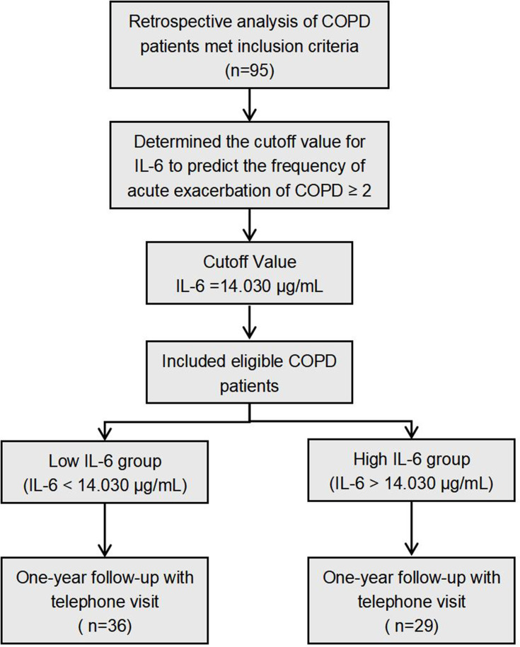
First, serum IL-6 concentration levels of stable COPD patients from January 2016 to December 2017 were reviewed and statistically analyzed to obtain the optimal threshold value to predict the frequency of COPD acute exacerbations. Outpatients with stable COPD were then recruited between January 2018 and December 2019 and were grouped into either the low IL-6 group or the high IL-6 group according to this identified optimal threshold value. All recruited patients were followed for 1 year. The study scheme is described in Figure 1.
Figure 1.
Study scheme.
Abbreviations: COPD, chronic obstructive pulmonary disease; IL-6, interleukin 6.
Participants and Data Collection Procedures in Retrospective Analysis
All studied subjects were recruited from the Department of Respiratory and Critical Care Medicine at Huizhou Municipal Central Hospital. All patients with COPD met the inclusion criteria: (1) the diagnosis of COPD was based on the Global initiative for Chronic Obstructive Lung Disease (GOLD) guidelines, (2) aged ≥ 40 years, (3) used inhaled corticosteroids (ICS)/bronchodilators regularly over 1 month, (4) no acute exacerbation required antibiotics and systemic hormones in the preceding 1 month, (5) no other lung disease or uncontrolled co-morbidities, and (6) able to sign informed consent and complete the questionnaire.
Data on demographics, smoking status, diagnosis, the frequency of acute exacerbation of COPD in 1 year, lung function, COPD assessment test (CAT), modified British medical research council (mMRC) dyspnea scale, and hematological indicators and treatment were collected from hospital electronic medical record system. Serum IL-6 of enrolled subjects would analyse to obtain the optimal threshold value to predict the frequency of COPD acute exacerbations, and then the value would use in the follow up study.
Participants and Data Collection Procedures in One-Year Follow-Up Study
The inclusion criteria of studied subjects were similarly with the case review. Lung function, CAT, mMRC and hematological indicators were measured in the first visit. Enrolled patients need to perform outpatient follow-up visit every 3 months to evaluate their condition. When their COPD symptoms exacerbated and need additional medical attention, they should contact the study doctor at any time and visit our hospital in time. Patients in the stable period who did not wish to perform outpatient follow-up visit were conducted telephone follow-up visit every 6 months to inquire about the frequency of acute exacerbation of COPD. In one-year follow-up study period, the frequency of AECOPD was assessed and confirmed according to the medical record and the treatment history.
Outcomes Measurement
CAT and mMRC were measured when subjects enrolled. Lung function was detected by lung function department in our hospital, and according to the lung function detection guideline. The results of lung function were admitted within 6 months. Hematological indicators comprising complete blood cell count, serum biochemical indexes and serum IL-6 were measured by the clinical laboratory in our hospital. Serum IL-6 concentration was determined by ELISA kit (PI330) which purchased from Beyotime Biotechnology.
Ethics
Our study complied with the Declaration of Helsinki Ethical Principles for Medical Research Involving Human Subjects. The study was also approved by the Ethics Committee of Huizhou Central People’s Hospital, and informed consent was obtained from all patients before enrollment in the study. The privacy of all patients was protected, and the data was anonymized and maintained with confidentiality.
Statistical Methods
The statistical analysis was performed using SPSS statistical package (SPSS version 22.0, IBM, Armonk, NY, USA). The normality of data was examined using the Shapiro–Wilk test. Continuous variables with normal distribution are presented as means ± standard deviation (SD), while continuous variables with non-normal distribution are presented as medians and interquartile range (IQR). Correlations among variables were analyzed using Spearman correlation test. Binary regression analyses were performed to evaluate the potential independent association between IL-6 and the frequency of acute exacerbation of COPD. The accuracy of IL-6 was assessed with receiver operating curve (ROC) analysis. The chi-squared test was used to compare categorical variables between the low IL-6 group and the high IL-6 group. The Mann–Whitney U-test was used to compare the non-normal distribution data between different groups. P<0.05 was considered statistically significant.
Results
Patient Characteristics
We reviewed data from 95 stable COPD patients who presented to our hospital between January 2016 and December 2017. The demographic, clinical, lung function and hematological indicators of patients are summarized in Table 1. The median number of acute exacerbations in the past year in the included COPD patients was 1.00; 35 of these patients had two or more. The overall median serum IL-6 concentration was 8.80 pg/mL.
Table 1.
Characteristics of COPD Patients (n=95)
| Variable | Mean/N/Median | SD/%/IQR |
|---|---|---|
| Demographic | ||
| Age (years-old; median, IQR) | 72.00 | 15.00 |
| Male (N, %) | 63 | 66.32 |
| BMI (kg/m2; median, IQR) | 19.60 | 2.37 |
| Smoking status (N, %) | ||
| Smoker | 69 | 72.63 |
| Ex-smoker | 12 | 12.63 |
| Clinical | ||
| Course of disease (years; median, IQR) | 10.00 | 5.00 |
| Frequency of acute exacerbation in the past 1 year (median, IQR) | 1.00 | 1.00 |
| Exacerbation frequency ≥ 2 | 35 | 36.84 |
| CAT score (median, IQR) | 18.00 | 7.00 |
| mMRC score (median, IQR) | 2.00 | 1.00 |
| Lung Function | ||
| FEV1 (L; median, IQR) | 1.20 | 0.60 |
| FEV1%pred (median, IQR) | 57.21 | 16.28 |
| FEV1/FVC% (mean, SD) | 53.94 | 7.85 |
| Hematological Indicators | ||
| WBC (×109; mean, SD) | 8.26 | 1.97 |
| NE (×109; median, IQR) | 5.63 | 2.28 |
| EO (×109; median, IQR) | 0.10 | 0.41 |
| hs-CRP (mg/L; median, IQR) | 5.50 | 6.64 |
| IL-6 (pg/mL; median, IQR) | 8.80 | 26.07 |
Notes: Normal range of WBC, NE, EO, hs-CRP and IL-6 (3.5~9.5×10⁹, 1.8~6.3×10⁹, 0.02~0.52×10⁹, 0~5 mg/L, 0~7 pg/mL).
Abbreviations: IQR, interquartile range (25th–75th percentile); N, number; SD, standard deviation; BMI, body mass index; CAT, chronic obstructive pulmonary disease assessment test; mMRC, modified British medical research council; FEV1, forced expiratory volume in 1 second; FEV1%pred, FEV1 as a percentage of the predicted value; FVC, forced vital capacity; WBC, white blood cell count; NE, absolute neutrophil count; EO, absolute eosinophil count; hs-CRP, hypersensitive C reactive protein; IL-6, interleukin-6.
Correlation Between hs-CRP, IL-6 and Clinical Data
We analyzed the correlation between hs-CRP, IL-6 and clinical data related to COPD prognosis. The results showed that IL-6 was positively correlated with frequency of acute exacerbation in the previous year, CAT score and mMRC score (r = 0.852, 0.850, and 0.684, respectively; all P< 0.001), but negatively correlated with FEV1%pred and FEV1/FVC% (r = −0.948, −0.522; both P< 0.001). Correlation analysis of hs-CRP also showed similar results, but the degree of correlation was lower than that of IL-6 (Table 2).
Table 2.
Correlation Between hs-CRP, IL-6 and Clinical Data
| Variables | hs-CRP | IL-6 | ||
|---|---|---|---|---|
| r | P | r | P | |
| Frequency of acute exacerbation in the past 1 year | 0.572 | 0.000 | 0.852 | 0.000 |
| CAT score | 0.559 | 0.000 | 0.850 | 0.000 |
| mMRC score | 0.363 | 0.000 | 0.684 | 0.000 |
| FEV1%pred | −0.556 | 0.000 | −0.948 | 0.000 |
| FEV1/FVC% | −0.388 | 0.000 | −0.522 | 0.000 |
Note: P<0.05 considered significant.
Abbreviations: CAT, chronic obstructive pulmonary disease assessment test; mMRC, modified British medical research council; FEV1, forced expiratory volume in 1 second; FEV1%pred, FEV1 as a percentage of the predicted value; FVC, forced vital capacity; hs-CRP, hypersensitive C reactive protein; IL-6, interleukin-6.
Risk Factors for ≥ 2 Acute Exacerbations of COPD
The frequency of acute exacerbation of COPD is associated with patients’ prognosis. Two or more exacerbations per year is one of the criteria for the comprehensive assessment of COPD in groups C and D of the GOLD classification, which often indicates more severe disease status. Because our results showed that IL-6 and hs-CRP correlated with frequency of acute exacerbation of COPD (Table 2), we analyzed these data using binary regression analysis to identify the risk factors for two or more acute exacerbations of COPD. The results suggested that higher IL-6 measurements were significantly associated with a higher risk of having two or more acute exacerbation within 1 year (OR 0.260, 95% CI 1.140–1.475), indicating that IL-6 measurements were capable of predicting the frequency of exacerbation (Table 3).
Table 3.
Risk Factors of the Frequency of Acute Exacerbation of COPD ≥ 2 Using Binary Regression Analysis
| Variables | OR | 95% CI | P |
|---|---|---|---|
| hs-CRP | 0.110 | 0.901–1.382 | 0.313 |
| IL-6 | 0.260 | 1.140–1.475 | 0.000 |
Abbreviations: hs-CRP, hypersensitive C reactive protein; IL-6, interleukin-6; OR, odds ratio; CI, confidence interval.
ROC Curve Analysis of IL-6 in Predicting the Frequency of Acute Exacerbation
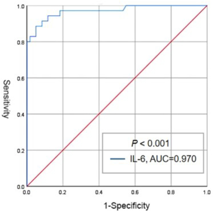
The validity of IL-6 in predicting two or more acute exacerbations of COPD was excellent and significant; the area under the curve (AUC) was 0.970 (P<0.001) (Figure 2). A cut-off value of IL-6 > 14.030 pg/mL had 88.6% sensitivity and 95% specificity.
Figure 2.
ROC analysis of IL-6 in predicting the frequency of acute exacerbation of COPD ≥ 2.
Abbreviations: IL-6, interleukin 6; AUC, the area under the curve.
Frequency of Acute Exacerbation by Group After 1-Year Follow-Up
Using this cut-off value, eligible COPD patients were divided into a low IL-6 group (IL-6 < 14.030 pg/mL) and a high IL-6 group (IL-6 >14.030 pg/mL). There were no differences between the two groups in terms of demographic and lung function data, CAT score, or mMRC score (Table 4). The frequency of acute exacerbation events in 1 year was higher in the high IL-6 group than in the low IL-6 group (2.00 vs 1.00, P<0.001), and those patients who had ≥ 2 exacerbation events were more likely to be in the high IL-6 group (79.31% vs 38.89%, P=0.001).
Table 4.
Compared the Frequency of Acute Exacerbation Between Different Groups After One-Year Follow-Up
| Variables | Low IL-6 Group (n=36) | High IL-6 Group (n=29) | P value |
|---|---|---|---|
| Demographic | |||
| Age (years-old; mean, SD) | 67.97 ± 1.47 | 68.27 ± 1.58 | 0.864 |
| Male (N, %) | 25, 69.44 | 19, 65.52 | 0.736 |
| BMI (kg/m2; mean, SD or median, IQR) | 20.40 ± 0.33 | 21.71, 5.32 | 0.078 |
| Smoker (N, %) | 31, 86.11 | 22, 75.86 | 0.290 |
| Clinical | |||
| Frequency of acute exacerbation in 1 year (median, IQR) | 1.00, 1.00 | 2.00, 1.00 | 0.000 |
| Exacerbation frequency ≥ 2 (N, %) | 14, 38.89 | 23, 79.31 | 0.001 |
| CAT Score (mean, SD) | 19.77 ± 0.54 | 20.14 ± 0.66 | 0.735 |
| mMRC Score (median, IQR) | 2.00, 1.00 | 3.00, 1.00 | 0.268 |
| Lung Function | |||
| FEV1%pred (mean, SD) | 42.46 ± 1.81 | 40.04 ± 1.90 | 0.391 |
| FEV1/FVC% (mean, SD) | 49.00 ± 1.31 | 48.75 ± 1.30 | 0.838 |
| Hematological Indicators | |||
| IL-6 (pg/mL; median, IQR) | 6.73, 0.62 | 32.07, 26.67 | 0.000 |
Abbreviations: IQR, interquartile range (25th–75th percentile); N, number; SD, standard deviation; BMI, body mass index; CAT, chronic obstructive pulmonary disease assessment test; mMRC, modified British medical research council; FEV1, forced expiratory volume in 1 second; FEV1%pred, FEV1 as a percentage of the predicted value; FVC, forced vital capacity; IL-6, interleukin-6.
Discussion
COPD is now recognized as a chronic systemic inflammatory disease. Persistent systemic inflammation has been found to be associated with exacerbation frequency in COPD.4 Frequent acute exacerbation of COPD not only affects patients’ quality of life, but also greatly shortens their life expectancy.14 Predicting the number of acute exacerbations in the following year is important because of the impact of these events on the natural history of COPD.5 IL-6 represents a keystone cytokine in inflammation which drives progression, and it is now regarded as a prominent target for clinical intervention.15,16 Increasing evidence shows that IL-6 is also increased in COPD patients and associated with the progression of COPD.17
In the present study, we reviewed the serum IL-6 concentration and clinical data of 95 stable COPD patients at our hospital. The median serum IL-6 concentration in these patients was 8.80 pg/mL, between the results found by Zeng (30.5±9.9 pg/mL) and by Gorska (3.4 pg/mL).18,19 The difference in results may be due to differences in the severity of COPD patients included, or to differences in suppliers of machines or reagents that measured IL-6 concentration. Most COPD patients included in our study had moderate-to-severe COPD (median FEV1%pred was 57.21%), but Zeng’s patients had more severe COPD (mean FEV1%pred was 45.2%) and Gorska’s patients had more mild-to-moderate COPD (median FEV1%pred was 71%).18,19 At present, there are no evidence-based studies with sufficient sample size to confirm serum IL-6 concentration in patients with COPD. Our results exceeded the normal range (0–7 pg/mL), which may indicate that most COPD patients had chronic systemic inflammation. Because one major effect of IL-6 is to stimulate the production of CRP, we also evaluated hs-CRP concentration. Hs-CRP is also used as a marker of systemic inflammation in COPD.20 In patients with stable stage 2 and stage 3 COPD, hs-CRP concentrations have been reported at median levels of 5.596±0.7982 mg/L and 5.093±0.7912 mg/L, respectively.21 Our data showed a similar result; median hs-CRP was 5.50 mg/L, which exceeded the normal range (0–5 mg/L). Because we included stable COPD patients who regularly used ICS or/and bronchodilators, other hematological indicators such as white blood cells (WBC), neutrophils (NE), and eosinophils (EO) were within normal limits. Some studies have also reported that WBC, NE, and EO were normal in stable COPD patients.22–24
Airway remodeling, one of the most prominent causes of airway obstruction in COPD, is often considered the result of chronic inflammation. It has been associated with decreases in lower lung function and frequent exacerbations of COPD.25 We analyzed the correlation between hs-CRP, IL-6 and clinical data from COPD patients, and found IL-6 and hs-CRP to be positively correlated with frequency of acute exacerbation in the preceding 1 year, CAT score, and mMRC score, and negatively correlated with FEV1%pred and FEV1/FVC%. These findings suggest that elevated levels of IL-6 and hs-CRP are related to poor outcomes from COPD. Binary regression analysis showed that elevated IL-6 (but not hs-CRP) was a risk factor for having two or more exacerbations of COPD within 1 year. CRP-gene expression has been shown to be regulated through IL-6 independent or dependent pathways, and the secretion of CRP requires induction of IL-6.26 We therefore speculate that IL-6 may better reflect the level of systemic inflammation in COPD patients compared with hs-CRP, potentially explaining why IL-6 was more effective in predicting the occurrence of two or more exacerbation events within 1 year. Based on ROC curve and AUC, the optimal prediction threshold for IL-6 was 14.030 pg/mL.
To further confirm the predictive power of IL-6 and to reduce the influence of confounding factors, we matched patients for age, sex, smoking status, CAT score, mMRC score and lung function at the time of inclusion. Finally, we enrolled 65 COPD patients and divided into low IL-6 group and high IL-6 group according to the predictive threshold for IL-6. We found more frequent exacerbations and a greater proportion of patients with multiple exacerbations (79.31% vs 38.89%) in the high IL-6 group than in the low IL-6 group.
The findings of the present study not only enhance the previous evidence on the relationship between IL-6 and outcomes of COPD, but also showed IL-6 to be a predictor of the frequency of acute exacerbation of COPD. Our study also has some limitations. The sample used for the retrospective analysis to obtain the predictive threshold of IL-6 was from a single center, and the sample size was small. Therefore, the predicted value obtained may not be applicable to all COPD patients. Likewise, the sample size used to verify the predictive power of IL-6 was limited. Although this study had some limitations, it also confirms the value of IL-6 in predicting the frequency of acute exacerbation of COPD, which has clinical significance for the prevention or early detection of susceptibility to these exacerbations.
Limitation
Our findings were based on a single center, the insufficient sample size was a objective fact and limitation of this study, which may affect the stability of the results. And it requires further verification through multi-center cooperation to expand the sample size.
Conclusion
Frequent exacerbations of COPD that are associated with persistent chronic systemic inflammation result in poor quality of life and shortened life expectancy. Measurement of IL-6 can help to predict the number of potential exacerbation events in the following year and guide patients or doctors in taking timely and appropriate steps to prevent occurrence of these events.
Acknowledgments
The authors would like to thank the staff of the Department of Respiratory and Critical Care Medicine in Huizhou Municipal Central Hospital for providing and collecting all the clinical data. We also thank John Daniel from Liwen Bianji (Edanz) for editing the English text of a draft of this manuscript.
Disclosure
The authors declare no conflicts of interest in this study.
References
- 1.Lopez-Campos JL, Soler-Cataluna JJ, Miravitlles M. Global strategy for the diagnosis, management, and prevention of chronic obstructive lung disease 2019 report: future challenges. Arch Bronconeumol. 2020;56(2):65–67. doi: 10.1016/j.arbres.2019.06.001 [DOI] [PubMed] [Google Scholar]
- 2.Collaborators GBDCoD. Global, regional, and national age-sex specific mortality for 264 causes of death, 1980–2016: a systematic analysis for the Global Burden of Disease Study 2016. Lancet. 2017;390(10100):1151–1210. [DOI] [PMC free article] [PubMed] [Google Scholar]
- 3.Disease GIfCOL. Global strategy for the diagnosis, management, and prevention of chronic obstructive pulmonary disease 2020 report. Available from: https://goldcopd.org/gold-reports/. Accessed November 9, 2019.
- 4.Agusti A, Edwards LD, Rennard SI, et al. Persistent systemic inflammation is associated with poor clinical outcomes in COPD: a novel phenotype. PLoS One. 2012;7(5):e37483. doi: 10.1371/journal.pone.0037483 [DOI] [PMC free article] [PubMed] [Google Scholar]
- 5.Decramer M, Janssens W, Miravitlles M. Chronic obstructive pulmonary disease. Lancet. 2012;379(9823):1341–1351. doi: 10.1016/S0140-6736(11)60968-9 [DOI] [PMC free article] [PubMed] [Google Scholar]
- 6.Tanaka T, Narazaki M, Kishimoto T. IL-6 in inflammation, immunity, and disease. Cold Spring Harb Perspect Biol. 2014;6(10):a016295. doi: 10.1101/cshperspect.a016295 [DOI] [PMC free article] [PubMed] [Google Scholar]
- 7.Ataie-Kachoie P, Pourgholami MH, Morris DL. Inhibition of the IL-6 signaling pathway: a strategy to combat chronic inflammatory diseases and cancer. Cytokine Growth Factor Rev. 2013;24(2):163–173. doi: 10.1016/j.cytogfr.2012.09.001 [DOI] [PubMed] [Google Scholar]
- 8.Rincon M, Irvin CG. Role of IL-6 in asthma and other inflammatory pulmonary diseases. Int J Biol Sci. 2012;8(9):1281–1290. doi: 10.7150/ijbs.4874 [DOI] [PMC free article] [PubMed] [Google Scholar]
- 9.Donaldson GC, Seemungal TA, Patel IS, et al. Airway and systemic inflammation and decline in lung function in patients with COPD. Chest. 2005;128(4):1995–2004. doi: 10.1378/chest.128.4.1995 [DOI] [PMC free article] [PubMed] [Google Scholar]
- 10.Grubek-Jaworska H, Paplinska M, Hermanowicz-Salamon J, et al. IL-6 and IL-13 in induced sputum of COPD and asthma patients: correlation with respiratory tests. Respiration. 2012;84(2):101–107. doi: 10.1159/000334900 [DOI] [PubMed] [Google Scholar]
- 11.Celli BR, Locantore N, Yates J, et al. Inflammatory biomarkers improve clinical prediction of mortality in chronic obstructive pulmonary disease. Am J Respir Crit Care Med. 2012;185(10):1065–1072. doi: 10.1164/rccm.201110-1792OC [DOI] [PubMed] [Google Scholar]
- 12.Ferrari R, Tanni SE, Caram LM, Correa C, Correa CR, Godoy I. Three-year follow-up of interleukin 6 and C-reactive protein in chronic obstructive pulmonary disease. Respir Res. 2013;14:24. doi: 10.1186/1465-9921-14-24 [DOI] [PMC free article] [PubMed] [Google Scholar]
- 13.Wu X, Yuan B, Lopez E, Bai C, Wang X. Gene polymorphisms and chronic obstructive pulmonary disease. J Cell Mol Med. 2014;18(1):15–26. doi: 10.1111/jcmm.12159 [DOI] [PMC free article] [PubMed] [Google Scholar]
- 14.Pavord ID, Jones PW, Burgel PR, Rabe KF. Exacerbations of COPD. Int J Chron Obstruct Pulmon Dis. 2016;11(Spec Iss):21–30. [DOI] [PMC free article] [PubMed] [Google Scholar]
- 15.Hunter CA, Jones SA. IL-6 as a keystone cytokine in health and disease. Nat Immunol. 2015;16(5):448–457. doi: 10.1038/ni.3153 [DOI] [PubMed] [Google Scholar]
- 16.Unver N, McAllister F. IL-6 family cytokines: key inflammatory mediators as biomarkers and potential therapeutic targets. Cytokine Growth Factor Rev. 2018;41:10–17. doi: 10.1016/j.cytogfr.2018.04.004 [DOI] [PMC free article] [PubMed] [Google Scholar]
- 17.Su B, Liu T, Fan H, et al. Inflammatory markers and the risk of chronic obstructive pulmonary disease: a systematic review and meta-analysis. PLoS One. 2016;11(4):e0150586. doi: 10.1371/journal.pone.0150586 [DOI] [PMC free article] [PubMed] [Google Scholar]
- 18.Zeng YY, Hu WP, Zuo YH, Wang XR, Zhang J. Altered serum levels of type I collagen turnover indicators accompanied by IL-6 and IL-8 release in stable COPD. Int J Chron Obstruct Pulmon Dis. 2019;14:163–168. doi: 10.2147/COPD.S188139 [DOI] [PMC free article] [PubMed] [Google Scholar]
- 19.Gorska K, Nejman-Gryz P, Paplinska-Goryca M, Korczynski P, Prochorec-Sobieszek M, Krenke R. Comparative Study of IL-33 and IL-6 levels in different respiratory samples in mild-to-moderate asthma and COPD. COPD. 2018;15(1):36–45. doi: 10.1080/15412555.2017.1416074 [DOI] [PubMed] [Google Scholar]
- 20.Ghobadi H, Fouladi N, Beukaghazadeh K, Ansarin K. Association of high sensitive CRP level and COPD assessment test scores with clinically important predictive outcomes in stable COPD patients. Tanaffos. 2015;14(1):34–41. [PMC free article] [PubMed] [Google Scholar]
- 21.Nillawar AN, Bardapurkar JS, Bardapurkar SJ. High sensitive C-reactive protein as a systemic inflammatory marker and LDH-3 isoenzyme in chronic obstructive pulmonary disease. Lung India. 2012;29(1):24–29. doi: 10.4103/0970-2113.92358 [DOI] [PMC free article] [PubMed] [Google Scholar]
- 22.Koo HK, Kang HK, Song P, Park HK, Lee SS, Jung H. Systemic white blood cell count as a biomarker associated with severity of chronic obstructive lung disease. Tuberc Respir Dis. 2017;80(3):304–310. [DOI] [PMC free article] [PubMed] [Google Scholar]
- 23.Bafadhel M, Pavord ID, Russell REK. Eosinophils in COPD: just another biomarker? Lancet Respir Med. 2017;5(9):747–759. doi: 10.1016/S2213-2600(17)30217-5 [DOI] [PubMed] [Google Scholar]
- 24.Bilir B, Altıntaş N, Aydın M, Oran M, Özsu S, Tutar Ü. The predictive role of neutrophil to lymphocyte ratio in chronic obstructive pulmonary disease. Eur J Gen Med. 2016;13(2):105–110. [Google Scholar]
- 25.Hirota N, Martin JG. Mechanisms of airway remodeling. Chest. 2013;144(3):1026–1032. doi: 10.1378/chest.12-3073 [DOI] [PubMed] [Google Scholar]
- 26.Luna JM, Moon YP, Liu KM, et al. High-sensitivity C-reactive protein and interleukin-6-dominant inflammation and ischemic stroke risk: the northern Manhattan study. Stroke. 2014;45(4):979–987. doi: 10.1161/STROKEAHA.113.002289 [DOI] [PMC free article] [PubMed] [Google Scholar]


