Abstract
Introduction
The current therapeutic armory for visceral leishmaniasis (VL) caused by Leishmania donovani complex is inadequate, coupled with serious limitations. Combination therapy has proved ineffective due to mounting resistance; however, the search for safe and effective drugs is desirable, in the absence of any vaccine. There is a growing interest in the application of nanoparticles for the therapeutic effectiveness of leishmaniasis. Aimed in this direction, we assessed the antileishmanial effect of gold nanoparticles (GNP) against L. donovani in vitro.
Methods
GNP were synthesized and characterized for particle size by dynamic light scattering (DLS) and atomic force microscopy (AFM) and for optical properties by UV-visible spectroscopy. Cytotoxicity of GNP was measured by the MTT proliferation assay. The antileishmanial activity of the nanoparticles was evaluated against L. donovani promastigotes and macrophage-infected amastigotes in vitro.
Results
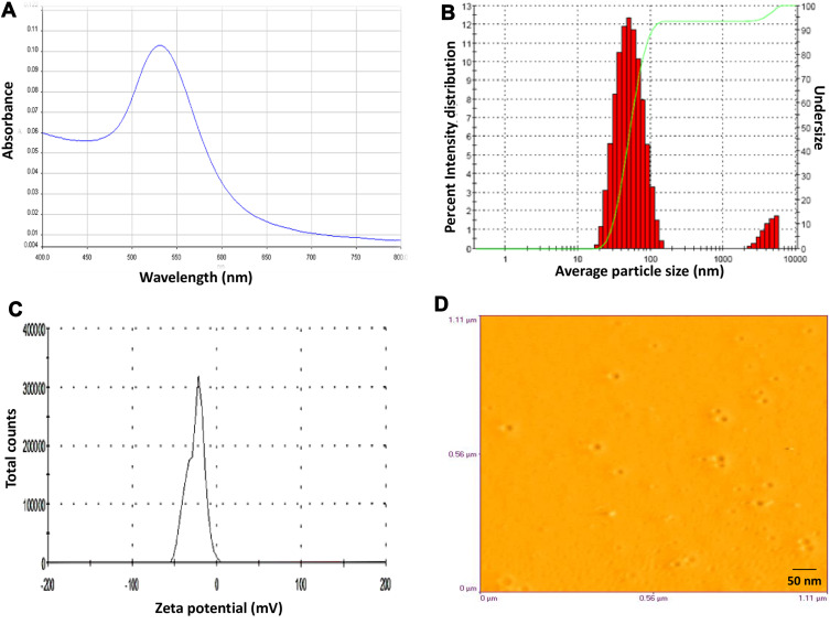
GNP showed a strong SPR peak at 520 nm and mean particle size, polydispersity index (PDI), and zeta potential of 56.0 ± 10 nm, 0.3 ± 0.1 and −27.0 ± 3 mV, respectively. The GNPs were smooth and spherical with a mean particle diameter of 20 ± 5 nm. Nanoparticles [1.2–100 µM] did not reveal any cytotoxicity on RAW 264.7 murine macrophage cell line, but exerted significant activity against both promastigotes and amastigote stages of L. donovani with 50% inhibitory concentrations (IC50) of 18.4 ± 0.4 µM and 5.0 ± 0.3 µM, respectively. GNP showed significant antileishmanial activity with deformed morphology of parasites and the least number of surviving promastigotes after growth reversibility analysis.
Conclusion
GNP may provide a platform to conjugate antileishmanial drugs onto the surface of nanoparticles to enhance their therapeutic effectiveness against VL. Further work is warranted, involving more in-depth mechanistic studies and in vivo investigations.
Keywords: gold nanoparticles, surface plasmon resonance, visceral leishmaniasis L. donovani, promastigotes, amastigotes
Introduction
Leishmaniasis encompasses a complex spectrum of diseases ranging from cutaneous, mucocutaneous to visceral forms depending on the infecting species. Visceral leishmaniasis (VL) is characterized by prolonged fever, hepatosplenomegaly, substantial weight loss, anemia, pancytopenia, and hypergammaglobulinemia, and is the most severe of the various clinical forms of leishmaniasis. This vector-borne disease is caused by Leishmania donovani complex, which includes L. donovani and Leishmania infantum (also known as Leishmania chagasi in South America). In India, VL or kala-azar is mainly caused by L. donovani species and represents 50% of the global burden of this neglected disease of poverty that can be 100% fatal in less than two years, if left untreated. In addition, there are reports of outbreaks in low- and non-endemic zones.1–3 Treatment of VL relies mainly on pentavalent antimonials, amphotericin B, liposomal amphotericin B, miltefosine, paromomycin or their combinations.4 The limitations of present chemotherapeutics include toxicity, noncompliance, prolonged and a cumbersome regimen that is unaffordable to the rural population, increasing antimonial resistance, complications of post kala-azar dermal leishmaniasis (PKDL), and HIV-coinfections.5 Combination therapy has proved to be useful over monotherapy in reducing the dose of the drug and hence toxicity;6 nonetheless, improved therapy is required in the absence of any vaccine. Furthermore, since the parasites reside mainly in phagolysosomal vacuoles within the macrophages, chemotherapy using present antiparasitics is difficult. Nanoparticulate materials are powerful tools to reach the therapeutic target that is otherwise difficult to attain through common procedures.7 Due to the low rate of discovery of antileishmanial drugs, researchers have focused mainly on modulating the delivery of existing drugs.8 In the case of leishmaniasis, nanoparticles such as nanoemulsions and liposomes have been shown to be useful as they increase the bioavailability and reduce the toxicity of loaded antileishmanial drugs.9,10
Metallic nanoparticles offer promise as an alternative approach to replace or complement the present antibiotics, especially in the therapy of infectious diseases.11,12 Studies on the antibacterial action of metal nanoparticles have provided encouraging results.13,14 The anti-trypanosomal activity of silver, gold, and platinum nanoparticles has also been explored.15 Research on metal nanoparticles and metallic compounds for the treatment of leishmaniasis is gaining ground due to the fact that these compounds inhibit trypanothione metabolism enzymes that are important for the survival of Leishmania.16 Auranofin, a drug containing gold has been reported to kill promastigotes of L. infantum by targeting trypanothione reductase.17 The inhibitory activity of silver nanoparticles against L. infantum has been reported to be mediated by its effect on trypanothione reductase.18 In one investigation, the clinical use of zinc sulfate was found to be as effective as glucantime for the treatment of cutaneous leishmaniasis.19 In another study, the antileishmanial activity of platinum compound has been reported against Leishmania major.20 The leishmanicidal activity of zinc oxide nanoparticles against Leishmania tropica has also been observed.21 Silver and titanium oxide nanoparticles have shown remarkable antileishmanial activity against a few species of Leishmania.22,23 The small size and high surface area to volume ratio of nanoparticles allow them to interact with important components of infectious agents especially DNA and enzymes, rendering them nonfunctional.24
Gold nanoparticles (GNP) have been widely used as important tools in therapy, drug delivery, targeting, and imaging.25 Gold nanoparticles have been reported to exhibit significant activity against L. major.26,27 Silencing of the gp63 gene in L. major followed by parasite killing has been observed with antisense oligonucleotides hybridized to GNP.28 Nanogold with antileishmanial molecules has been reported against CL29 as well as VL.30 Different drugs or bioactive compounds have been conjugated with GNP to enhance their delivery to the target site. Surface functionalized GNP via quercetin,31 gallic acid32 and kaempherol33 have shown improved activity against L. donovani. However, in these studies, the activity was tested against axenic amastigotes and macrophage-infected axenic amastigotes. Axenic amastigotes do not represent true intracellular amastigotes within the macrophages. When macrophages are infected with promastigotes that transform into amastigotes, the natural infection is mimicked. Macrophage-infected amastigotes are a more elaborate and physiologic model for drug testing compared to axenic amastigotes.34,35 Moreover, activity against axenic amastigotes does not always correlate with efficacy against the parasite in its intracellular niche.34
Furthermore, in earlier studies, side effects were observed against peritoneal macrophages harvested from Swiss outbred mice after induction of thioglycollate. Inflammatory thioglycollate potentiates the activation of macrophages. Therefore, RAW 264.7 murine macrophage cells were used as model phagocytes in our study. There are also reports on the activity of green synthesized GNP against L. donovani36 but, the activity was evaluated against the promastigote forms alone. An effective antileishmanial drug should be active against both the morphological stages of the parasite. Furthermore, loss of activity of biosynthesized nanoparticles has also been reported.37,38 At present, there are no reports on the antileishmanial effect of GNP alone against L. donovani promastigotes and amastigotes in vitro. In this study, we evaluated the antileishmanial potential of GNP against L. donovani promastigotes and macrophage-infested amastigotes.
Materials and Methods
Animals and Cell Lines
Six-eight weeks old female BALB/c mice (weighing about 25–30 g), obtained from the Central Animal House Facility, Jamia Hamdard, were used to maintain the Leishmania parasites. Ethics approval for the study (Approval No. 458) was sought from Jamia Hamdard Animal Ethics Committee, which followed the guidelines laid down by the Committee for the Purpose of Control and Supervision of Animal Experiments, Ministry of Empowerment and Social Justice, Government of India. The use of the murine macrophage cell line, RAW 264.7 (a kind gift from Dr. B.S. Dwarakanath, INMAS, New Delhi), had approval from the ethics review board.
Parasite Culture
The Indian strain of L. donovani (MHOM/IN/83/AG83) was routinely passaged in BALB/c mice and, after transformation, was cultured at 22°C in M199 supplemented with 10% heat-inactivated FBS, HEPES (25 mM), L-glutamine (2 mM), penicillin G (100 IU mL−1) and streptomycin (100 µg m−1) at an average density of 2×10⁶ cells mL−1.39
Synthesis of Gold Nanoparticles (GNPs)
GNPs were synthesized by the microwave-assisted heating method via a chemical reduction wherein, trisodium citrate dihydrate was used to reduce the HAuCl4.3H2O solution.40 Briefly, 1mM tetrachloroaurate (HAuCl4.3H2O) solution was brought to a rolling boil in a microwave to which 1% trisodium citrate dihydrate was quickly added in a 10:1 ratio. The change in the colour of the suspension from pale yellow to brilliant red indicated the successful synthesis of GNP. The solution was cooled and then subjected to ultra-centrifugation to wash off the free citrate. The nanoparticle pellet was resuspended in nano-pure water and characterized by UV-visible spectroscopy, dynamic light scattering (DLS), and atomic force microscopy (AFM).
Characterization of Gold Nanoparticles (GNPs)
Surface Plasmon Resonance (SPR) Determination by UltraViolet (UV)-Visible Spectroscopy
The synthesized GNPs were analysed spectrophotometrically at wavelength ranging from 200 to 800 nm using a UV-Visible spectrophotometer (Lamda 20, Perkin-Elmer) to determine the characteristic properties of surface plasmon resonance (SPR).41 The GNP concentration was adjusted to give an absorbance of less than 1. The synthesized GNPs were further used for antileishmanial studies.
Particle Size Distribution and Zeta Potential by Dynamic Light Scattering (DLS)
The mean particle size and polydispersity index (PDI) of GNPs were estimated in a Malvern Zeta Sizer (Nano ZS, Malvern Instruments Inc. Worcestershire, UK) using the DLS technique at a scattering angle of 90° at 25°C. The sample of GNP was diluted with Milli-Q water and analyzed in triplicate.
The zeta potential (ζ) of the nanoparticles was also measured by a zetasizer at room temperature. The diluted GNPs were placed in an electrophoretic cell and the data were acquired in triplicate.
Surface Morphology of Gold Nanoparticles (GNP) Using Atomic Force Microscopy
The surface morphology, shape and size of GNP were further visualized by the atomic force microscope (AFM, Veeco, Innova) in contact mode with the help of the silicon nitride nano probe cantilever, at a spring constant of 49 N m−1.41 The obtained images were further analyzed and processed by Veeco SPM Lab analysis software.
In vitro Analysis of Cytotoxicity of Gold Nanoparticles (GNPs)
The cytotoxicity of GNP was evaluated in the RAW 264.7 murine macrophage Cell line using MTT assay.42 In brief, 2×10⁵ Cells per well in RPMI 1640 medium were seeded in a flat bottom 96-well tissue culture plate for 24 h at 37°C in a CO2 incubator. The cells were incubated in triplicate with serial three-fold dilutions of GNP or pentamidine, starting from 100 µM at 37°C, 5% CO2. Macrophages without GNP or pentamidine were left as negative control. After 48 h of incubation, MTT (5 mg mL−1) was added to each well and on the formation of formazan crystals, the plate was centrifuged (2100 × g, 20 min, 4°C), the supernatant carefully aspirated, and the crystals so formed were dissolved using DMSO:isopropanol (1:1). Using a microplate reader (Spectra max 450), the plate was read at 570 nm, with 690 nm as background. The result was represented as a percentage of viable Cells compared to the negative control and was calculated as follows:
 |
In vitro Antileishmanial Activity of Gold Nanoparticles (GNPs) Against L. donovani Promastigotes
The growth kinetics of L. donovani promastigotes was studied by incubating promastigotes (2×10⁶ cells mL−1) with GNP (75 µM) in M199 medium supplemented with 10% FBS (complete medium). Pentamidine was added as a reference drug, while parasites in medium alone, without any drug or GNP, served as a control. Viable parasites were microscopically counted for 5 days and parasites without any motility were marked dead.43
To further confirm the leishmanicidal effect of GNPs, promastigotes after 7 days in culture, with or without treatment, were analyzed using a growth reversibility assay. Briefly, promastigotes were subjected to drug withdrawal by washing twice with incomplete M199 medium (without FBS) and then resuspended in fresh complete M199 medium at 22°C for a further 4 days. The viable parasites were counted using a phase-contrast microscope with a 40x objective.43
Evaluation of the 50% Inhibitory Concentration (IC50) of Gold Nanoparticles (GNPs) for L. donovani Promastigotes
L. donovani promastigotes were incubated with serial two-fold dilutions of GNP (0–25 µM) and cultured at 22°C. Pentamidine was added as a standard antileishmanial drug. The untreated L. donovani promastigotes served as a parasite control. After 4 days, the parasites were enumerated microscopically using a phase contrast microscope and the percent viability determined43 according to the formula:
 |
The IC50 of GNP on promastigotes was calculated using a linear regression analysis.
Evaluation of Promastigote Morphology After Gold Nanoparticles (GNPs) Treatment
Morphology of L. donovani promastigotes with and without GNP treatment was studied after 4 days in culture by enumeration under a phase-contrast microscope with 40x objective. For each sample, a minimum of 10 microscopic fields were observed and the images were taken using NIS-Elements imaging software.44
Anti-Amastigote Effect of Gold Nanoparticles (GNPs) ex vivo
L. donovani promastigotes were cultured in RPMI 1640 medium prior to infecting the RAW 264.7 macrophage cell line. Briefly, 5×10⁵ macrophages per cover slip were seeded in a 24-well tissue culture plate and left to adhere overnight in a CO2 incubator at 37°C. Adherent macrophages were incubated with stationary phase L. donovani promastigotes in a cell to parasite ratio of 1:10, for at least 12 h at 37°C, 5% CO2. Non-phagocytosed promastigotes were gently aspirated by washing with incomplete RPMI 1640 medium (without FBS) and infected macrophages were further incubated with GNP (1–15 µM) for 48 h at 37°C, 5% CO2. Infected macrophages that were not treated were left as a control. The cover slips were then fixed, Giemsa stained and a minimum of 200 macrophages per cover slip were microscopically enumerated to determine the number of infected macrophages and the number of intracellular amastigotes.42 The mean percent infection was calculated using the following formula:
 |
The IC50 of GNP on amastigotes was determined using a linear regression analysis. The percentage of infected macrophages was also analyzed according to the following formula:
 |
Statistical Analysis
All data were represented as mean ± standard error (SE) of samples in triplicate and were from one of three independent experiments. Statistical analysis was performed by one-way analysis of variance (ANOVA) using the graph pad InStat. P-values of <0.05 were considered statistically significant.
Results
Synthesis and Characterization of Gold Nanoparticles (GNPs)
GNPs were successfully synthesized via a simple chemical reduction by mixing an aqueous solution of trisodium citrate dihydrate with a microwave-heated HAuCl4 solution. A brilliant red suspension of GNPs was formed after complete reduction of Au3+ ions to Au0 during the reaction. UV-visible absorption spectra revealed the presence of a prominent SPR peak at 520 nm, indicating the formation of spherical GNPs (Figure 1A). The position of SPR peak varies depending on the particle size, shape, and dielectric constant of the surrounding medium. The mean hydrodynamic diameter and PDI of GNP were 56.0 ± 10 nm and 0.3 ± 0.1, respectively, as determined by DLS (Figure 1B). The zeta potential, which dictates the stability of GNP, was found to be −27 ± 3 mV (Figure 1C). The presence of uniform negative charge on the surface of synthesized GNP imparts high stability to these nanoparticles, thus preventing their aggregation. For further morphological analysis, GNPs were vacuum dried onto clean glass round cover slips (18 mm) and observed under AFM. The AFM images revealed that the synthesized GNP had an average particle size of 20 ± 5 nm and were spherical with a smooth surface (Figure 1D).
Figure 1.
Characterization of GNP (A) absorption spectra showing the surface plasmon resonance peak, (B) size distribution pattern, (C) zeta potential measurement graph, (D) microscopic image showing the morphological depictions of nanoparticles by AFM at contact mode.
In vitro Cytotoxicity of Gold Nanoparticles (GNPs)
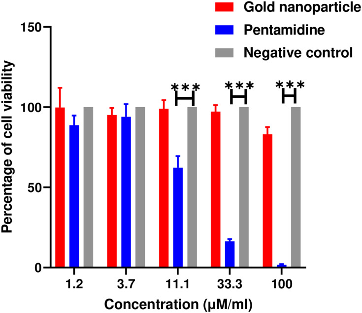
The dose-dependent cytotoxicity of the synthesized GNP in macrophage cell lines (RAW 264.7) was assessed using the MTT assay. The cells were incubated with different concentrations of GNP for 48 h at 37°C, 5% CO2 while the pentamidine (as a reference antileishmanial drug) treated cells served as a positive control. The untreated RAW 264.7 cells (negative control) showed 100% formazan crystal formation upon subsequent addition of MTT. GNP exhibited negligible toxicity even at 100 µM unlike pentamidine, resulting in a significant decrease in cell viability (Figure 2). The cytotoxicity data revealed that GNPs have excellent biocompatibility in macrophage cell lines.
Figure 2.
Cytotoxicity of gold nanoparticle on RAW 264.7 macrophage cell line by MTT assay. Pentamidine was used as a reference antileishmanial drug. Macrophages alone served as a negative control. Data (n=3) are represented as mean ± SE. Significance is calculated as ***P < 0.001, pentamidine versus negative control.
Antileishmanial Effect of Gold Nanoparticles (GNP) on L. donovani Promastigotes
Time-Dependent Killing of L. donovani Promastigotes
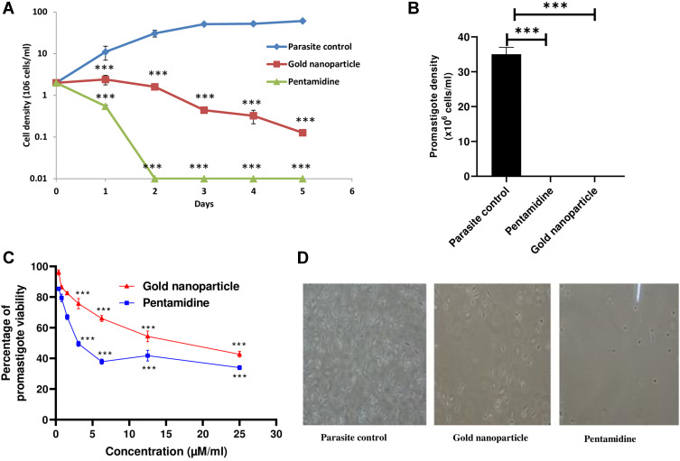
The growth kinetics of L. donovani promastigotes were studied after incubation with GNPs and pentamidine for 5 days. L. donovani growth decreased significantly and cell density reached zero within 5 days of incubation with GNPs (75 µM), whereas all parasites were not-motile within 48 h after pentamidine treatment. The untreated parasites proliferated at a normal rate (Figure 3A).
Figure 3.
Antileishmanial activity of gold nanoparticles against L. donovani. (A) Time-dependent activity of gold nanoparticle or pentamidine (75 µM) against promastigotes compared to untreated parasite control, (B) growth reversibility assay at 4 days in fresh medium after the withdrawal of spent culture medium, (C) dose-dependent killing of L. donovani promastigotes by gold nanoparticles or pentamidine, (D) cellular morphology of treated promastigotes at 40× magnification. All data (n=3) are represented as mean ± SE. Significance is represented as ***P < 0.001 versus untreated parasite control.
Growth Reversibility Assay
Seven days after incubation with GNPs, the parasites were washed and cultured in fresh M199 medium for a further 4 days. The GNP-treated parasites showed a slight reversal of growth, whereas the untreated control promastigotes exhibited visible reversibility. This confirmed the antileishmanial effect of GNP as also observed with pentamidine (Figure 3B).
Dose-Dependent Killing of L. donovani Promastigotes
GNPs demonstrated a dose-dependent activity on L. donovani promastigotes. The 50% inhibitory concentration (IC50) was calculated by linear regression analysis of the plot between percent viability versus nanoparticle concentration after 4 days of incubation of promastigotes with GNPs or pentamidine (Figure 3C). The IC50 of GNP and pentamidine in promastigotes was found to be 18.4 ± 0.4 µM and 3.5 ± 0.2 µM, respectively (Table 1).
Table 1.
Effect of GNP on L. donovani Promastigotes and Amastigotes
| Test Sample | IC50 (µM) Promastigotes | IC50 (µM) Amastigotes |
|---|---|---|
| GNP | 18.4 ± 0.4 | 5.0 ± 0.3 |
| Pentamidine | 3.5 ± 0.2 | 1.5 ± 0.7 |
Notes: IC50 values of GNP and pentamidine (a reference antileishmanial drug) on the promastigote and amastigote forms of L. donovani are shown. Data (n=3) is represented as mean ± SE.
Morphology of Gold Nanoparticles (GNP)-Treated Promastigotes
The cellular morphology of the parasites studied by phase contrast microscopy confirmed that the promastigotes were shrunken, round, and non-motile upon incubation with GNPs and pentamidine in contrast to the untreated control parasites that were elongated, slender, and motile (Figure 3D).
Antileishmanial Activity of Gold Nanoparticles (GNP) Against L. donovani Amastigotes
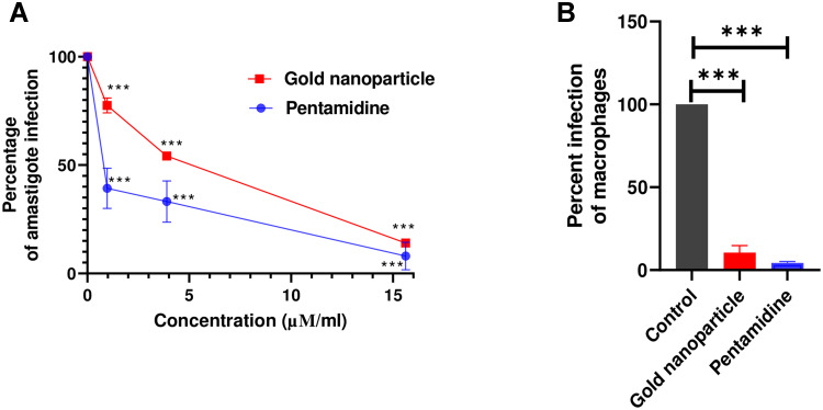
The effect of GNP on macrophage-infected amastigotes was investigated and compared with the standard antileishmanial drug pentamidine. The percent infection by amastigote (with respect to the infected control) upon treatment with GNPs is presented in Figure 4A. GNPs were effective against the amastigote form of the parasites with IC50 of 5.0 ± 0.3 µM, which was close to that of pentamidine having IC50 of 1.5 ± 0.7 µM (Table 1). GNP at a concentration of 15 µM reduced infected macrophages to around 10% compared to untreated infected control (Figure 4B).
Figure 4.
Antileishmanial activity of the gold nanoparticles against L. donovani amastigotes (A) dose-dependent activity of gold nanoparticle or pentamidine on L. donovani amastigotes presented as amastigote infectivity (percentage of infected control), (B) infected macrophages (percentage of infected control) at 15µM GNP. All the data (n = 3) are represented as mean ± SE. ***P < 0.001 with respect to the untreated infected control.
Discussion
There is a dire need to develop new therapeutic modalities for the treatment of VL. Exploring nanoparticles is one way to overcome the problem of drug resistance and toxicity of antileishmanial compounds on healthy cells.10,45 The high surface area to volume ratio of nanoparticles may lead to an increase in the area of contact with parasites to mediate the antileishmanial effect.24 The limitations of current therapeutic drugs coupled with the absence of any effective vaccine and the remarkable activity of GNP against various Leishmania species27,28,46 prompted us to investigate its potential against L. donovani. Few studies have investigated the efficacy of GNP against the visceralizing species, L. donovani. In one study, it has been tested against the axenic amastigotes32 and in another against the promastigotes.36 However, the intracellular macrophage-amastigote model is so far the gold standard in the search for in vitro antileishmanial drug screening, as it involves the host cell-mediated effects,47 including macrophage’s microbicidal activities.48 With axenic amastigotes, the natural niche of the parasite is absent, unlike the macrophage-infected amastigotes.49 Pentavalent antimonials have been reported to show antileishmanial activity only against intracellular amastigotes, as reduction to trivalent antimonials, critical for leishmanicidal activity, occurs only with fully differentiated intramacrophagic amastigotes.50 On the contrary, free-living axenic amastigotes have been reported to retain some promastigote-like traits and therefore require continuous monitoring for amastigote-like features and have also been found to contribute to high false-positive rates in drug sensitivities.50 Furthermore, the IC50 values for intramacrophagic amastigotes have been reported to be much higher than those for the axenic amastigotes.34,35
In this study, we synthesized and characterized citrate-stabilized GNP and assessed its potential as an antileishmanial agent in vitro against L. donovani. The GNP showed a strong surface plasmon band at 520 nm that falls in the visible range, thus confirming the synthesis of nanoparticles.51 The hydrodynamic diameter, PDI and zeta potential of the synthesized GNP were 56.0 ± 10 nm, 0.3 ± 0.1 and −27.0 ± 3 mV, respectively. The zeta sizer measures the hydrodynamic size; therefore, to reveal the accurate size of GNPs, atomic force microscopy was carried out. The AFM data revealed that the GNPs were spherical in shape, having a smooth morphology and the average size of the GNPs was around 20 ± 5 nm, that is, about two times smaller when analyzed by AFM compared to DLS.
The important finding is that GNP exerted significant antileishmanial activity against extracellular promastigotes and intracellular amastigotes. There was a decrease in the proliferation of L. donovani promastigotes upon incubation with increasing concentrations of nanoparticles. GNPs inhibited promastigote growth in a time-dependent manner and the number of parasites approached zero within 5 days, in contrast to untreated control promastigotes that proliferated at a normal rate. The treated promastigotes after the withdrawal of GNPs and culture in fresh M199 medium for 4 days showed scrimpy growth compared to the untreated promastigotes, corroborating the antileishmanial effect of GNP, as also observed with pentamidine. Pentamidine, the second line of treatment for the various forms of leishmaniasis, was used as the reference drug in our studies. Phase-contrast microscopic images of L. donovani promastigotes treated with GNP demonstrated certain morphological changes that were very similar to the signs of programmed cell death.23 The treated promastigotes became aflagellated, oval, or round and shrunken in contrast to the untreated parasites. We also demonstrated a potent activity of GNPs against intracellular amastigotes of L. donovani (IC50 5.0 ± 0.3 µM), which was 3.6 times less compared to that on promastigotes (IC50 18 ± 0.4 µM). Thus, the amastigote forms of the parasites were more susceptible to GNPs.
It is a well-established fact that the uptake of GNPs, as well as toxicity within the cells, depends on the size, shape, and surface functionality. In the case of macrophages, endocytosis is the most effortless and energetically favored pathway for uptake of nanoparticles. Receptor-mediated endocytosis has been reported for the entry and transport of GNPs into cells.52 The “rule of thumb” states that particles larger than 200 nm are internalized through phagocytosis, while particles smaller than 200 nm enter through micropinocytosis.53 Since the synthesized GNPs in our study are less than 100 nm, they are likely to enter macrophages via pinocytosis.54 However, phagocytosis or pinocytosis, or both, may have been involved in the uptake of GNPs.55
Additionally, it was observed that macrophages exposed to GNPs did not generate nitric oxide (NO), the major microbicidal molecule (data not shown). Gold atoms are known to exhibit a low reactivity, in particular, regarding oxidation by dissolved oxygen. Thus, less free ions are released from GNPs and fewer reactive oxygen species (ROS) are generated. Consequently, ROS generation and ion release are supposed to play a subordinate role in activity of GNPs, while direct interaction with the cell membrane and binding to intracellular components may represent the key mechanisms of GNP activity.56 The absence of ROS has also been observed in the GNP-mediated killing of bacterial cells.57 On the contrary, the NO-mediated immunomodulatory effect of L. amazonensis-infected macrophages has been reported to induce leishmanicidal activity by silver nanoparticles.23 Bimetallic gold-silver nanoparticles have been found to induce antileishmanial activity against L. donovani by apoptosis-like death and ROS generation.58 We conclude ROS-independent killing of Leishmania parasites by GNPs, which may be mediated by apoptosis.
Direct adherence of nanoparticles to the cell surface causing disruption of cell membrane has also been suggested.56 GNPs have been reported to damage proteins and organelles after cellular entry. GNPs may also induce the secretion of hypoxic macrophages. In the absence of oxygen, GNPs can aggregate within macrophages, lowering cell proliferation and can induce the activation of macrophage inflammasome, which promotes the inflammatory response.59 The high surface area-to-volume ratio and the small size of the nanoparticles enhances their interaction with microbes and thus mediate a wide range of antimicrobial effects.60 The probable mechanism of antileishmanial activity of GNP may be due to the surface area of GNP in contact with L. donovani promastigotes and amastigotes, which can damage the membrane or bind to proteins or DNA, making them non-functional.61 The enhanced activity of GNP towards amastigotes may be due to more interaction of GNP with the cell surface receptors of amastigotes than with those of promastigotes. Our results also demonstrated that GNPs are devoid of cytotoxicity in the RAW 264.7 macrophage cell line. The ROS-independent mechanism of action of GNP partly explains the non-toxicity of GNP to mammalian cells.62,63
Thus, our study confirms the dose and time-dependent antileishmanial effect of GNP against both forms of L. donovani: the promastigotes in vitro and intracellular amastigotes ex vivo. GNP stands out as a new therapeutic option that can be considered for future in vivo investigations, depicting a potential treatment for VL. The leishmanicidal potential of functionalized silver nanoparticles also opens the door for the functionalization of GNPs.64 The construction of nanogold with antileishmanial molecules could represent another effective strategy, as has been reported for CL.29 Further,; the synergistic antileishmanial effect of nanoassemblies of chitosan-titanium dioxide loaded with glucantime,46 and Amphotericin B conjugated GNPs65 also open the possibility of exploring GNPs to complement existing antileishmanial chemotherapy at lower doses of drugs.
Conclusions
The GNP synthesized by the microwave-assisted heating method was evaluated for its activity against L. donovani and toxicity on the macrophage cell line. The ROS-independent antileishmanial activity of GNP may have been mediated via parasite membrane disruption and/or apoptosis, which needs to be explored. Our in vitro results encourage the study of GNP against L. donovani in vivo and may have prospects in therapeutic applications of functionalized and/or drug conjugated GNP. However, a deeper understanding of the biological effects of GNPs is required to develop safe and effective nanomedicines.
Acknowledgment
The authors thank Prof. T. Basu at the Department of Biochemistry, University of Kalyani, West Bengal, for providing the facility of the Zeta sizer and AFM. The authors are grateful to the Deanship of Scientific Research, King Saud University for funding through Vice Deanship of Scientific Research Chairs. This research was also funded by the Deanship of Scientific Research at Princess Nourah bint Abdulrahman University through the Fast-track Research Funding Program.
Disclosure
The authors report no conflicts of interest in this work.
References
- 1.Kumar A, Saurabh S, Jamil S, Kumar V. Intensely clustered outbreak of visceral leishmaniasis (kala-azar) in a setting of seasonal migration in a village of Bihar, India. BMC Infect Dis. 2020;20(1):10. doi: 10.1186/s12879-019-4719-3 [DOI] [PMC free article] [PubMed] [Google Scholar]
- 2.Mandal R, Kesari S, Kumar V, Das P. Trends in spatio-temporal dynamics of visceral leishmaniasis cases in a highly-endemic focus of Bihar, India: an investigation based on GIS tools. Parasit Vectors. 2018;11(1):220. doi: 10.1186/s13071-018-2707-x [DOI] [PMC free article] [PubMed] [Google Scholar]
- 3.Kumari S, Dhawan P, Panda PK, Bairwa M, Pai VS. Rising visceral leishmaniasis in Holy Himalayas (Uttarakhand, India) – a cross-sectional hospital-based study. J Family Med Prim Care. 2020;9(3):1362. doi: 10.4103/jfmpc.jfmpc_1174_19 [DOI] [PMC free article] [PubMed] [Google Scholar]
- 4.van Griensven J, Diro E. Visceral leishmaniasis: recent advances in diagnostics and treatment regimens. Infect Dis Clin. 2019;33(1):79–99. [DOI] [PubMed] [Google Scholar]
- 5.Srivastava S, Mishra J, Gupta AK, Singh A, Shankar P, Singh S. Laboratory confirmed miltefosine resistant cases of visceral leishmaniasis from India. Parasit Vectors. 2017;10(1):49. doi: 10.1186/s13071-017-1969-z [DOI] [PMC free article] [PubMed] [Google Scholar]
- 6.Goswami RP, Rahman M, Das S, Tripathi SK, Goswami RP. Combination therapy against Indian visceral Leishmaniasis with Liposomal Amphotericin B (FungisomeTM) and short-course miltefosine in comparison to miltefosine monotherapy. Am J Trop Med Hyg. 2020;103:308. [DOI] [PMC free article] [PubMed] [Google Scholar]
- 7.Bruni N, Stella B, Giraudo L, Della Pepa C, Gastaldi D, Dosio F. Nanostructured delivery systems with improved leishmanicidal activity: a critical review. Int J Nanomed. 2017;12:5289. doi: 10.2147/IJN.S140363 [DOI] [PMC free article] [PubMed] [Google Scholar]
- 8.de Souza A, Marins DSS, Mathias SL, et al. Promising nanotherapy in treating leishmaniasis. Int J Pharm. 2018;547(1–2):421–431. doi: 10.1016/j.ijpharm.2018.06.018 [DOI] [PubMed] [Google Scholar]
- 9.Dar MJ, Din FU, Khan GM. Sodium stibogluconate loaded nano-deformable liposomes for topical treatment of leishmaniasis: macrophage as a target cell. Drug Deliv. 2018;25(1):1595–1606. doi: 10.1080/10717544.2018.1494222 [DOI] [PMC free article] [PubMed] [Google Scholar]
- 10.Ray L, Karthik R, Srivastava V, et al. Efficient antileishmanial activity of amphotericin B and piperine entrapped in enteric coated guar gum nanoparticles. Drug Deliv Transl Res. 2021;11:118–130. [DOI] [PubMed] [Google Scholar]
- 11.Allahverdiyev AM, Abamor ES, Bagirova M, et al. Antileishmanial effect of silver nanoparticles and their enhanced antiparasitic activity under ultraviolet light. Int J Nanomedicine. 2011;6:2705–2714. doi: 10.2147/IJN.S23883 [DOI] [PMC free article] [PubMed] [Google Scholar]
- 12.Liao S, Zhang Y, Pan X, et al. Antibacterial activity and mechanism of silver nanoparticles against multidrug-resistant Pseudomonas aeruginosa. Int J Nanomed. 2019;14:1469. doi: 10.2147/IJN.S191340 [DOI] [PMC free article] [PubMed] [Google Scholar]
- 13.Zhou Y, Kong Y, Kundu S, Cirillo JD, Liang H. Antibacterial activities of gold and silver nanoparticles against Escherichia coli and bacillus Calmette-Guerin. J Nanobiotechnol. 2012;10:19. doi: 10.1186/1477-3155-10-19 [DOI] [PMC free article] [PubMed] [Google Scholar]
- 14.Araujo EA, Andrade NJ, da Silva LH, et al. Antimicrobial effects of silver nanoparticles against bacterial cells adhered to stainless steel surfaces. J Food Prot. 2012;75(4):701–705. doi: 10.4315/0362-028X.JFP-11-276 [DOI] [PubMed] [Google Scholar]
- 15.Adeyemi OS, Molefe NI, Awakan OJ, et al. Metal nanoparticles restrict the growth of protozoan parasites. Artif Cells Nanomed Biotechnol. 2018;46(sup3):S86–S94. doi: 10.1080/21691401.2018.1489267 [DOI] [PubMed] [Google Scholar]
- 16.Colotti G, Ilari A, Fiorillo A, et al. Metal‐based compounds as prospective antileishmanial agents: inhibition of trypanothione reductase by selected gold complexes. ChemMedChem. 2013;8(10):1634–1637. [DOI] [PubMed] [Google Scholar]
- 17.Ilari A, Baiocco P, Messori L, et al. A gold-containing drug against parasitic polyamine metabolism: the X-ray structure of trypanothione reductase from Leishmania infantum in complex with auranofin reveals a dual mechanism of enzyme inhibition. Amino Acids. 2012;42(2–3):803–811. doi: 10.1007/s00726-011-0997-9 [DOI] [PMC free article] [PubMed] [Google Scholar]
- 18.Baiocco P, Ilari A, Ceci P, et al. Inhibitory effect of silver nanoparticles on trypanothione reductase activity and Leishmania infantum proliferation. ACS Med Chem Lett. 2010;2(3):230–233. doi: 10.1021/ml1002629 [DOI] [PMC free article] [PubMed] [Google Scholar]
- 19.Farajzadeh S, Parizi MH, Haghdoost AA, et al. Comparison between intralesional injection of zinc sulfate 2% solution and intralesional meglumine antimoniate in the treatment of acute old world dry type cutaneous leishmaniasis: a randomized double-blind clinical trial. J Parasit Dis. 2016;40(3):935–939. doi: 10.1007/s12639-014-0609-1 [DOI] [PMC free article] [PubMed] [Google Scholar]
- 20.Ghasemi E, Ghaffarifar F, Dalimi A, Sadraei J. In-vitro and in-vivo antileishmanial activity of a compound derived of platinum, oxaliplatin, against Leishmania major. Iran J Pharm Res. 2019;18(4):2028. [DOI] [PMC free article] [PubMed] [Google Scholar]
- 21.Afridi MS, Hashmi SS, Ali GS, Zia M, Abbasi BH. Comparative antileishmanial efficacy of the biosynthesised ZnO NPs from genus Verbena. IET Nanobiotechnol. 2018;12(8):1067–1073. doi: 10.1049/iet-nbt.2018.5076 [DOI] [PMC free article] [PubMed] [Google Scholar]
- 22.Allahverdiyev AM, Abamor ES, Bagirova M, et al. Investigation of antileishmanial activities of Tio2@Ag nanoparticles on biological properties of L. tropica and L. infantum parasites, in vitro. Exp Parasitol. 2013;135(1):55–63. doi: 10.1016/j.exppara.2013.06.001 [DOI] [PubMed] [Google Scholar]
- 23.Fanti JR, Tomiotto-Pellissier F, Miranda-Sapla MM, et al. Biogenic silver nanoparticles inducing Leishmania amazonensis promastigote and amastigote death in vitro. Acta Trop. 2018;178:46–54. doi: 10.1016/j.actatropica.2017.10.027 [DOI] [PubMed] [Google Scholar]
- 24.Gold K, Slay B, Knackstedt M, Gaharwar AK. Antimicrobial activity of metal and metal‐oxide based nanoparticles. Adv Ther. 2018;1(3):1700033. doi: 10.1002/adtp.201700033 [DOI] [Google Scholar]
- 25.Kong FY, Zhang JW, Li RF, Wang ZX, Wang WJ, Wang W. Unique roles of gold nanoparticles in drug delivery, targeting and imaging applications. Molecules. 2017;22(9):1445. doi: 10.3390/molecules22091445 [DOI] [PMC free article] [PubMed] [Google Scholar]
- 26.Torabi N, Mohebali M, Shahverdi AR, et al. Nanogold for the treatment of zoonotic cutaneous leishmaniasis caused by Leishmania major (MRHO/IR/75/ER): an animal trial with methanol extract of Eucalyptus camaldulensis. JPHS. 2011;1:15–18. [Google Scholar]
- 27.Sazgarnia A, Taheri AR, Soudmand S, Parizi AJ, Rajabi O, Darbandi MS. Antiparasitic effects of gold nanoparticles with microwave radiation on promastigotes and amastigotes of Leishmania major. Int J Hyperth. 2013;29(1):79–86. doi: 10.3109/02656736.2012.758875 [DOI] [PubMed] [Google Scholar]
- 28.Jebali A, Anvari-Tafti MH. Hybridization of different antisense oligonucleotides on the surface of gold nanoparticles to silence zinc metalloproteinase gene after uptake by Leishmania major. Colloids Surf B Biointerfaces. 2015;129:107–113. doi: 10.1016/j.colsurfb.2015.03.029 [DOI] [PubMed] [Google Scholar]
- 29.Abamor ES, Allahverdiyev AM. A nanotechnology based new approach for chemotherapy of cutaneous leishmaniasis: TIO2@ AG nanoparticles–Nigella sativa oil combinations. Exp Parasitol. 2016;166:150–163. doi: 10.1016/j.exppara.2016.04.008 [DOI] [PubMed] [Google Scholar]
- 30.Corpas-López V, Díaz-Gavilán M, Franco-Montalbán F, et al. A nanodelivered Vorinostat derivative is a promising oral compound for the treatment of visceral leishmaniasis. Pharmacol Res. 2019;139:375–383. doi: 10.1016/j.phrs.2018.11.039 [DOI] [PubMed] [Google Scholar]
- 31.Das S, Roy P, Mondal S, Bera T, Mukherjee A. One pot synthesis of gold nanoparticles and application in chemotherapy of wild and resistant type visceral leishmaniasis. Colloids Surf B Biointerfaces. 2013;107:27–34. doi: 10.1016/j.colsurfb.2013.01.061 [DOI] [PubMed] [Google Scholar]
- 32.Das S, Halder A, Roy P, Mukherjee A. Biogenic gold nanoparticles against wild and resistant type visceral leishmaniasis. Mater Today: Proc. 2018;5(1):2912–2920. [Google Scholar]
- 33.Halder A, Das S, Bera T, Mukherjee A. Rapid synthesis for monodispersed gold nanoparticles in kaempferol and anti-leishmanial efficacy against wild and drug resistant strains. RSC Adv. 2017;7(23):14159–14167. doi: 10.1039/C6RA28632A [DOI] [Google Scholar]
- 34.Berry SL, Hameed H, Thomason A, et al. Development of NanoLuc-PEST expressing Leishmania mexicana as a new drug discovery tool for axenic-and intramacrophage-based assays. PLoS Negl Trop Dis. 2018;12(7):e0006639. doi: 10.1371/journal.pntd.0006639 [DOI] [PMC free article] [PubMed] [Google Scholar]
- 35.Mahmoud AB, Danton O, Kaiser M, Khalid S, Hamburger M, Mäser P. HPLC-based activity profiling for antiprotozoal compounds in Croton gratissimus and Cuscuta hyalina. Front Pharmacol. 2020;11:1246. doi: 10.3389/fphar.2020.01246 [DOI] [PMC free article] [PubMed] [Google Scholar]
- 36.Ghosh S, Jagtap S, More P, et al. Dioscorea bulbifera mediated synthesis of novel Au core Ag shell nanoparticles with potent antibiofilm and antileishmanial activity. J Nanomater. 2015;16(1):161. [Google Scholar]
- 37.Botteon C, Silva L, Ccana-Ccapatinta G, et al. Biosynthesis and characterization of gold nanoparticles using Brazilian red propolis and evaluation of its antimicrobial and anticancer activities. Sci Rep. 2021;11(1):1–16. doi: 10.1038/s41598-021-81281-w [DOI] [PMC free article] [PubMed] [Google Scholar]
- 38.Gatea F, Teodor ED, Seciu AM, et al. Antitumour, antimicrobial and catalytic activity of gold nanoparticles synthesized by different pH propolis extracts. J Nanopart Res. 2015;17(7):1–13. doi: 10.1007/s11051-015-3127-x [DOI] [Google Scholar]
- 39.Want MY, Islammudin M, Chouhan G, et al. Nanoliposomal artemisinin for the treatment of murine visceral leishmaniasis. Int J Nanomed. 2017;12:2189. doi: 10.2147/IJN.S106548 [DOI] [PMC free article] [PubMed] [Google Scholar]
- 40.Tournebize J, Sapin-Minet A, Schneider R, Boudier A, Maincent P, Leroy P. Simple spectrophotocolorimetric method for quantitative determination of gold in nanoparticles. Talanta. 2011;83(5):1780–1783. doi: 10.1016/j.talanta.2010.12.005 [DOI] [PubMed] [Google Scholar]
- 41.Ramezani F, Jebali A, Kazemi B. A green approach for synthesis of gold and silver nanoparticles by Leishmania sp. Appl Biochem Biotechnol. 2012;168(6):1549–1555. doi: 10.1007/s12010-012-9877-3 [DOI] [PubMed] [Google Scholar]
- 42.Want MY, Islamuddin M, Chouhan G, Dasgupta AK, Chattopadhyay AP, Afrin F. A new approach for the delivery of artemisinin: formulation, characterization, and ex-vivo antileishmanial studies. J Colloid Interface Sci. 2014;432:258–269. doi: 10.1016/j.jcis.2014.06.035 [DOI] [PubMed] [Google Scholar]
- 43.Afrin F, Chouhan G, Islamuddin M, Want MY, Ozbak HA, Hemeg HA. Cinnamomum cassia exhibits antileishmanial activity against Leishmania donovani infection in vitro and in vivo. PLoS Negl Trop Dis. 2019;13(5):e0007227. doi: 10.1371/journal.pntd.0007227 [DOI] [PMC free article] [PubMed] [Google Scholar]
- 44.Chouhan G, Islamuddin M, Want MY, et al. Apoptosis mediated leishmanicidal activity of Azadirachta indicabioactive fractions is accompanied by Th1 immunostimulatory potential and therapeutic cure in vivo. Parasit Vectors. 2015;8(1):183. doi: 10.1186/s13071-015-0788-3 [DOI] [PMC free article] [PubMed] [Google Scholar]
- 45.Mondal S, Roy P, Das S, Halder A, Mukherjee A, Bera T. In vitro susceptibilities of wild and drug resistant Leishmania donovani amastigote stages to andrographolide nanoparticle: role of vitamin E derivative TPGS for nanoparticle efficacy. PLoS One. 2013;8(12):e81492. doi: 10.1371/journal.pone.0081492 [DOI] [PMC free article] [PubMed] [Google Scholar]
- 46.Varshosaz J, Arbabi B, Pestehchian N, Saberi S, Delavari M. Chitosan-titanium dioxide-glucantime nanoassemblies effects on promastigote and amastigote of Leishmania major. Int J Biol Macromol. 2018;107:212–221. doi: 10.1016/j.ijbiomac.2017.08.177 [DOI] [PubMed] [Google Scholar]
- 47.Vermeersch M, da Luz RI, Toté K, Timmermans J-P, Cos P, Maes L. In vitro susceptibilities of Leishmania donovanipromastigote and amastigote stages to antileishmanial reference drugs: practical relevance of stage-specific differences. Antimicrob Agents Chemother. 2009;53(9):3855–3859. doi: 10.1128/AAC.00548-09 [DOI] [PMC free article] [PubMed] [Google Scholar]
- 48.Dias CN, Nunes TAL, de Sousa JMS, et al. Methyl gallate: selective antileishmanial activity correlates with host-cell directed effects. Chem Biol Interact. 2020;320:109026. [DOI] [PubMed] [Google Scholar]
- 49.De Muylder G, Ang KK, Chen S, Arkin MR, Engel JC, McKerrow JH. A screen against Leishmania intracellular amastigotes: comparison to a promastigote screen and identification of a host cell-specific hit. PLoS Negl Trop Dis. 2011;5(7):e1253. doi: 10.1371/journal.pntd.0001253 [DOI] [PMC free article] [PubMed] [Google Scholar]
- 50.De Rycker M, Hallyburton I, Thomas J, et al. Comparison of a high-throughput high-content intracellular Leishmania donovani assay with an axenic amastigote assay. Antimicrob Agents Chemother. 2013;57(7):2913–2922. doi: 10.1128/AAC.02398-12 [DOI] [PMC free article] [PubMed] [Google Scholar]
- 51.Yang Y, Matsubara S, Nogami M, Shi J, Huang W. One-dimensional self-assembly of gold nanoparticles for tunable surface plasmon resonance properties. Nanotechnology. 2006;17(11):2821. doi: 10.1088/0957-4484/17/11/015 [DOI] [Google Scholar]
- 52.Zhang Q, Hitchins VM, Schrand AM, Hussain SM, Goering PL. Uptake of gold nanoparticles in murine macrophage cells without cytotoxicity or production of pro-inflammatory mediators. Nanotoxicology. 2011;5(3):284–295. doi: 10.3109/17435390.2010.512401 [DOI] [PubMed] [Google Scholar]
- 53.Xiang SD, Scholzen A, Minigo G, et al. Pathogen recognition and development of particulate vaccines: does size matter? Methods. 2006;40(1):1–9. doi: 10.1016/j.ymeth.2006.05.016 [DOI] [PubMed] [Google Scholar]
- 54.Naha PC, Chhour P, Cormode DP. Systematic in vitro toxicological screening of gold nanoparticles designed for nanomedicine applications. Toxicol In Vitro. 2015;29(7):1445–1453. doi: 10.1016/j.tiv.2015.05.022 [DOI] [PMC free article] [PubMed] [Google Scholar]
- 55.França A, Aggarwal P, Barsov EV, Kozlov SV, Dobrovolskaia MA, González-Fernández Á. Macrophage scavenger receptor A mediates the uptake of gold colloids by macrophages in vitro. Nanomedicine. 2011;6(7):1175–1188. doi: 10.2217/nnm.11.41 [DOI] [PMC free article] [PubMed] [Google Scholar]
- 56.Godoy-Gallardo M, Eckhard U, Delgado LM, et al. Antibacterial approaches in tissue engineering using metal ions and nanoparticles: from mechanisms to applications. Bioact Mater. 2021;6(12):4470–4490. doi: 10.1016/j.bioactmat.2021.04.033 [DOI] [PMC free article] [PubMed] [Google Scholar]
- 57.Cui Y, Zhao Y, Tian Y, Zhang W, Lü X, Jiang X. The molecular mechanism of action of bactericidal gold nanoparticles on Escherichia coli. Biomaterials. 2012;33(7):2327–2333. doi: 10.1016/j.biomaterials.2011.11.057 [DOI] [PubMed] [Google Scholar]
- 58.Alti D, Veeramohan Rao M, Rao DN, Maurya R, Kalangi SK. Gold–silver bimetallic nanoparticles reduced with herbal leaf extracts induce ROS-mediated death in both promastigote and amastigote stages of Leishmania donovani. ACS Omega. 2020;5(26):16238–16245. doi: 10.1021/acsomega.0c02032 [DOI] [PMC free article] [PubMed] [Google Scholar]
- 59.Zhao P, Li T, Li Z, et al. Preparation of gold nanoparticles and its effect on autophagy and oxidative stress in chronic kidney disease cell model. J Nanosci Nanotechnol. 2021;21(2):1266–1271. doi: 10.1166/jnn.2021.18655 [DOI] [PubMed] [Google Scholar]
- 60.Zhang W, Li Y, Niu J, Chen Y. Photogeneration of reactive oxygen species on uncoated silver, gold, nickel, and silicon nanoparticles and their antibacterial effects. Langmuir. 2013;29(15):4647–4651. doi: 10.1021/la400500t [DOI] [PubMed] [Google Scholar]
- 61.Chaloupka K, Malam Y, Seifalian AM. Nanosilver as a new generation of nanoproduct in biomedical applications. Trends Biotechnol. 2010;28(11):580–588. doi: 10.1016/j.tibtech.2010.07.006 [DOI] [PubMed] [Google Scholar]
- 62.Shukla R, Bansal V, Chaudhary M, Basu A, Bhonde RR, Sastry M. Biocompatibility of gold nanoparticles and their endocytotic fate inside the cellular compartment: a microscopic overview. Langmuir. 2005;21(23):10644–10654. doi: 10.1021/la0513712 [DOI] [PubMed] [Google Scholar]
- 63.Pan Y, Neuss S, Leifert A, et al. Size-dependent cytotoxicity of gold nanoparticles. Small. 2007;3(11):1941–1949. doi: 10.1002/smll.200700378 [DOI] [PubMed] [Google Scholar]
- 64.Isaac-Márquez A, Talamás-Rohana P, Galindo-Sevilla N, et al. Decanethiol functionalized silver nanoparticles are new powerful leishmanicidals in vitro. World J Microbiol Biotechnol. 2018;34(3):38. doi: 10.1007/s11274-018-2420-0 [DOI] [PubMed] [Google Scholar]
- 65.Kumar P, Shivam P, Mandal S, et al. Synthesis, characterization, and mechanistic studies of a gold nanoparticle–amphotericin B covalent conjugate with enhanced antileishmanial efficacy and reduced cytotoxicity. Int J Nanomedicine. 2019;14:6073. doi: 10.2147/IJN.S196421 [DOI] [PMC free article] [PubMed] [Google Scholar]


