Figure 2.
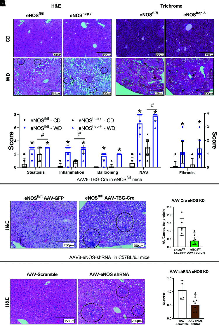
Hepatocellular eNOS deficiency exacerbates histological hepatic steatosis and inflammation. From eNOSfl/fl and eNOShep−/− mice on either a CD or WD for 16 weeks. A: Representative liver H&E and trichrome staining. B: Histological and fibrosis scoring based on H&E and trichrome images (n = 10–17/group). Hepatocyte eNOS knockdown via AAV8-TGB-Cre injection in eNOSfl/fl animals at 10 weeks of age. C: Representative liver H&E staining after 6 weeks of CD feeding and (D) mRNA expression of eNOS in isolated primary hepatocytes (n = 3–5/group). AAV8-TTR-eNOS-shRNA knockdown of hepatocyte eNOS in C57BL/6J mice at 10 weeks of age. E: Representative liver H&E staining after 6 weeks of CD feeding and (F) mRNA expression of eNOS in isolated primary hepatocytes from shRNA eNOS knockdown mice (n = 2-3/group). Data are presented as mean ± SD. *Main effect of diet (P < 0.05 vs. CD). #Main effect of genotype (P < 0.05 vs. eNOSfl/fl). $Significantly different from GFP or AAV-scramble controls (P < 0.05). CD, control diet; H&E, hemotoxylin and eosin; KD, knockdown; NAS, NAFLD activity score; PPIB, cyclophilin B; RQ, relative quotient; WD, Western diet.

