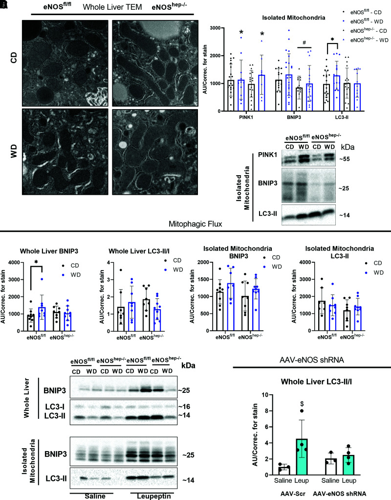
Figure 4.
Hepatocellular eNOS deficiency impairs mitochondrial morphology, quality, and turnover. In eNOSfl/fl and eNOShep−/− mice on either a CD or WD for 16 weeks. A: Representative TEM images of whole liver. B: Protein expression of markers of mitophagy in isolated liver mitochondria (n = 13–23/group) and their representative Western blot images. C: Protein expression of the accumulation of mitophagy proteins after in vivo leupeptin injections in eNOSfl/fl and eNOShep−/− mice on either a CD or WD for 16 weeks: whole-liver BNIP3 and LC3-II/I, and isolated liver mitochondria BNIP3 and LC3-II (n = 5–9/group), and their representative Western blot images. C57BL/6J mice were injected with AAV8-TTR-eNOS-shRNA to knockdown hepatocyte eNOS at 10 weeks of age and then fed a WD for 2 weeks. D: Protein expression of the accumulation of whole-liver LC3-II/I after in vivo leupeptin injection in AAV-scramble and AAV-eNOS-shRNA–injected animals after 2 weeks of WD feeding (n = 3–4/group). Data are presented as mean ± SD. *Effect of diet (P < 0.05 vs. CD). #Main effect of genotype (P < 0.05 vs. eNOSfl/fl). $Significantly different from saline (P < 0.05). AU/correc., arbitrary unit/correction; CD, control diet; TEM, transmission electron microscopy; WD, Western diet.

