Abstract
Aim
The present work aimed to evaluate age-related variations in the dental pulp chamber volume of second molars using cone beam computed tomography (CBCT) imaging, in order to establish a specific mathematical model for second molars and measure its accuracy, especially in the case of Egyptian adults.
Subjects and methods
From 187 subjects between 21–50 years of age, CBCT images of 257 maxillary and 248 mandibular second molars were included. A mathematical model for human age estimation was established. An independent additional set of CBCT images was obtained to test the model's accuracy.
Results
For maxillary and mandibular teeth, R2 for the pooled sexes were 0.51 and 0.52, and SEE were 5.92 and 5.71, respectively. A model for each sex was established, due to the significant difference between them, where R2 was equal to 0.668 and 0.650 in males and 0.46 and 0.48 in females, concerning maxillary and mandibular teeth, respectively. When testing the validation samples, the mean absolute error (MAE) between the actual and estimated ages from the pooled sex model were 4.89 and 4.61 for maxillary and mandibular teeth, respectively.
Conclusion
The pulp chamber volume of second molars is a relatively accurate indicator for age estimation in Egyptian adults.
Keywords: Forensic Odontology; Anthropology population data,
Keywords: Cone beam computed tomography,
Keywords: Second molar,
Keywords: Pulp chamber volume
INTRODUCTION
Forensic odontology deals with the proper evaluation and presentation of dental findings for many purposes, including the identification of living persons and corpses, mainly for age, sex, and race identification. (1, 2) Teeth have gained the attention in personal identification as they follow a well-defined sequential developmental pattern. In addition, they are highly resistant to mechanical, chemical, and physical impacts and time. (3, 4)
Estimating the chronological age of unidentified individuals is a practice based on gradual structural changes in enamel, dentine, and cementum, which occur in teeth throughout life. Factors like attrition and cementum apposition are highly influenced by the life-style of an individual, so they cannot be regarded as reliable parameters. However, secondary dentine deposition and root dentine translucency have been found to be more reliable by many authors. (5)
Secondary dentine formation by odontoblasts starts when the apical part of the root has completely developed, and it is regular when not under the influence of dental caries or other physical/chemical insults to the given tooth. Secondary dentine formation produces age-related decrease in pulp chamber volume that is variable among different populations and both sexes. (6-8) Many radiological techniques have been introduced for quantification of dental pulp chamber volume, where some of them have been two-dimensional studies (e.g., common periapical X-rays and orthopantomograms—OPGs) and others have been three-dimensional studies (computed tomography—CT, micro-CT, and cone beam CT, and x-ray free imaging like Magnetic Resonance). (9) CBCT offers the advantage of causing no magnification errors due to geometric distortion, giving more detailed information, less artefacts, and having a lower radiation dose than CT. (10) Quantification of the pulpal chamber volume from a dental modelling based on CBCT analysis uses a geometrical approximation of the different parts of the tooth, namely root, pulp and crown, which can be assimilated to elliptical-based cones (root and pulp) or elliptical-based truncated cones (crown). This allows a quick evaluation of the volumes of interest on the examined tooth. (11)
On analysis of previous studies, most of them focused on single rooted teeth for assessment of secondary dentine deposition for dental age estimation. In addition, the previous studies on an Egyptian population were performed mostly using a two- dimensional technique and no Egyptian specific equation was established for any type of tooth.
AIM OF THE WORK
This study aimed to evaluate the age-related variations in the dental pulp chamber volume of second molars using CBCT imaging, in order to establish a mathematical model specific for second molars, especially in the case of Egyptian adults, and measure its accuracy.
SUBJECTS AND METHODS
Sample size calculation:
Sample size calculation was performed using G power 3.1.9.2 software with linear multiple regression model, study power 95%, α value 0.05, the R2 value for maxillary and mandibular second molars (0.498, 0.487 respectively). (12) The sample size determined was 40 cases.
Subjects:
Archived CBCT scans from 101 females and 86 males whose age ranged between 21-50 years old were collected retrospectively in duration from January 2018 till January 2019 (table 1). All procedures performed in studies involving human participants were in accordance with the 1964 Helsinki declaration and its later amendments or comparable ethical standards. The work has been approved by the Institutional Review Board (R.19.12.692). All the CBCT images were taken for diagnosis or treatment purposes, thus, there was no unnecessary or additional radiation exposure to the subjects. All participants have given informed consent to conduct radiological examination for different diagnostic reasons. To observe ethical considerations, all information provided was treated as confidential by the researchers, and the names of individuals were not released at the analysis and reporting stage.
Table 1. Age and sex distribution of the samples used for the establishment of the method.
| Age (years) | Male | Female | Maxillary Second Molar | Mandibular Second Molar | ||||
|---|---|---|---|---|---|---|---|---|
| Male | Female | Total | Male | Female | Total | |||
| 21–30 | 38 | 56 | 63 | 70 | 133 | 59 | 65 | 124 |
| 31–40 | 24 | 28 | 27 | 42 | 69 | 34 | 37 | 71 |
| 41–50 | 24 | 17 | 30 | 25 | 55 | 30 | 23 | 53 |
| Total | 86 | 101 | 120 | 137 | 257 | 123 | 125 | 248 |
The inclusion criteria of the second molars were completely formed roots, no caries, minimal tooth attrition or abrasion, and no pulpal calcification. [4] The exclusion criteria of the second molars were fractured roots, teeth with any signs of periapical surgery, endodontic access, crown preparation, and fixed prosthodontics. To ensure minimal abrasion or attrition to teeth, all tooth surfaces were covered by enamel on the radiograph. Both maxillary and mandibular second molars were included for analysis. Although some authors used only one tooth per subject (13) others have selected more than one tooth of the same type per subject. (14)
Image acquisition:
CBCT unit Planmeca (Quantitative Radiology, Helsinki, Finland) was used for obtaining the image. The exposure parameters of the CBCT image were 120 kVp, 4.19–107.39 mAs, in accordance with the subjects’ size and field of view (FOV). The selected FOV was 5x5 cm. The acquired images were subsequently reconstructed with a voxel-size of 0.15 × 0.15 × 0.15 mm and exported as DICOM datasets.
Image segmentation:
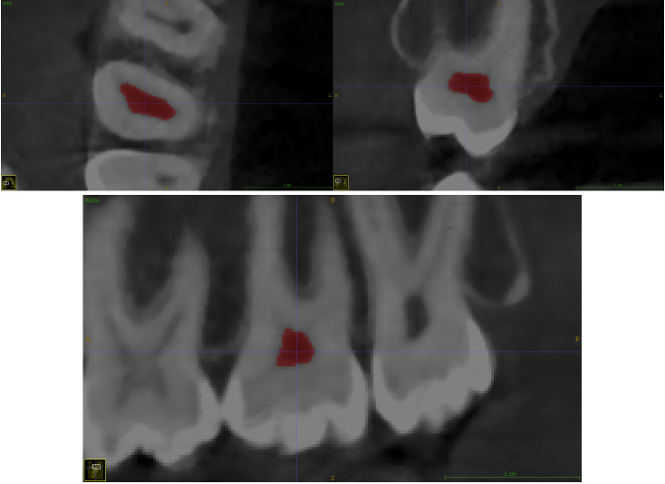
The software ITK-SNAP 3.8 (open source software, www.itksnap.org) was used for measuring pulp chamber volumes using automatic segmentation or seed region-growing To apply it, the observer sets up a “seed” inside the structure to reconstruct, in this case the pulp chamber. This seed grows and a volume is finally obtained. (15)
Segmenting the chamber space is based on difference of the gray levels corresponding to surrounding dentine tissue (high intensity structure) and the present pulp tissue (low intensity tissue). Succeeding the image opening in software, the intensity of gray level values of the pulp chamber space and the surrounding dentine was checked at different levels and places to determine the least value at both structures to determine “lower and upper thresholds”. For the lower threshold, the darkest point at the pulp chamber was selected as it represents the area with no calcification and represents the gray level of the soft tissue. For the upper threshold, the lowest value at dentine that visually surrounds the pulp space at the CBCT was recorded. Determination of thresholds allows the software to select voxels with gray levels lying in between the lower and the upper thresholds automatically which is corresponding only to pulp chamber space. To decrease the calculation errors, the “regions of interest” (ROIs) tool was used to limit the image boundaries to the lateral walls, pulp floor, and pulp roof only to prevent the segmentation tool to select voxels at the radicular pulp space in case of presence of wide radicular canals. After proper evaluation, each segmented area was three-dimensionally reconstructed and the volume was digitally calculated by the software (figure 1).
Figure 1.
Mean value of parameter C3 and C4 from the CV body by chronological age group
Model establishment:
In order to establish an Egyptian specific mathematical model for the estimation of human age, linear regression analysis was conducted with age as the dependent variable and pulp chamber volume as the independent variable, The age used in the model was approximated to a lower age if less than 6 months and to higher age if more than 6 months.
Inter-observer and intra-observer variability:
All the measurements were carried out by the same examiner. To test inter-observer reproducibility, random samples of 40 teeth were re-examined by another examiner. To test intra-observer reproducibility, random samples of 40 teeth were re-examined by the same examiner after a one month interval.
Model validation:
Another group of CBCT images of 38 maxillary and 28 mandibular second molars (age 23-49 years old) was collected in order to validate the established models. After obtaining the pulp chamber volume, the mathematical model was used to obtain the estimated age. From both the estimated and actual age, the mean absolute error (MAE) and root mean square error (RMSE) were calculated to assess the accuracy of the model.
Statistical analysis:
The data were analyzed using SPSS program Standard version 21. The normality of the data was first tested with a one sample Kolmogorov–Smirnov test. Continuous variables were presented as mean ± SD (standard deviation). The intra-class correlation coefficient (ICC) was calculated for assessment of the inter- and intra-observer variability. A paired t-test was used to compare the pulp chamber volume between the right and left second molars in the same subject. Pulp chamber volumes were compared with Student’s t-test for the two groups (i.e., between both sexes) or via a one-way ANOVA for multiple groups. Pearson correlation was used to assess the relationship between dental pulp chamber volume and age. The significant variables of the analysis were entered into linear regression models to predict the most significant determinants and to control for possible interactions and confounding effects. For all the statistical tests mentioned above, the threshold of significance was fixed at a 5% level (p-value).
RESULTS
The ICC was strong (0.917, 0.979) for the inter-observer and intra-observer cases, respectively. No significant differences were found between the right- and left-sided teeth (p-values of 0.333 and 0.731 for maxillary and mandibular second molars, respectively), thus, both of them were included.
Results showed significant difference in pulp chamber volume between both sex (P-value 0.001 in maxillary teeth and 0.004 in mandibular teeth) and between different age groups (p-value 0.001) (tables 2 and 3)
Table 2. Mean value of the pulp chamber volume of maxillary second molars.
| Age (years) | Volume of Maxillary Second Molar (mm3) | P-value | |||||
|---|---|---|---|---|---|---|---|
| Male | Female | ||||||
| Min | Max | Mean ± SD | Min | Max | Mean ± SD | ||
| 21–30 | 19.46 | 46.63 | 29.35 ± 5.21 | 13.00 | 41.98 | 25.25 ± 5.79 | 0.002*c |
| 31–40 | 18.00 | 29.03 | 22.91 ± 3.48 | 13.38 | 32.14 | 19.83 ± 4.45 | |
| 41–50 | 10.47 | 23.34 | 15.6 ± 3.17 | 7.43 | 19.67 | 13.44 ± 3.50 | |
| P-value | <0.001*a | <0.001*b | |||||
(a) Comparison between different age groups in males (ANOVA test); (b) comparison between different age groups in females (ANOVA test); (c) comparison between both sexes (t-test). * significant results were found where p <0.05.
Table 3. Pulp chamber volume of mandibular second molars.
| Age (years) | Volume of Mandibular Second Molar (mm3) | P-value | |||||
|---|---|---|---|---|---|---|---|
| Male | Female | ||||||
| Min | Max | Mean ± SD | Min | Max | Mean ± SD | ||
| 21–30 | 20.40 | 48.39 | 29.59 ± 5.84 | 13.89 | 40.55 | 25.24 ± 6.154 | 0.004*c |
| 31–40 | 14.94 | 30.63 | 20.82 ± 3.56 | 13.61 | 31.42 | 18.14 ± 3.97 | |
| 41–50 | 10.26 | 24.24 | 15.1 ± 3.12 | 7.68 | 22.61 | 13.24 ± 4.00 | |
| P-value | <0.001*a | <0.001*b | |||||
(a) Comparison between different age groups in males (ANOVA test); (b) comparison between different age groups in females (ANOVA test); (c) comparison between both sexes (t-test). * significant results were found where p <0.05.
The r value for maxillary teeth were –0.82, –0.69, and –0.71, and for mandibular teeth they were –0.81, –0.70 –0.72 in males, females, and the pooled sex, respectively (Table 4).
Table 4. Correlation coefficient (r), coefficient of determination (R2) and standard error of estimate (SEE) for the male, female, and pooled sex samples.
| Pooled Sex | Male | Female | |||||||
|---|---|---|---|---|---|---|---|---|---|
| r | R2 | SEE | r | R2 | SEE | r | R2 | SEE | |
| Maxillary | -0.71 | 0.51 | 5.92 | –0.82 | 0.67 | 4.89 | - 0.68 | 0.46 | 6.20 |
| Mandibular | -0.72 | 0.52 | 5.71 | –0.81 | 0.65 | 4.87 | -0.70 | 0.48 | 5.87 |
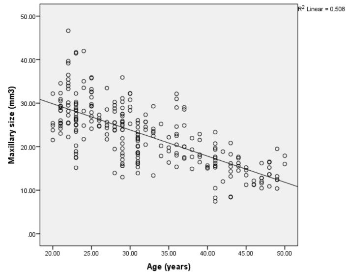
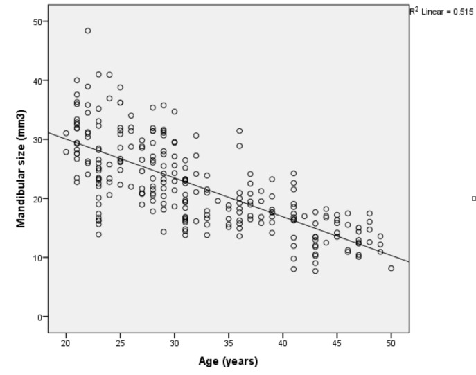
The regressions analysis was statistically significant (<p = 0.001). Figure 2 and 3 shows a scatterplot distribution between age and the pulp chamber volume of maxillary and mandibular second molars. In the establishment of the linear regression model, the coefficient of determination (R2) and standard error of estimate were calculated R2 was for maxillary teeth were 0.68, 0.46, and 0.51, and for mandibular teeth they were 0.65, 0.48, 0.52 in males, females, and the pooled sex, respectively (Table 4). The established mathematical models are shown in Table 5.
Figure 2.
Scatterplot distribution between age and the pulp chamber volume of maxillary second molars.
Figure 3.
Scatterplot distribution between age and the pulp chamber volume of mandibular second molars.
Table 5. Established mathematical model for age estimation via the pulp chamber volume of maxillary and mandibular second molars.
| Maxillary Second Molar | Unknown sex | Age = 50.985 – 0.846 × pulp chamber volume |
|---|---|---|
| Male | Age = 55.626 – 0.956 × pulp chamber volume | |
| Female | Age = 49.375 – 0.851 × pulp chamber volume | |
| Mandibular Second Molar | Unknown sex | Age = 49.261 – 0.783 × pulp chamber volume |
| Male | Age = 52.933 – 0.858 × pulp chamber volume | |
| Female | Age = 47.697 – 0.798 × pulp chamber volume |
After the establishment of the mathematical models, another group of CBCT were used for their validation. The MAE and RMSE of the estimated and actual ages are shown in Table 6.
Table 6. Mean absolute error (MAE) and root mean square error (RMSE) between the actual age and estimated age, using established models.
| Maxillary Second Molar | Mandibular Second Molar | ||
|---|---|---|---|
| Pooled Sex | Number (teeth) | 38 | 28 |
| MAE (mm3) | 4.89 | 4.61 | |
| RMSE (mm3) | 6.08 | 5.97 | |
| Male | Number (teeth) | 15 | 9 |
| MAE (mm3) | 3.40 | 1.33 | |
| RMSE | 4.24 | 1.94 | |
| Female | Number (teeth) | 23 | 19 |
| MAE (mm3) | 4.77 | 4.89 | |
| RMSE (mm3) | 5.86 | 5.871 |
DISCUSSION
From human teeth, molars were selected in the present study as they have a wider pulp chamber cavity, allowing better delineation of the pulp chamber borders in three-dimensional image-based segmentation as compared to other teeth. Moreover, the anatomical position of molars at a planar field in the posterior part of the mouth enables the attainment of clear and accurate three-dimensional images after reconstruction. Specifically, the second molar was chosen due to its morphological stability and less common cases of caries and attrition when compared to first molar. [16] In addition, in the study conducted by Ge et al. in 2016, they found that among the 13 types of teeth, second molars were the best for age identification. (12)
When assessing a pulp chamber, volume is more preferred over assessing area or linear measurements, as secondary dentine apposition is not homogenously distributed along all pulp surfaces. In molar teeth, it appears in greater amounts on the roof and floor of the coronal pulp chamber. [17] When the pulp chamber and tooth volumes were examined in relation to age in a previous study, pulp chamber volume variability was found to better correlate with age than tooth volume. (18)
Regarding the selected age group, previous studies have reported that a change in pulp chamber volume is significant in subjects below 50 years old, after which change becomes relatively slow and insignificant. [16] Therefore, the age group selected was 21–50 years old. In addition, statistically significant shrinkage in pulp chambers occurs after every 10 years. [4] Thus stratification on a 10-year age difference in the present study was performed.
In regard to the assessment method, a three-dimensional technique is preferred for volume assessment. CBCT was selected as it is an easy, low cost, low radiation dose, accurate, and highly reproducible approach that allows for the accurate calculation of tooth volumes. In addition, the technique provides a larger scanning area, in the form of a multi-planar cross-sectional dataset and three-dimensional reconstructions (from a single scan). (14, 19, 20)
According to our knowledge, there are few studies in Egypt assessing dental age estimation and this is the first study that uses pulp chamber volume changes in second molars as an indicator of secondary dentine apposition in relation to age.
The pulp volume was ranging between 7.43-48.39 and SD 3.12-6.154. these values are less than that recorded by Ge et al., 2016 but this is due to the more confined age group selected in our study. (12) When assessing differences in pulp chamber volume by sex, the mean value was higher in males in both maxillary and mandibular teeth, and this is explained by the fact that the size of the teeth may be smaller in females, which could result in a smaller pulp chamber size. (16) In addition, hormonal differences in both sexes may be a reason for such variations. (21) The difference was statistically significant here, necessitating the derivation of a sex-specific equation to predict age. This difference can be explained by Stroud et al., 1994, who found that there is a degree of sexual dimorphism in dentine thickness controlled by the Y chromosome. (22) Our finding is consistent with previous studies that have found a difference between sexes, (4, 12, 16, 23) but goes against other studies (10, 13, 17, 24) which have stated no sex difference.
The previous studies on Egyptian subjects (25-28) reported no sex difference, unlike the present research. This could be attributed to the imaging techniques used, as they used two-dimensional radiographs. When the three-dimensional pulp is reproduced in a two-dimensional radiograph, the edges of the pulp may be blurred due to the cylindrical shape of the pulp of multi-rooted teeth.
As regards pulp chamber volume, the present study showed statistically significant changes in pulp chamber volume with increasing age with moderate to strong negative correlation (r value for maxillary teeth were –0.82, –0.69, and –0.71, and for mandibular teeth they were –0.81, –0.70 –0.72 in males, females, and the pooled sex, respectively). This finding is higher than the previous study on the Davangere population, assessing pulp to tooth area ratio in mandibular second molars using a two-dimensional imaging technique, where r = –0.441, –0.406, and –0.419 among males, females, and pooled sexes respectively. (16) Also, it is higher than a previous study assessing the tooth coronal index (TCI) of mandibular second molars in Egyptian adults using a two-dimensional technique, where r ranged between 0.079–0.17 in different sexes. [27] The correlation was found to be more relevant in males than females and this is similar to previous studies on mandibular second molars in the Davangere population. (16)
The R2 is often used to indicate the association between chronological age and pulp chamber volume. In previous studies, R2 was highly variable depending on the tooth type, sample size, age distribution range, imaging system, and population source. In the present study, the R2 is relatively large, ranging between 0.46-0.68. These values were very close to the previous study that assessed the pulp chamber volume of 13 types of teeth and reported that the second molars showed the best R2 value with respect to age (0.458–0.642), especially in the case of the maxillary teeth. (12)
The observed R2 was stronger for males than females. These findings are similar to the previous study on mandibular second molars in the Davangere population, in which the R2 of the pulp to tooth area ratio in second molars was 0.19, 0.16, and 0.174 for males, females, and pooled sexes, respectively. (16) However, the relationship goes against the other study by Ge et al., 2016, in which males showed a higher R2 value in both mandibular and maxillary second molars, where the R2 values of maxillary teeth were 0.498. 0.491, 0.642, and for mandibular teeth they were 0.487, 0.458, and 0.614, in pooled sexes, males, and females, respectively. (12)
In the present study, the R2 of mandibular teeth was better than maxillary teeth. This goes against the previous study assessing the pulp chamber volume of second molars. (17) However, other studies assessing other types of teeth are similar to our study, with mandibular teeth showing a better R2. Kazmi et al., 2019, found that the R2 values of mandibular and maxillary canine pulp chamber volumes of identified sexes were 0.33 and 0.31, respectively. (23) Also, Biuki et al., 2017, assessed the pulp chamber volumes of anterior teeth and reported an R2 value ranging from 0.65 to 0.75 for maxillary teeth and from 0.60 to 0.76 for mandibular teeth. (29) Similarly, another previous study on first molars reported an R2 value of pulp chamber volume with age of 0.586 and 0.609 for maxillary and mandibular teeth, respectively. (30)
The SEE predicts the deviation of estimated age from the actual age. In the present work, the SEE was calculated and was around 6 years for maxillary teeth and around 5 years for mandibular teeth. These findings are much better than the SEE reported for the pulp to tooth area ratios of mandibular second molars in the Davangere population, which were 11.9 and 12.0 years in males and females, respectively. (16) Also, this is much lower than the SEE reported by Ge et al., 2016, in maxillary (6.75–8) and mandibular (7–8.11) second molars. (17) This difference in SEE could be attributed to the localized age group selected in the current study (21–50 years).
After the establishment of mathematical models, another group of CBCT images was used for validation. The MAE between the actual and estimated ages from the regression model was less than 10, which is accepted in forensic practice. However, the MAE of mandibular teeth in males was relatively small. The RMSE is the standard deviation of the residuals (prediction errors). Residuals are a measure of how far from the regression line data points are found. The RMSE was very close to that of the SEE from regression model, except for mandibular teeth in males, in which the RMSE was relatively small. This extremely low value of both the MAE and RMSE in mandibular teeth in males can be attributed to the relatively small number of teeth used from this group in the validation procedure.
One of the major limitations of the present study is unavailability of micro-CT for the assessment of the segmentation accuracy of CBCT. In addition, a smaller number of teeth was obtained from older people due to tooth loss and difficulty in retrieving teeth meeting the inclusion criteria. However, this selection was necessary as the volume measurement is the sole purpose. Another limitation was the small number of teeth in the validation sample; thus, it is recommended to perform validation of the established model on a wider scale.
CONCLUSIONS
The present study has investigated the relationship between age and the pulp chamber volume of maxillary and mandibular second molars. There was significant difference between both sexes and between maxillary and mandibular second molars. The established model, intended for age estimation in Egyptian adults, has reasonable accuracy, with a relatively large correlation coefficient. It is recommended to use a sex specific equation if possible due its higher accuracy. The study of the pulp chamber volume of multi-rooted teeth, specifically second molars, for age estimation is promising and is worthy of analysis using a larger sample size.
Footnotes
The authors declare that they have no conflict of interest.
REFERENCES
- 1.Bing L, Wu X-P, Shangguan H, Xiao W, Yun K-M. Morphology and volume of maxillary canine pulp cavity for individual age estimation in forensic dentistry. Int J Morphol. 2017;35(3):1058–62. 10.4067/S0717-95022017000300039 [DOI] [Google Scholar]
- 2.Nagi R, Jain S, Agrawal P, Prasad S, Tiwari S, Naidu GS. Tooth coronal index: Key for age estimation on digital panoramic radiographs. J Indian Acad Oral Med Radiol. 2017;30(1):64–7. 10.4103/jiaomr.jiaomr_139_17 [DOI] [Google Scholar]
- 3.Shah PH, Venkatesh R. Pulp/tooth ratio of mandibular first and second molars on panoramic radiographs: An aid for forensic age estimation. J Forensic Dent Sci. 2016;8(2):112–6. 10.4103/0975-1475.186374 [DOI] [PMC free article] [PubMed] [Google Scholar]
- 4.Ge Z-P, Ma R-H, Li G, Zhang J-Z, Ma X-C. Age estimation based on pulp chamber volume of first molars from cone-beam computed tomography images. Forensic Sci Int. 2015;253:133.e1–7. 10.1016/j.forsciint.2015.05.004 [DOI] [PubMed] [Google Scholar]
- 5.Gupta P, Kaur H, Shankari GSM, Jawanda MK, Sahi N. Human age estimation from tooth cementum and dentin. J Clin Diagn Res. 2014;8(4):ZC07–10. 10.7860/JCDR/2014/7275.4221 [DOI] [PMC free article] [PubMed] [Google Scholar]
- 6.Talabani RM, Baban MT, Mahmood MA. Age estimation using lower permanent first molars on a panoramic radiograph: A digital image analysis. J Forensic Dent Sci. 2015;7(2):158–62. 10.4103/0975-1475.154597 [DOI] [PMC free article] [PubMed] [Google Scholar]
- 7.Porto LV, Celestino da Silva J, Pontual AD, Catunda RQ. Evaluation of volumetric changes of teeth in a Brazilian population by using cone beam computed tomography. J Forensic Leg Med. 2015;36:4–9. 10.1016/j.jflm.2015.07.007 [DOI] [PubMed] [Google Scholar]
- 8.Aboshi H, Takahashi T, Komuro T. Age estimation using microfocus X-ray computed tomography of lower premolars. Forensic Sci Int. 2010;200(1-3):35–40. 10.1016/j.forsciint.2010.03.024 [DOI] [PubMed] [Google Scholar]
- 9.Pinchi V, Pradella F, Buti J, Baldinotti C, Focardi M, Norelli G-A. A new age estimation procedure based on the 3D CBCT study of the pulp cavity and hard tissues of the teeth for forensic purposes: A pilot study. J Forensic Leg Med. 2015;36:150–7. 10.1016/j.jflm.2015.09.015 [DOI] [PubMed] [Google Scholar]
- 10.Bjørk M.B., Kvaal S.I. CT. and MR imaging used in age estimation: A systematic review. J Forensic Odontostomatol. 2018;36(1):14–25. [PMC free article] [PubMed] [Google Scholar]
- 11.Sironi E, Taroni F, Baldinotti C, Nardi C, Norelli GA, Gallidabino M, et al. Age estimation by assessment of pulp chamber volume: a Bayesian network for the evaluation of dental evidence. Int J Legal Med. 2018. July;132(4):1125–38. 10.1007/s00414-017-1733-0 [DOI] [PubMed] [Google Scholar]
- 12.Ge Z-P, Yang P, Li G, Zhang J-Z. MA X-C. Age estimation based on pulp cavity/chamber volume of 13 types of tooth from cone beam computed tomography images. Int J Legal Med. 2016;130:1159–67. 10.1007/s00414-016-1384-6 [DOI] [PubMed] [Google Scholar]
- 13.Asif MK, Nambiar P, Mani SA, Ibrahim NB, Khanc IM, Lokman NB. Dental age estimation in Malaysian adults based on volumetric analysis of pulp/tooth ratio using CBCT data. Leg Med (Tokyo). 2019;36:50–8. 10.1016/j.legalmed.2018.10.005 [DOI] [PubMed] [Google Scholar]
- 14.Tardivo D, Sastre J, Ruquet M, Thollon L, Adalian P, Leonetti G, et al. Three-dimensional modeling of the various volumes of canines to determine age and sex: a preliminary study. J Forensic Sci. 2011;56(3):766–70. 10.1111/j.1556-4029.2011.01720.x [DOI] [PubMed] [Google Scholar]
- 15.Kumar NN, Panchaksharappa MG, Annigeri RG. Digitized morphometric analysis of dental pulp of permanent mandibular second molar for age estimation of Davangere population. J Forensic Leg Med. 2016;39:85–90. 10.1016/j.jflm.2016.01.019 [DOI] [PubMed] [Google Scholar]
- 16.Marroquin Penaloza TYM, Karkhanis S, Kvaal SI, Vasudavan S, Castelblanco E, Kruger E, et al. Reliability and repeatability of pulp volume reconstruction through three different volume calculations. J Forensic Odontostomatol. 2016;34(2):35–46. [PMC free article] [PubMed] [Google Scholar]
- 17.Star H, Thevissen P, Jacobs R, Fieuws S, Solheim T, Willems G. Human dental age estimation by calculation of pulp-tooth volume ratios yielded on clinically acquired cone beam computed tomography images of monoradicular teeth. J Forensic Sci. 2011;56 Suppl 1:S77–82. 10.1111/j.1556-4029.2010.01633.x [DOI] [PubMed] [Google Scholar]
- 18.Akay G, Gungor K, Gurcan S. The applicability of Kvaal methods and pulp/tooth volume ratio for age estimation of the Turkish adult population on cone beam computed tomography images. Aust J Forensic Sci. 2017;51(3):251–65. 10.1080/00450618.2017.1356872 [DOI] [Google Scholar]
- 19.Rathore A, Suma GN, Sahai S, Sharma ML, Srivastava S. Pulp chamber volume estimation using CBCT- an in vitro pilot study on extracted monoradicular teeth. J Dent Specialities. 2016;4(2):124–30. [Google Scholar]
- 20.Agbaje JO, Jacobs R, Maes F, Michiels K, van Steenberghe D. Volumetric analysis of extraction sockets using cone beam computed tomography: a pilot study on ex vivo jaw bone. J Clin Periodontol. 2007;34(11):985–90. 10.1111/j.1600-051X.2007.01134.x [DOI] [PubMed] [Google Scholar]
- 21.Singal K, Sharma N, Kumar V, Singh P. Coronal pulp cavity index as noble modality for age estimation: a digital image Analysis. Egypt J Forensic Sci. 2019;9:42–9. 10.1186/s41935-019-0150-6 [DOI] [Google Scholar]
- 22.Stroud JL, Buschang PH, Goaz PW. Sexual dimorphism in mesiodistal dentin and enamel thickness. Dentomaxillofac Radiol. 1994;23(3):169–71. 10.1259/dmfr.23.3.7835519 [DOI] [PubMed] [Google Scholar]
- 23.Kazmi S, Mânica S, Revie G, Shepherd S, Hector M. Age estimation using canine pulp chamber volume s in adults: A CBCT image analysis. Int J Legal Med. 2019;133(6):1967–76. 10.1007/s00414-019-02147-5 [DOI] [PMC free article] [PubMed] [Google Scholar]
- 24.Cameriere R, Cunha E, Sassaroli E, Nuzzolese E, Ferrante L. Age estimation by pulp/tooth area ratio in canines: Study of a Portuguese sample to test Cameriere’s method. Forensic Sci Int. 2009;193(1-3):128.e1–6. 10.1016/j.forsciint.2009.09.011 [DOI] [PubMed] [Google Scholar]
- 25.Zaher JF, Fawzy IA, Habib SR, Ali MM. Age estimation from pulp/tooth area ratio in maxillary incisors among Egyptians using dental radiographic images. J Forensic Leg Med. 2011;18(2):62–5. 10.1016/j.jflm.2010.12.004 [DOI] [PubMed] [Google Scholar]
- 26.Afify MM, Zayet MK, Mahmoud NF, Ragab AR. Age Estimation from Pulp/Tooth Area Ratio in Three Mandibular Teeth by Panoramic Radiographs: Study of an Egyptian Sample. J Forensics Res. 2014;5(3):231–5. 10.4172/2157-7145.1000231 [DOI] [Google Scholar]
- 27.El Morsi DA, Rezk HM, Aziza A, El Sherbiny M. Tooth Coronal Pulp Index as a Tool for Age Estimation in Egyptian Population. J Forensic Sci Criminol. 2015;3(2):201–8. [Google Scholar]
- 28.Elmoazen R, Salah Eldin MS, Abd Elkhalek WM. Three Dimensional observation of Age related change in pulp chamber volume using CBCT among Egyptians’ Mandibular Canines. Egypt Dent J. 2017;63(2):597–602. [Google Scholar]
- 29.Biuki N, Razi T, Faramarzi M. Relationship between pulp-tooth volume ratios and chronological age in different anterior teeth on CBCT. J Clin Exp Dent. 2017;9(5):e688–93. 10.4317/jced.53654 [DOI] [PMC free article] [PubMed] [Google Scholar]
- 30.Sue M, Oda T, Sasaki Y, Ogura I. Age-related changes in the pulp chamber of maxillary and mandibular molars on cone-beam computed tomography images. Oral Radiol. 2018;34(3):219–23. 10.1007/s11282-017-0300-1 [DOI] [PubMed] [Google Scholar]


