PLOS ONE logoLink to PLOS ONE
. 2021 Nov 4;16(11):e0258204. doi: 10.1371/journal.pone.0258204

Characterizing the distributions of IDO-1 expressing macrophages/microglia in human and murine brains and evaluating the immunological and physiological roles of IDO-1 in RAW264.7/BV-2 cells

Rong Ji 1,#, Lixiang Ma 2,#, Xinyu Chen 2, Renqiang Sun 3, Li Zhang 1,*, Hexige Saiyin 4,*, Wenshi Wei 1,*
Editor: Nagaraj Kerur5
PMCID: PMC8568167  PMID: 34735466

Abstract

Indoleamine 2,3-dioxygenase 1 (IDO-1) is an immunosuppressive enzyme expressed in the placenta, neoplastic cells, and macrophages to reject T cells by converting tryptophan into kynurenine. However, the role of IDO-1 in brain immunity, especially in the meninges, is unclear. We aim to elucidate the distribution pattern of IDO-1+ macrophages/microglia in the human brain tissues, human glioblastoma, APP/PS1 mouse brains, and quinolinic acid model brains and explore the physiological and immunological roles of IDO-1+ macrophages/microglia. Here, we find that both human and mouse macrophages/microglia of the perivascular and subarachnoid space and in glioblastoma (GBM) expressed IDO-1 but not macrophages/microglia of parenchyma. Using IDO-1 inhibitors including 1-MT and INCB24360, we observed that inhibiting IDO-1 reduced the cellular size and filopodia growth, fluid uptake, and the macropinocytic and phagocytic abilities of human blood monocytes and RAW264.7/BV-2 cells. Inhibiting IDO-1 with 1-MT or INCB24360 increased IL-1β secretion and suppressed NLRP3 expression in RAW264.7/BV-2 cells. Our data collectively show that IDO-1 expression in perivascular and meninges macrophages/microglia increases cellular phagocytic capacity and might suppress overactivation of inflammatory reaction.

Introduction

Indoleamine 2,3-dioxygenase 1 (IDO-1), an immunosuppressive metabolic enzyme, prevents maternal T cell-driven immune rejection during pregnancy by metabolizing tryptophan into kynurenine [1]. IDO-1 in neoplastic cells, macrophages, and dendritic cells in neoplasia suppress T-cell proliferation and natural killer cells, promotes regulatory T-cell (Treg) and myeloid-derived suppressor cell (MDSC) development via tryptophan depletion and kynurenine production, and prevents the overactivation of the immune response [24]. Meninges, which cover brain parenchyma, are populated by immune sentinels, including macrophages and dendritic cells, mast cells, T cells, and B cells [57]. However, the brain parenchyma rejects T-cell infiltrates [8]. The barriers formed by the pia matter and glia limitans are thought to contribute to T-cell rejection. Whether immune cells in the meninges are involved in T-cell rejection by the parenchyma is unknown.

Although debated for many years, activated macrophages/microglia are classified into the M1 or M2 phenotype for functional annotation [9]. The M1 macrophage is proinflammatory, while the M2 macrophage is anti-inflammatory [911]. M1 macrophages increase iNOS and secrete proinflammatory factors, such as TNF-α, IL-1β, IL-6, superoxide, nitric oxide, reactive oxygen species, and proteases [11, 12]. IDO-1 is reported to be highly expressed in M1 macrophages and drives macrophages to express M2 markers such as IL-10 and CXCR4 [13]. IL-4, IL-13, and IL-10 are induced to form typical M2 macrophages [12]. M2 macrophages engulf cellular debris or misfolded proteins, facilitate extracellular matrix (ECM) reconstruction and tissue repair, and support neuronal survival by secreting neurotrophic factors [14, 15]. In viral infections, inhibiting IDO-1 in macrophages leads to a surge in the secretion of proinflammatory cytokines such as IFN-γ, IL-1β, IL-6, and TNF-α [16]. IDO-1-/- mice also secrete type I IFN in LP-BM5 infection and restrict viral replication [17]. If the brain parenchyma utilizes IDO-1 to form an immune barrier and reject T-cell infiltrates, it has not yet been reported. The microglia database (http://research-pub.gene.com/BrainMyeloidLandscape) has shown that in Alzheimer’s disease (AD) and Huntington’s disease (HD) patients and murine neurodegenerative models (APP/PS1 and PS2/APP), microglia do not upregulate IDO-1, but the microglia in glioblastoma, murine ischemia, cuprizone and LPS models exhibited upregulated IDO-1 [18]. IDO-1 expression in microglia does not depend on aging in either healthy murine models or humans. Isolated human monocytes from the brain expressed a higher level of IDO-1 compared to microglia. Nonparenchymal macrophages in the perivascular space, subdural meninges, and the choroid plexus partially or mainly originate from monocytes [18]. These data provided us with the idea that IDO-1+ might preferentially reside in perivascular space, meninges, and the choroid plexus.

In this work, we detected IDO-1 expression in murine neurodegenerative models and human brain tissue macrophage/microglia and inhibited IDO-1 activities with 1-MT and INCB24360 from testing the roles that IDO-1 plays in macrophage/microglia physiological and immunological activities. Here, we show that IDO-1 is exclusively expressed in macrophage/microglia of the perivascular space, subarachnoid space, and glioblastoma but not in the parenchymal microglia of the brain in humans or murine models. Inhibiting IDO-1 with 1-MT or INCB24360 significantly affects physiological and immunological behavior but not the migration or proliferation of macrophages/microglia, indicating that IDO-1-expressing macrophages in the brain are distinct with the intensive phagocytic ability and lower proinflammatory activity.

Materials and methods

Ethics statements

The study’s human sample ethics were approved by the Human Ethics Committee of Huashan Hospital at Fudan University (Approval Series Number, 2018–310). Dr. Li Li from Huashan Hospital collected the freshly surged brain samples from GBM patients after a pathologist check, fixed them by 4% PFA, and sectioned them for immunostaining. All mice were cultured in the animal culturing conditions with air conditioning at the School of Basic Medical Science of Fudan University Animal Cores. All animal procedures were reviewed and approved by the School of Basic Medical Science of Fudan University and Use Committee (Approval Series Number, 2020-0306-002). All mice freely access enough food and sterilized water. A maximum of 5–6 mice was maintained per cage. The infection or injury after surgery were monitored by the veterinarians at the animal cores daily. Mice were deeply anesthetized with isoflurane and perfused by 4% PFA for collecting the brain for immunostaining. 10-month-old APP/PS1 mice were purchased from SLAC. Quinolinic acid models were constructed by following our previously described protocols [19]. After three weeks of quinolinic acid injection to the striatum, the mouse brains were harvested for staining.

Human peripheral blood monocyte isolation and culture

10 mL of blood were drawn from a healthy male, and the monocytes were isolated by Solarbio Human Blood Monocyte Isolation Kit (P8680, Solarbio, China). The isolated monocytes were plated in 24 well’s dishes that contain a poly-o-nithine coated cell culturing slide and then cultured 37°C incubator with 5% CO2.

Cell line and drug treatment and Flow cytometry

RAW264.7 cells (Applied Biological Materials Inc.) and BV-2 cells (China Center for Type Culture Collection) were maintained in Dulbecco’s modified Eagle’s medium (DMEM, Gibco, Carlsbad, CA, USA) with 10% fetal bovine serum (FBS, Gibco, Carlsbad, CA, USA) in 37°C/5% CO2. Contamination-free cell lines were ensured through testing with a mycoplasma PCR detection kit (BioThrive, Myco-P-20, Shanghai, China). After 5–6 passages, cells were used for experiments.

Human isolated blood monocytes, RAW264.7 and BV-2 cells were separately incubated with IFN-γ, 1-MT, and INCB24360 for 24 hours. The inhibition of IFN-γ-induced IDO-1 was accomplished as follows: After incubating cells with IFN-γ for 12 hours, 1-MT/INCB24360 was added to the culture medium without removing IFN-γ and then incubated for another 12 hours. RAW264.7 cells were separately incubated with MCC950 and oridonin for 24 hours to inhibit NLRP3 inflammasome. The doses of the treatment agents were as follows: IFN-γ, 20 ng/mL (interferon-γ, mouse, Sigma, USA), 1-MT, 20 μM (26988-72-7, Sigma, USA), INCB24360, 20 μM (S7910, Selleck, Shanghai, China), MCC950, 10 μM (inh-MCC, Invivogen, USA), oridonin, 2 μM (Dr. RB Zhou Hefei, China).

After treatments, the cells were digested with 0.25 Trypsin EDTA, stained by PI and a Flow Cytometry (BD FACSCalibur).

Western blot analysis

The cells were lysed in ice-cold RIPA buffer supplemented with phosphatase inhibitor PMSF for 30 min. The supernatant’s protein concentration was measured by a BCA assay kit (PD-BCA-125, BioThrive, Shanghai, China). Proteins (25 μg) were loaded in 12% gels for SDS-PAGE electrophoresis, and then, the proteins were transferred from the gel to PVDF membranes (Millipore, MA, USA). The membranes were blocked with 5% skim milk in TBST (PBS and 0.1% Tween), and incubated at 4°C overnight with antibodies. After incubation with primary antibodies, the membranes were rinsed with TBST 3 times for 5 min each time and then incubated with species-specific horseradish peroxidase-conjugated secondary antibodies (1:5000, Santa Cruz, Germany) for 60 min at room temperature. After 3 washes with TBST for 10 min each time, the membranes were developed with a super-sensitive enhanced chemiluminescence substrate kit (Biothrive Ltd, ECL-P-100, Shanghai, China) for visualization with a Tanon- 4600 imaging system.

The following antibodies were used: rabbit anti-iNOS (1:500, ab3523, Abcam, USA), rabbit anti-CD206 (1:500, BM4881, Boster Biological Technology, CA, USA), rat anti-IDO and rabbit anti-IDO (1:500, 654002, BioLegend, San Diego, CA, USA), rabbit anti-IDO (HPA023149, Thermo Scientific, Lafayette, CO, USA), mouse anti-NLRP3 (1:1000, AG-20B-0014, Adipogen, CA, USA), rabbit anti-caspase-1 (1:500, 3866S, Cell Signaling Technology, MA, USA), rabbit-anti-S6K (polyclonal, AF8962, R&D Systems, MN, USA), rabbit anti-p-S6K (Recombinant Monoclonal Antibody, MAB8963, R&D Systems, MN, USA), anti-α-tubulin (1:10000, HRP-66031, Proteintech, Wuhan, China), and anti-GAPDH (1:10000, HRP-60004, Proteintech, Wuhan, China), Goat anti-IBA-1 antibody (ab5076, Abcam, USA), Rabbit IBA-1 antibody (019–19741, FUJIFILM, VA, USA), CD11b antibody (EPR1344, Cambridge, UK).

Real-time PCR analysis

EZ-press RNA Purification Kit (B0004DP, USA) was used to extract total RNA by following the manufacturer’s protocol. Verso cDNA kit (Thermo Scientific, Lafayette, CO, USA) is applied to cDNA reverse transcription. Quantitative real-time PCR was performed on a Bio-Rad Cx96 Detection System (Bio-Rad, USA) using an SYBR green PCR kit (Applied Biosystems, USA). Primers: 1; iNOS, forward primer (5’-3’)GGAGTGACGGCAAACATGACT, reverse primer(5’-3’)TCGATGCACAACTGGGTGAAC; 2; CD206, forward primer (5’-3’)CTCAACCCAAGGGCTCTTCTAA, reverse primer(5’-3’)AGGTGGCCTCTTGAGGTATGTG; 3; TNF-α, forward primer (5’-3’)CTGTGAAGGGAATGGGTGTT, reverse primer(5’-3’)GGTCACTGTCCCAGCATCTT; 4; Arg-1, forward primer(5’-3’)CTCCAAGCCAAAGTCCTTAGAG, reverse primer(5’-3’)GGAGCTGTCATTAGGGACATCA; 5; NLRP3, forward primer(5’-3’)ATGCTGGCTTCGACATCTCCT, reverse primer(5’-3’)GTTTCTGGAGGTTGCAGAGC; 6; Caspase-1, forward primer(5’-3’)AGATGCCCACTGCTGATAGG, reverse primer(5’-3’)TTGGCACGATTCTCAGCATA; 7; GAPDH, forward primer(5’-3’)ATACGGCTACAGCAACAGGG, reverse primer(5’-3’)GCCTCTCTTGCTCAGTGTCC.

Immunofluorescence staining of cell and tissues

Immunostaining was performed as previously described. Briefly, cells were fixed with 4% paraformaldehyde (PFA), incubated with primary antibody overnight, and incubated with fluorescent secondary antibodies and DAPI for 1 hour. After staining, slides were mounted in medium and observed by microscopy. The following antibodies were used: rabbit anti-iNOS (1:200, ab3523, Abcam, USA), rabbit anti-CD206 (1:200, BM4881, Boster Biological Technology, CA, USA), rat anti-F4/80 (1:100, ab16911, Abcam, USA), mouse anti-NLRP3 (1:400, AG-20B-0014, Adipogene, CA, USA), goat and rabbit anti-IBA-1 (1:100, Abcam, USA; 1:1000, FUJIFILM, Japan), rabbit anti-GFAP (1:2000, Abcam, USA). 4′,6-diamidino-2-phenylindole (DAPI, 1:2000, D9542, Sigma, MO, USA). Images were obtained using a Leica SP8 confocal microscope (Leica Microsystems, Japan). Cell counting and morphological analyses were performed using Fiji (ImageJ) software.

ELISAs

The supernatants of RAW264.7 and BV2 cells were collected to detect the level of interleukin-1β (IL-1β) and interleukin-18 (IL-18). An enzyme-linked immunosorbent assay kit (MeiLian, Shanghai, China) was used according to the manufacturer’s instructions. Briefly, the samples were diluted to 1:1 by a standard sample diluent, add 50 μl standard samples and 50 μl detecting samples to the wells, and then supplement the wells with 50 μl of biotin-labeled IL-1β or IL-18 antibodies. After mixing, the wells were incubated at 37°C for 1 h. Discard the liquid, wash the wells for 3 times, add 80 μl streptavidin-conjugated HRP to each well, and incubate at 37°C for 30 min, rinse the wells with the washing solutions for 3 times. After washing, add 50μl substrate to each well, incubate at 37°C for 10 min, stop the reaction by 100 μl termination buffer, and read OD values by 450nm.

Transwell migration assay

Cells were suspended in serum-free DMEM and placed in a 150 μl (E5/ml) cell suspension in the upper chamber of a Transwell apparatus. Then, 800 μl of DMEM containing 10% FBS was added to the lower chamber. After incubating the RAW264.7 cells for 48 h or BV-2 cells for 24 h, the upper Transwell chambers were removed, and the culture medium in the upper chambers was removed by aspiration. The cells in the chambers were fixed with 4% PFA for 15 min, washed three times with PBS, and then stained in the chambers with crystalline violet solution for 30 min. After staining, the upper layer cells were gently swabbed with a cotton swab moistened in PBS, rinsed with PBS three times and dried for use in microscopy.

Edu essay

Edu assay (Invitrogen) was performed by following the supplier’s protocol. After drug treatments, an Edu working solution (10 mM, 1:2000, with green fluorescence) was added to the medium and then incubated for 12 h. After incubation, the cells were fixed with 4% PFA for 20 min, rinsed with PBS, permeabilized with 0.2% Triton X-100 for 20 min, and then added to freshly prepared Click-iT reaction solution and incubated for 30 min. After washing, the cells were mounted on slides for visualization by microscopy.

Imaging and image analysis

A Leica SP8 microscope (Leica, German), Structured illumination microscopy (SIM) (Nikon, Japan) and 880 confocal micros-copy (ZEISS, Jena, Germany) was used to scan all images. ImageJ software (Fiji, NIH, Bethesda, MD, USA) was used to count cells and perform morphological analyses. Imaris 9.5 (Bitplane AG, Zürich, Switzerland) was used for the particle size analysis and figure display.

Statistical analyses

SPSS version 21.0 (SPSS Inc, Chicago, IL, USA) and GraphPad software were used for data analysis. One-way ANOVA and t–test were used to test the differences.

Results

Meninges and GBM macrophages/microglia expressed IDO-1 in murine models and humans

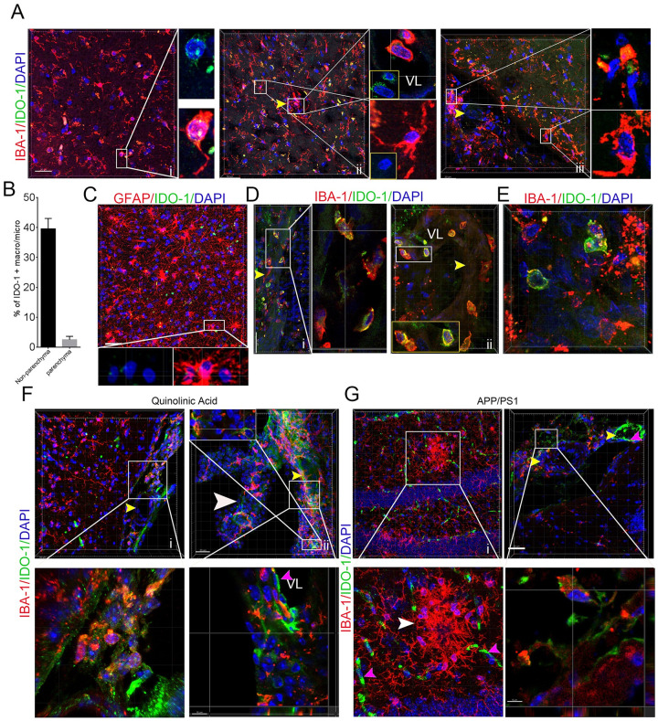
To see if the IDO-1+ macrophages/microglia preferentially reside in specific sites in murine model and human brains, we adopted a thick-section staining method to localize IDO-1+ macrophages/microglia in the human brain, human GBM tissues, and the brains from the APP/PS1 mouse and quinolinic acid models with IBA-1 or GFAP and IDO-1 antibodies. The results showed a few IDO-1+ microglia/macrophages with small bodies and few processes in the brain parenchyma and microglia with larger bodies and longer processes not expressing IDO-1 in the brain parenchyma (Fig 1A-i and 1A-ii). We also observed a considerable number of IDO-1+ microglia/macrophages in the vascular lumen, vascular wall, and perivascular space (Fig 1A-ii) and subarachnoid space in the human brain tissues (Fig 1A-iii and 1B). However, GFAP + astrocytes did not express IDO-1 (Fig 1C). In the tumor region and the region close to tumors, we observed that multiple IDO-1+ microglia/macrophages also closely surrounded blood vessels and appeared in the vascular walls or lumen (Fig 1D). Consistent to other observations [20], we also observed that some brain neoplastic cells are highly expressed IDO-1 (Fig 1E). To see IDO-1+ expression patterns in the microglia of murine neurodegenerative models, we stained APP/PS1 mouse and quinolinic acid model brains with anti-IDO-1 and anti-IBA-1 antibodies and found that IDO-1 is not expressed in the microglia of the cortex, hippocampus or striatum in the quinolinic acid injury model brains (Fig 1F-i) or in the activated microglia surrounding amyloid deposits in the APP/PS1 mouse hippocampus and cortex (Fig 1G-i). We observed a significant fraction of microglia in the subarachnoid space and the choroid plexus of the third ventricle highly expressed IDO-1 (Fig 1F-ii and 1G-ii). The vascular endothelial cells, including capillaries and small arteries, expressed IDO-1 in both models. These data implied that IDO-1+ macrophages/microglia might have distinct physiological and immunological properties.

Fig 1. IDO-1+ macrophages/microglia reside in the meninges and perivascular space of the human and mouse brain and in human GBM tissues.

Fig 1

(A) IBA-1 andIDO-1 antibodies staining in noncancerous brain tissues from GBM patients (i, cortex; ii, cortex with intravascular IDO-1+ cells; iii, the meninges and cortex; yellow arrow, blood vessel). i and ii are from noncancerous parenchyma of a male GBM patient with age 63 and grade IV tumor. iii is from the noncancerous parenchyma of a female GBM patient with age 32 and grade III tumor. (B) The percentage of IDO-1+ nonparenchymal macrophages/microglia in the meninges/perivascular space and parenchymal macrophages/microglia in brain (sample numbers, 4). (C) IDO-1 and GFAP antibodies staining of noncancerous brain tissues. Noncancerous parenchyma of GBM a female GBM patient with age 55 and grade IV tumor. (D) IDO-1 and IBA-1 antibodies staining of the vascular region in the surrounding region of neoplastic lesions. Noncancerous parenchyma of a female GBM patient with age 32 and grade III tumor. (E) IDO-1 and IBA-1 staining of GBM tissues. The tumor tissue of a female GBM patient with age 32 and grade III tumor. (F, G) IBA-1 and IDO-1 antibodies staining of the microglia of APP/PS1 and quinolinic acid mouse model brains. Boxed area, magnified region. White arrow, choroid plexus. VL, vascular lumen. Pink arrows, endothelial cells.

Inhibiting IDO-1 with INCB24360 reduced most M1 markers but not M2 markers in the RAW264.7 and BV-2 cells

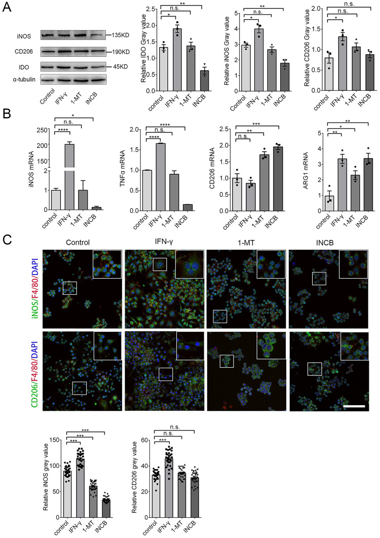
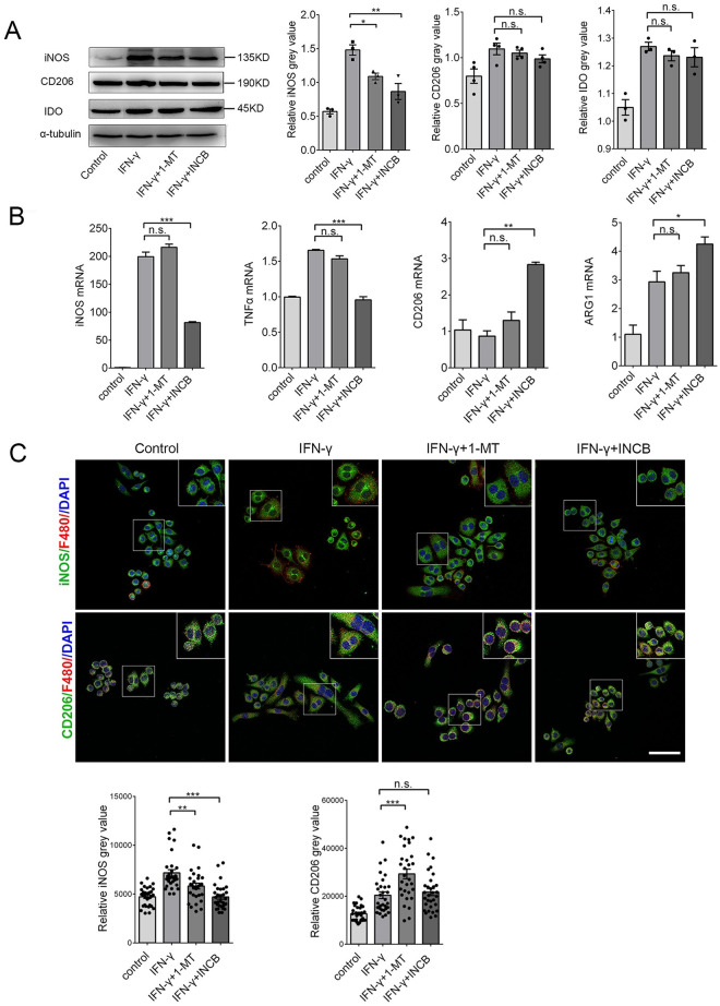
To test how IDO-1 effects macrophage/microglia activities, we treated RAW264.7 and BV-2 cells with 20 ng/mL IFN-γ to increase IDO-1 levels and used 20 μM 1-MT and 20 μM INCB24360 to inhibit IDO-1 activity for 24 h. We detected CD206, an M2 marker, iNOS, an M1 marker, and IDO-1 by western blotting. Consistent with other findings [21], IFN-γ treatment increased IDO-1 and iNOS expression in the RAW264.7 and BV-2 cells (Fig 2A and S1A Fig). Inhibiting endogenous IDO-1 with INCB24360 or 1-MT reduced iNOS levels but did not affect CD206 in the RAW264.7 or BV-2 cells, and the effects of INCB24360 were more significant than those of 1-MT (Fig 2A and S1A Fig). We also detected M1/M2 markers, including iNOS, TNF-α, CD206, and Arginase 1 (Arg1) by qRT-PCR, and we found that INCB24360 decreased iNOS and TNF-α RNA in the RAW264.7 and BV-2 cells (Fig 2B and S1B Fig); 1-MT treatment decreased iNOS and TNF-α RNA levels in the BV2 cells but not in the RAW264.7 cells (Fig 2B and S1B Fig). Interestingly, both 1-MT and INCB increased the CD206 and Arg-1 RNA levels in the RAW264.7 cells. 1-MT did not affect CD206 RNA in BV-2 cells; both 1-MT and INCB did not affect the Arg-1 in BV-2 (Fig 2B and S1B Fig). Immunofluorescence staining of RAW264.7 and BV-2 cells treated with INCB24360 and 1-MT revealed that iNOS expression was significantly decreased in INCB24360 groups but CD206 was not changed compared to the levels in the control group (Fig 2C and S1C Fig). The data indicate that IDO-1 might induce an increase in M1-like macrophages/microglia in the brain.

Fig 2. Inhibition of IDO-1 with 1-MT and INCB24360 decreased iNOS and TNF-α levels.

Fig 2

(A) The IDO-1, iNOS, and CD206 expression in RAW264.7 cells treated with IFN-γ, 1-MT or INCB24360 for 24 h. The relative intensity of IDO-1, iNOS, and CD206 in RAW264.7 cells as measured by ImageJ software. (B) The transcription levels of iNOS, TNFα, CD206 and Arg1 in RAW264.7 cells treated with IFN-γ, 1-MT or INCB24360 for 24 h. (C) The immunostaining images of iNOS and CD206 in RAW264.7 cells treated by IFN-γ, 1-MT or INCB24360 for 24 h. The relative intensity of iNOS or CD206 in RAW264.7 cells after treatment with IFN-γ, 1-MT or INCB24360, which was measured by ImageJ software. n≥30. Scale bars, 100 μm. One-way ANOVA; all data are expressed as the means ± SEM. *, P<0.05, **, P<0.01; ns, no significant difference.

Inhibiting IDO-1 decrease the cellular body and membrane filopodia in RAW264.7 and BV-2 cells

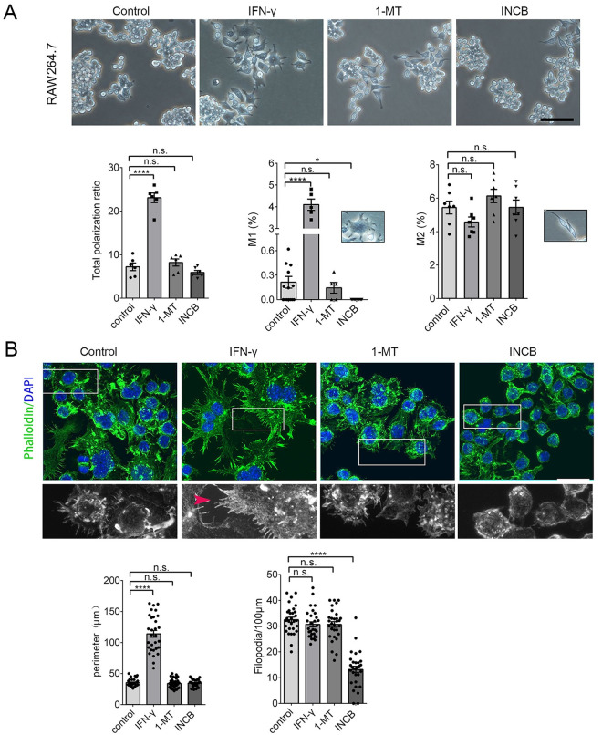
Non-polarized RAW264.7 cells are small and round with few processes [22]. When polarized, RAW264.7 grow large and round with a pancake-like shape, representative of the M1 phenotype, or they become slimmer cells with longer processes, representative of the M2 phenotype [22]. To determine whether iNOS reduction in RAW264.7 and BV-2 cells after INCB24360 treatment caused cellular morphological and filopodium changes, we assessed the morphology of the RAW264.7 and BV-2 cells after IFN-γ, 1-MT, and INCB24360 treatment (Fig 3A and S2A Fig). IFN-γ treatment dramatically increased the number of polarized RAW264.7 and BV-2 cells, while neither the 1-MT nor INCB24360 treatment significantly changed the number of polarized RAW264.7 cells (Fig 3A) or BV-2 cells (S2A Fig). We divided the polarized macrophages into the M1 or M2 types based on their morphology. Consistent with other observations [22], IFN-γ treatment preferentially increased the proportion of M1-like RAW264.7 and BV-2 cells and slightly decreased the proportion of M2 type cells (Fig 3A and S2A Fig); INCB24360 treatment significantly decreased the proportion of M1 RAW264.7 and BV-2 cells compared to the control (Fig 3A and S2A Fig). Further, 1-MT treatment did not change the M1/M2 ratio in the RAW264.7 cells (Fig 3A). However, both 1-MT and INCB24360 slightly increased the proportion of M2 BV-2 cells (S2A Fig).

Fig 3. Inhibiting IDO-1 with 1-MT or INCB24360 reduced the proportion of M1-like but not M2-like RAW264.7 cells.

Fig 3

(A) The typical morphology of RAW264.7 cells treated with IFN-γ, 1-MT and INCB24360 for 24 h. More round and small RAW264.7 cells were observed in the 1-MT and INCB24360 groups compared to the IFN-γ and control groups. The percentage of the total polarized, M1-type-cells (ramified), and M2-type-cells (slender) in the control, IFN-γ, 1-MT and INCB24360 groups. N ≥ 5. Scale bars, 100 μm. (B) The phalloidin Alexa-488 staining of RAW264.7 cells treated with IFN-γ, 1-MT or INCB24360 for 24 h. The cellular perimeters in the control, IFN-γ, 1-MT and INCB24360 groups. The density of the filopodia on the membrane of RAW264.7 cells in the control, IFN-γ, 1-MT and INCB24360 groups. n ≥30. Scale bars, 30 μm. One-way ANOVA; all data are expressed as the means ± SEM. *, P<0.05, **, P<0.01; ns, no significant difference.

M1 macrophages showed multiple filopodia that facilitate the phagocytosis of fluid or foreign substances, including inorganic particles [23]. We treated RAW264.7 and BV-2 cells with IFN-γ, 1-MT, or INCB24360 for 24 h, fixed the cells and stained them with phalloidin Alexa-488, an actin-binding dye, and DAPI. We then observed that the macrophages treated with IFN-γ formed a ruffled border with abundant filopodia (Fig 3B and S2B Fig). INCB24360-treated macrophages showed fewer filopodia than the control, IFN-γ, and 1-MT RAW264.7 and BV-2 cell groups (Fig 3B and S2B Fig). The cellular perimeter and filopodia density data showed that the 1-MT and INCB24360 treatment did not change the cellular perimeter but decreased the filopodia density on the cellular membrane (Fig 3B and S2B Fig). Collectively, these findings implied that inhibition of IDO-1 preferentially blocks cellular size increases and filopodia growth.

Inhibiting IDO-1 reduces TMR-dextran uptake and phagocytosis of RAW264.7 and BV-2 cells

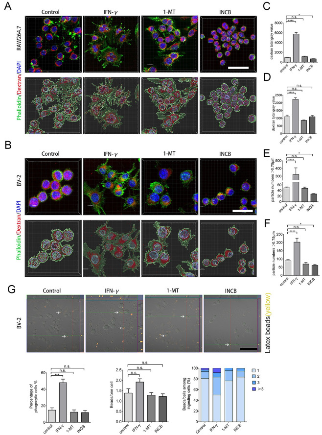
To determine whether, in addition to a decrease in iNOS and filopodia and the acquisition of a ruffled cellular border, INCB24360 treatment also reduces the phagocytosis ability of the RAW264.7 and BV-2 cells, we administered TMR-dextran to RAW264.7 and BV-2 cells after treating them with IFN-γ, 1-MT, and INCB24360 for 24 h. After 1 h of TMR-dextran treatment, we fixed the cells and counterstained them with phalloidin-Alex-488 and DAPI, and we found that INCB24360 treatment significantly reduced DMR-dextran uptake compared to the amount internalized by the control and IFN-γ+ RAW264.7 cell groups compared to the BV-2 cell groups (Fig 4A–4D).

Fig 4. INCB24360 treatment reduced DTR-dextran uptake by RAW264.7 and BV-2 cells.

Fig 4

(A, B) The typical images of engulfed DTR-dextran in IFN-γ induced RAW264.7 or BV-2 cells after administration of dextran for 1 h in the control, IFN-γ, 1-MT and INCB24360 groups. The phagocytic particles larger than 0.75 μm were analyzed by IMARIS 9.6. Scale bars, 40 μm. (C, D) Quantification of DTR-dextran particles in RAW264.7 cells (B) and BV-2 cells (E). Single-cell area X Dextran average grayscale was used as statistical data; n ≥ 15. (E, F) DTR-dextran particles larger than 0.75 μm in the control, IFN-γ, 1-MT and INCB24360 groups of IFN-γ induced RAW264.7 cells (C) or BV2 cells (F). n ≥ 15. (G) Phagocytosis of latex beads in BV-2 cells treated with IFN-γ, 1-MT or INCB24360 for 24 h. The three statistical charts represent that the phagocytic ratio, the number of beads in each cell, the number of cells with one, two, three, or more beads. Scale bars, 100 μm. One-way ANOVA; all data are expressed as the means ± SEM. *, P<0.05, **, P<0.01; ns, no significant difference.

Macropinocytosis, which depends on membrane ruffling, is the mechanism by which macrophages internalize large amounts of extracellular fluid [24]. The decrease in filopodia on the ruffled border after IDO-1 inhibition implied that IDO-1 might enhance macropinocytosis. The macropinosome ranged from 0.2 μm to 5 μm in diameter [25]. The vesicles, which are larger than 0.75 μm, have been identified as apparent macropinocytic vesicles in DTR-dextran uptake assays performed in some studies [26]. We found that INCB24360 treatment decreased the number of phagocytic vesicles larger than 0.75 μm in RAW264.7 and BV-2 cells (Fig 4E and 4F).

Macrophages, especially tissue-resident macrophages, can phagocytize foreign-derived particulates such as alum and silica. M1 macrophages can also phagocytize foreign-derived particulates [27, 28]. We treated RAW264.7 and BV-2 cells with Latex beads, a spherical polymer particle, and fluorescence red, after incubating the cells with IFN-γ, 1-MT, or INCB24360 for 24 h. After fixing the cells with 4% PFA, we analyzed the phagocytized Latex beads and observed that 1-MT and INCB24360 treatment slightly decrease the Latex beads uptake by the BV-2 cells compared to the control cells, but the decrease was not statistically significant (Fig 4G).

In contrast to M2 macrophages, M1 macrophages show low motility [29, 30]. We used 1-MT and INCB24360 to test their effects on the migration and proliferation of macrophages in a Transwell migration assay and in an Edu staining assay and Flow cytometry to test cell proliferative potential. Consistent with the formation of M1 macrophages, the results showed that IFN-γ treatment dramatically decreased the migration ability of the RAW264.7 and BV-2 cells (S3A Fig) but it not affect the proliferation of the RAW264.7 or BV-2 cells (S3B and S3C Fig); 1-MT and INCB24360 do not affect the migration or proliferation of the RAW264.7 or BV-2 cells (S3A–S3C Fig). Taken together, 1-MT and INCB24360, especially INCB24360, preferentially inhibited the phagocytic ability and macropinocytosis of the macrophages but not their capacities for migration or proliferation.

Inhibiting IDO-1 with 1-MT or INCB24360 suppresses the IFN-γ-induced iNOS upregulation in RAW264.7 cells

To determine whether IDO-1 inhibitors affected IFN-γ-induced M1 or M2 markers, we treated RAW264.7 and BV-2 cells with IFN-γ for 12 h, and then we added 1-MT or INCB24360 for 24 h. Both 1-MT and INCB24360 significantly decreased iNOS levels but did not change CD206 levels in the IFN-γ-treated RAW264.7 cells; a greater decrease in iNOS was observed in the INCB24360 group (Fig 5A). However, neither 1-MT nor INCB24360 changed the iNOS and CD206 expression in the IFN-γ-treated BV-2 cells (S4A Fig). We also detected iNOS, TNF-α, CD206, and Arg1 RNA levels by RT-PCR in the four cell groups and found that INCB24360 treatment reduced the iNOS and TNF-α RNA levels in the IFN-γ-induced RAW264.7 and BV-2 cells (Fig 5B and S4B Fig). INCB24360 treatment significantly increased the transcription level of CD206 RNA but not Arg1 RNA in the IFN-γ-treated RAW264.7 cells, but 1-MT treatment did not significantly affect the transcription of iNOS, TNF-α, CD206, or Arg1 genes (Fig 5B). In agreement with the western blot analysis results, immunofluorescence staining results showed that iNOS expression significantly decreased in the IFN-γ-treated cells treated with INCB24360 or 1-MT compared to the IFN-γ-treated RAW264.7 and BV-2 cells (Fig 5C and S4C Fig). Further, 1-MT treatment significantly increased CD206 expression in the IFN-γ-treated RAW264.7 cells, while 1-MT and INCB24360 increased CD206 expression in the IFN-γ-induced BV-2 cells (Fig 5C and S4C Fig).

Fig 5. Inhibiting IDO-1 with INCB24360 or 1-MT suppresses IFN-γ induced iNOS and TNFα increases in RAW264.7 cells.

Fig 5

(A) iNOS, CD206 and IDO-1 expression in RAW264.7 cells treated with IFN-γ, IFN-γ+1-MT or IFN-γ + INCB24360 for 24 h (12 h with IFN-γ, following another 12 hours with 1-MT or INCB24360) and the relative intensity of iNOS, CD206, and IDO-1 in the RAW264.7 cells as measured by ImageJ. (B) The transcription levels of iNOS, TNFα, CD206 and Arg1 in RAW264.7 cells treated with IFN-γ, IFN-γ+1-MT or IFN-γ + INCB24360 for 24 h. (C) The immunostained images of iNOS and CD206 in RAW264.7 cells treated by IFN-γ, IFN-γ+1-MT or IFN-γ + INCB24360 for 24 h. The quantification of iNOS and CD206 intensity in RAW264.7 cells as measured by ImageJ software. Scale bars, 50 μm. One-way ANOVA; all data are expressed as the means ± SEM. *, P<0.05, **, P<0.01; ns, no significant difference.

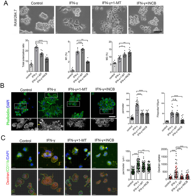
To determine whether IDO-1 inhibitors also inhibit IFN-γ-induced cellular size and filopodia growth, we treated RAW264.7 and BV-2 cells with IFN-γ for 12 h and then added 1-MT or INCB24360 for 24 h. After imaging, we found that INCB24360 treatment not only decreased the total number of polarized RAW264.7 cells (Fig 6A) and BV-2 cells driven by IFN-γ (S5A Fig), but INCB24360 treatment preferentially decreased the number of IFN-γ-induced M1 RAW264.7 and BV-2 cells (Fig 6A and S5A Fig). Interestingly, INCB24360 treatment significantly increased the IFN-γ-induced M2 macrophage proportion (Fig 6A and S5A Fig). Further, 1-MT treatment decreased the number of total IFN-γ-induced polarized macrophages, and this decrease affected M1 or M2 macrophages equally (Fig 6A and S5A Fig). The findings support the supposition that INCB24360 can reverse IFN-γ-driven M1 macrophage increases.

Fig 6. Inhibiting IDO-1 with INCB24360 reduced the M1-like macrophage proportion in the RAW264.7 cells induced by IFN-γ.

Fig 6

(A) The typical morphology of RAW264.7 cells after treatment with IFN-γ, IFN-γ+1-MT and IFN-γ + INCB24360. A greater number of round and small RAW264.7 cells were observed in the IFN-γ+1-MT and IFN-γ+INCB24360 groups compared to the IFN-γ group. The percentage of the polarized M1-like macrophages (ramified) and M2-like macrophages (slender) in the control, IFN-γ, IFN-γ+1-MT and IFN-γ + INCB24360 groups. N ≥ 5. Scale bars, 60 μm. (B) The phalloidin Alexa-488 staining of RAW264.7 cells treated with IFN-γ, IFN-γ+1-MT and IFN-γ + INCB24360 for 24 h. The cellular perimeters in the control, IFN-γ, IFN-γ+1-MT and IFN-γ + INCB24360 groups. The density of the filopodia of RAW264.7 cell in the control, IFN-γ, IFN-γ+1-MT and IFN-γ + INCB24360 groups. One-way ANOVA; all data are expressed as the means ± SEM. *, P<0.05, **, P<0.01; ns, no significant difference. Scale bars, 40 μm. (C) INCB24360 and 1-MT decreased IFN-γ induced cellular size increase and dextran uptake. Human peripheral blood monocytes of 47 healthy male was isolated by Solarbio Human Peripheral Monocyte Isolation Kit, and cells were treated with IFN-γ, IFN-γ+1-MT and IFN-γ + INCB24360 for 24 h. High-resolution images were taken by Zeiss 880 Airscan Microscopy after staining with CD11B antibody. Endocytic dextran particles were analyzed by Spot of Imaris 9.7. Images in each group, n>10; Cellular perimeter analysis, cells>100; dextran particles analysis, cells>40.

To determine whether IDO-1 inhibitors block IFN-γ-driven ruffled border formation and filopodia growth, we treated RAW264.7 and BV-2 cells with IFN-γ for 12 h, and then added 1-MT or INCB24360 for 24 h, fixed the cells and stained them with phalloidin Alexa-488 and DAPI. Our data showed that INCB24360 inhibited the formation of the IFN-γ-induced ruffled border and filopodia formation in the RAW264.7 and BV-2 cells (Fig 6B and S5B Fig). Measuring and counting data showed that both 1-MT and INCB24360 treatments significantly inhibited the increase in RAW264.7 cell size induced by IFN-γ (Fig 6B) but did not affect BV-2 cells (S5B Fig) INCB24360 treatment also inhibited the increase in filopodia density on the cellular membrane induced by IFN-γ (Fig 6B and S5B Fig), but 1-MT treatment did not restrict the increase in filopodia density on the cellular membrane induced by IFN-γ (Fig 6B and S5B Fig). Inhibiting IDO-1 with INCB24360 or 1-MT reduced both cell size and filopodia growth in the macrophages/microglia.

Inhibiting IDO-1 suppresses the IFN-γ induced endocytic, macropinocytic and phagocytic abilities of RAW264.7 and BV-2 cells

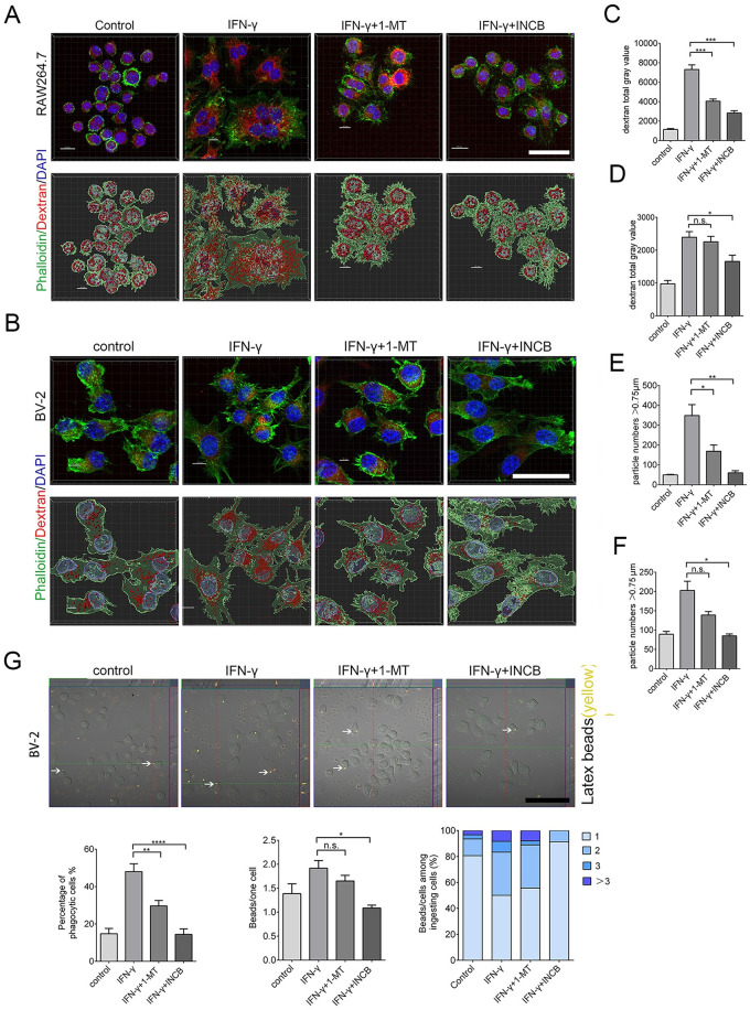
To determine whether 1-MT and INCB24360 can decrease the endocytic and macropinocytic ability induced by IFN-γ, we treated freshly isolated human peripheral blood monocytes, BV-2 and RAW264.7 cells with IFN-γ for 12 h and then treated them with 1-MT and INCB24360 for another 12 h. We then added DTR-dextran to the culture medium. After treating with DTR-dextran for 1 h, we fixed the cells and counterstained them with phalloidin Alex-48h. In human blood monocytes, both INCB24360 and I-MT decreased IFN-γ-induced cellular size increase and the TMR-dextran uptake ability (Fig 6C). INCB24360 decreased the TMR-dextran uptake ability of both the RAW264.7 and BV-2 cells; 1-MT significantly decreased the TMR-dextran uptake ability of the RAW264.7 cells but not of the BV-2 cells (Fig 7A–7D). We analyzed the DTR-dextran phagocytic vesicles that were larger than 0.75 μm, which is reflective of macropinocytosis, by Imaris9.6 software. The findings indicated that INCB24360 treatment decreased the number of vesicles larger than 0.75 μm phagocytosed by RAW264.7 and BV-2 cells, and 1-MT significantly decreased the number of vesicles larger than 0.75 μm phagocytosed by RAW264.7 cells but not by BV-2 cells (Fig 7E and 7F).

Fig 7. Inhibiting IDO-1 with 1-MT or INCB24360 reduced DTR-dextran uptake by RAW264.7 and BV-2 cells induced by IFN-γ.

Fig 7

(A, B) The typical images of engulfed DTR-dextran in IFN-γ induced RAW264.7 (A) or BV-2 (B) cells after administration of dextran for 1 h in the control, IFN-γ, IFN-γ+1-MT and IFN-γ + INCB24360 groups. The phagocytic particles larger than 0.75 μm were analyzed by IMARIS 9.6. Scale bars, 40 μm. (C, D) Quantification of DTR-dextran particles in RAW264.7 cells (C) and BV-2 cells (D). Single-cell area X Dextran average grayscale was used as statistical data; n ≥ 15. (E, F) DTR-dextran particles larger than 0.75 μm in the control, IFN-γ, IFN-γ+1-MT and IFN-γ + INCB24360 groups of IFN-γ induced RAW264.7 cells (E) or BV2 cells (F). n ≥ 15. (G) Phagocytosis of latex beads in BV-2 cells treated with IFN-γ, IFN-γ+1-MT or IFN-γ + INCB24360 for 24 h. The three statistical charts represent that the phagocytic ratio, the number of beads in each cell, the number of cells with one, two, three, or more beads. Scale bars, 100 μm. One-way ANOVA; all data are expressed as the means ± SEM. *, P<0.05, **, P<0.01; ns, no significant difference.

To determine the effect of 1-MT and INCB24360 on macrophage phagocytic ability, we treated RAW264.7 and BV-2 with IFN-γ for 12 h. We added 1-MT or INCB24360 and incubated the cells for 24 h, then introduced Latex beads to the culture medium. After fixing the cells, we analyzed the number of phagocytized Latex beads in the cells and found that INCB24360 and 1-MT treatment significantly decreased the number of Latex beads phagocytosed by BV-2 cells. The number of latex beads phagocytosed by IFN-γ-induced BV-2 cells treated with INCB24360 was significantly lower than that phagocytosed by the untreated IFN-γ-induced BV-2 cells. However, the number of latex beads phagocytosed by IFN-γ-induced BV-2 cell treated with 1-MT was not significantly lower than that phagocytosed by untreated IFN-γ-induced BV-2 cells (Fig 7G). In summary, I-MT and INCB24360, but especially INCB24360, inhibited the endocytic, phagocytic, and macropinocytic ability of IFN-γ-treated macrophages.

Inhibiting IDO-1 with 1-MT and INCB24360 reduces NLRP3 expression in RAW264.7 and BV-2 cells

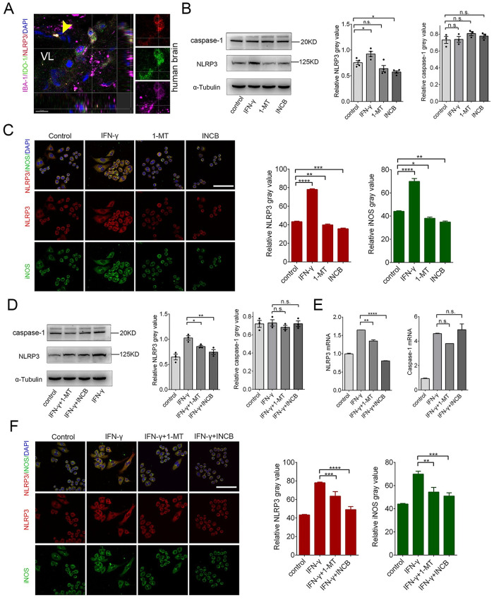
The NLRP3 inflammasome, a large protein complex, includes NLRP3, ASC, and caspase 1 [31]. The NLRP3 inflammasome is involved in M1 macrophage formation [32]. Curcumin, an IDO-1 inhibitor, suppresses NLRP3 expression in chronic unpredictable mild stress (CUMS) and reduces depression-like behaviors [33]. To determine whether NLRP3 and IDO-1 are co-upregulated in macrophages/microglia in vivo, we stained the human brain tissues with anti-IBA-1, anti-IDO-1 and anti-NLRP3 antibodies and observed that IDO-1+ macrophage/microglia in the perivascular space (Fig 8A). To determine how IDO-1 affects NLRP3 expression, we treated RAW264.7 and BV-2 cells with IFN-γ, 1-MT, and INCB24360 for 24 h. We detected NLRP3 expression by western blotting and found that both 1-MT and INCB24360 decreased NLRP3 but not caspase-1 expression in the RAW264.7 and BV2 cells (Fig 8B). The decreases in NLRP3 were statistically significant in the INCB24360 and 1-MT groups (Fig 8B). We also used immunostaining of anti-NLRP3 and anti-iNOS antibodies to detect changes in NLRP3 and iNOS expression. Consistent with immunoblotting and RT-PCR results, INCB24360 treatment significantly decreased NLRP3 expression in the RAW264.7 cells (Fig 8C) and BV-2 cells (S6C Fig).

Fig 8. Inhibiting IDO-1 with 1-MT or INCB24360 reduced NLRP3 expression in RAW264.7 cells.

Fig 8

(A) NLRP3, IDO-1 and IBA-1 triple-staining in the meninges of a human noncancerous brain (Yellow arrow, vessels; VL, vascular lumen). (B) The changes of NLRP3 and caspase-1 in RAW264.7 cells treated with IFN-γ, 1-MT or INCB24360 for 24 h. The relative intensity of NLRP3 and caspase-1 in RAW264.7 cells as measured by ImageJ software. (C) The immunostained results of NLRP3 and iNOS in RAW264.7 cells treated with IFN-γ, 1-MT or INCB24360 for 24 h. The relative NLRP3 or iNOS intensity in RAW264.7 cells as measured by ImageJ software. n≥10. Scale bars, 80 μm. (D) The changes of NLRP3 and caspase-1 in RAW264.7 cells treated with IFN-γ, IFN-γ+1-MT or IFN-γ + INCB24360 for 24 h. The relative intensity of NLRP3, caspase-1 in RAW264.7 cells as measured by ImageJ software. (E) The transcription levels of NLRP3 and caspase-1 in RAW264.7 cells after treatment with IFN-γ, IFN-γ+1-MT or IFN-γ + INCB24360 for 24 h. (F) The immunostained images of NLRP3 and iNOS in RAW264.7 cells treated with IFN-γ, IFN-γ+1-MT or IFN-γ + INCB24360 for 24 h. The relative NLRP3 or iNOS intensity in RAW264.7 cell as measured by ImageJ software. Scale bars, 80 μm. One-way ANOVA; all data are expressed as the means ± SEM. *, P<0.05, **, P<0.01; ns, no significant difference. Scale bars, 80 μm.

To determine whether 1-MT and INCB24360 can also decrease IFN-γ-induced NLRP3 expression, we also treated RAW264.7 cells with IFN-γ for 12 h, and then added 1-MT or INCB24360 for 24 h to inhibit IDO-1 activity. The data showed that both 1-MT and INCB24360 inhibited IFN-γ-induced NLRP3 expression but not caspase-1 expression (Fig 8D). The RT-PCR data also showed that INCB24360 and 1-MT reduced IFN-γ-induced NLRP3 gene transcription in both RAW264.7 and BV-2 cells but did not reduce caspase-1 transcription (Fig 8E and S6F Fig). We immunostained these treated cells with anti-NLRP3 and anti-iNOS antibodies, and the results of the immunofluorescence staining of RAW264.7 cells were consistent with the protein blotting results (Fig 8F and S6F Fig).

1-MT and INCB24360 treatments increase IL-1β secretion in RAW264.7 and BV-2 cells

The NLRP3 inflammasome activates caspase 1, which cleaves pro-IL-1β and pro- IL-18, and leads to the secretion of IL-1β and IL-18 [31]. Inhibiting IDO with 1-MT dramatically caused a surge in IFN-γ, IL-1β, IL-6, and TNF-α secretions from the macrophages infected with the influenza virus [9, 16]. To determine whether the decrease in NLRP3 by 1-MT and INCB24360 affects IL-1β and IL-18 secretion, we treated RAW264.7 cells with 20 ng/mL IFN-γ, 20 μM 1-MT, and 20 μM INCB24360 for 24 h. We detected IL-1β and IL-18 secretion levels in the medium by ELISA and found that IL-1β and IL-18 levels in the culture medium of IFN-γ-, 1-MT-, and INCB24360-treated RAW264.7 and BV-2 cells were significantly increased (S7A and S7B Fig).

Tryptophan deprivation by IFN-γ-induced IDO-1 inhibits mTORC1 kinase, and tryptophan or 1-MT treatment reversed mTORC1 inhibition [2, 34]. To determine whether INCB24360 reverses mTORC1 inhibition, we used an anti- phosphorylated (p) S6K (T389) antibody to evaluate mTORC1 activation levels in RAW264.7 cells. Interestingly, we did not observe a direct effect of 1-MT or INCB24360 treatment on pS6K (T389) levels in the RAW264.7 cells (S7C Fig). We observed that inhibiting IFN-γ-induced IDO-1 with 1-MT and INCB24360 increased IL-1β and IL-18 secretion by RAW264.7 and BV-2 cells (S7A and S7B Fig). The increase in IL-1β secretion in the INCB24360-treated RAW264.7 and BV-2 cells induced by IFN-γ was significantly higher than that in the RAW264.7 and BV-2 cells induced with IFN-γ alone (S7B Fig). Interestingly, 1-MT significantly increased IL-18 secretions in IFN-γ-induced RAW264.7 cells by but not IL-1β (S7A and S7B Fig). Additionally, in agreement with other findings [27, 34], inhibiting IFN-γ-induced RAW264.7 cells with 1-MT or INCB24360 dramatically increased pS6K (T389) levels (S7D Fig). Activation of mTORC1 by 1-MT and INCB24360 in IFN-γ-induced cells might partially explain why 1-MT and INCB24360 increase IL-1β secretion by RAW264.7 cells.

NLRP3 deficiency inhibits the IDO-1 upregulation induced by LPS in the hippocampal microglia [35]. To determine whether NLRP3 inhibition affects endogenous IDO-1 levels in RAW264.7 cells, we suppressed NLRP3 in RAW264.7 cells with MCC950, an inhibitor of NLRP3 by targeting ATP-hydrolysis [36], and oridonin, a covalent NLRP3 inhibitor [37]. After exposing RAW264.7 cells to MCC950 or oridonin for 24 h, we detected NLRP3 and IDO-1 expression (S7E Fig), and found that inhibiting NLRP3 with oridonin and MCC950 increased IDO-1 expression in the RAW264.7 cells (S7F Fig). Our data indicate that NLRP3 and IDO-1 might co-regulate each other in macrophages.

Discussion

IDO-1 expression in placental epithelial cells, neoplastic cells, and macrophage reject T-cell infiltrates and diminishes the immune response [38, 39]. We showed that IDO-1+ macrophages/microglia reside in the perivascular and subarachnoid spaces at the brain parenchyma interface. Using IDO-1 inhibitors, we found that IDO-1 enhanced macrophage/microglia endocytic, phagocytic, and macropinocytic capacities via increases in cell size and filopodia growth. Inhibition of IDO-1 in macrophages/microglia reduced NLRP3 expression but increased the secretion of IL-1β. Consistent with a lower migrating ability of M1 macrophages and morphometric analysis [29], IFN-γ treatment significantly reduced the migration of RAW264.7 and BV-2 but not 1-MT, INCB24360. The multiple filopodia of macrophage in IFN-γ treatment might contribute to phagocytic or endocytic ability but not migrating. Our findings showed that IDO-1+ macrophages/microglia have strong endocytic, phagocytic, and macropinocytic capacities and weaker proinflammatory properties in the meninges.

Macrophages/microglia scavenge cellular debris, effete cells, invading microbes, and metabolites in the cerebrospinal fluid (CSF) and parenchyma to maintain homeostasis [5, 7]. The meninges, perivascular, and choroid-plexus macrophages patrol the CSF to surveil waste and invasion [5, 7]. The observation of vascular wall-traversing or vascular luminal IDO-1+ macrophages indicates that some IDO-1+ macrophages might originate from a monocyte, which is consistent with the transcriptome database on macrophages/microglia [18]. In contrast to the meninges with abundant immune infiltrates, T cells are almost absent in healthy brain parenchyma. T-cell infiltration in brain parenchyma is a hallmark of multiple sclerosis (MS) [40]. Ectopic T-cell infiltration is also observed in the parenchyma of Parkinson’s and Alzheimer’s patients [4143]. The only pathway to parenchyma for T cells involves crossing the blood-brain barrier (BBB) and the pia matter. Thus, whether IDO-1+ macrophages/microglia partially or significantly participate in the rejection of T cells at the brain parenchyma is worth exploring in the future.

Consistent with attenuated ruffled border and filopodia formation, inhibiting IDO-1 decreased the endocytic, macropinocytic, and phagocytic abilities of macrophages/microglia. Surprisingly, we also observed that IDO-1+ macrophages in metastatic lymph nodes and tumors possess multiple processes and larger body sizes in pancreatic cancer patients (unpublished data). These observations further support the supposition that IDO-1 is related to large cell size and greater macrophage/microglia phagocytic and macropinocytic abilities. The decrease in endocytic, macropinocytic, and phagocytic ability and iNOS and TNF-ɑ levels after inhibiting IDO-1 showed that IDO-1 drives the formation of M1-like macrophages. IL-1β is a critical proinflammatory cytokine in M1 macrophages [44]. Previous data showed that IDO-1-/- mice suppressed LP-BM5 replication by secreting excessive type I IFN, and 1-MT treatment dramatically led to a surge in IL-1β secretion by macrophages in conjunction with virus-induced inflammation [9, 16, 17]. The enhancement of IL-1β secretion after inhibiting IDO-1 hints at an anti-inflammatory role for IDO-1 in macrophages/microglia. IDO-1 is a downstream enzyme of the NLRP3 inflammasome, and IDO-1 upregulation induced by lipopolysaccharide (LPS) is diminished in the glial cells of Nlrp3-/- mice, including microglia [32, 35]. The inhibition of IDO-1 with curcumin decreased NLRP3 expression [33]. We showed that both 1-MT and INCB24360 reduced endogenous or IFN-γ-induced NLRP3 expression, and inhibiting NLRP3 also increased IDO-1 expression, indicating that the relationship of NLRP3 and IDO-1 is bidirectional, and the direction depends on the activation status of macrophages/microglia.

In the future, to what extent IDO-1 expressing microglia/macrophages contribute to immune barriers of the brain parenchyma and if the IDO-1 expressing endothelial cells also contribute to the T cell rejection by parenchyma are worth exploring. Another interesting point is that if IDO-1 deregulation in macrophage/microglia correlated with brain autoimmunity diseases caused by the abnormal entrance of T cells to parenchyma, such as Multiple Sclerosis [45].

Our findings collectively show that IDO-1+ macrophages/microglia in meninges are phagocytes with higher scavenging ability and lower proinflammatory activity, implying that IDO-1+ macrophages/microglia combined with IDO-1 + endothelial cells might be involved in the prevent T cells from the meninges/perivascular space from entering the parenchyma or crossing the BBB and may prevent the overactivation of immune response.

Supporting information

S1 Fig. Inhibition of IDO-1 in BV-2 with 1-MT and INCB 24360 decreased iNOS and TNF-α levels.

(A) The iNOS and CD206 expression in BV-2 cells treated with IFN-γ, 1-MT or INCB24360 for 24 h. (B) The transcription levels of iNOS, TNFα, CD206 and Arg1 in BV-2 cells treated with IFN-γ, 1-MT or INCB24360 for 24 h. (C) The immunostaining images of iNOS and CD206 in BV-2 cells treated with IFN-γ, 1-MT or INCB24360 for 24 h. The relative intensity of iNOS or CD206 in BV-2 cells after treatment with IFN-γ, 1-MT or INCB24360, which was measured by ImageJ software. n≥20. Scale bars, 100μm. One-way ANOVA; all data are expressed as the mean ± SEM. *, P<0.05, **, P<0.01; ns, no statistical difference.

(TIF)

S2 Fig. 1-MT and INCB-24360 treatment reduced M1-like macrophage while increased M2-like macrophage in BV-2.

(A) The typical morphology of BV-2 cells treated with IFN-γ, 1-MT and INCB24360 for 24 h. The percentage of M1-like macrophage (ramified); M2-like macrophage (slender) in the control, IFN-γ, 1-MT and INCB24360 groups. N ≥ 5. Scale bars, 80μm. (B) The phalloidin Alexa-488 staining of BV-2 cells treated with IFN-γ, 1-MT or INCB24360 for 24 h. The cellular perimeters in the control, IFN-γ, 1-MT and INCB24360 groups. The density of the filopodia on the membrane of BV-2 cells in the control, IFN-γ, 1-MT and INCB24360 groups. n ≥10. Scale bars, 40μm. One-way ANOVA; all data are expressed as the mean ± SEM. *, P<0.05, **, P<0.01; ns, no statistical difference.

(TIF)

S3 Fig. 1-MT, and INCB treatment did not change the migrating and proliferating capacity of RAW264.7 and BV-2.

(A) The representative images of RAW264.7 or BV-2 cells treated with IFN-γ, 1-MT or INCB24360 in Transwell assay. Scale bars, 150μm. Quantifying migrating RAW264.7 or BV2 cells in Transwell assay. N = 3. Counts were done in ImageJ software. (B) The representative images of EDU assays in RAW264.7 or BV-2 cells treated with IFN-γ, 1-MT or INCB24360 for 24 h. Scale bars, 150μm. The percentage of the Edu- positive RAW264.7 or BV-2 cells treated with IFN-γ, 1-MT or INCB24360 for 24 h. N = 3, repeats. Count were done by ImageJ software. Scale bars, 150μm. One-way ANOVA; all data are expressed as the mean ± SEM. *, P<0.05, **, P<0.01; ns, no statistical difference. (C) The cell cycles of RAW264.7 cells treated with IFN-γ, 1-MT or INCB24360 for 24 h by Flow cytometry after PI staining. N = 4, repeats.

(TIF)

S4 Fig. Inhibiting IDO-1 with INCB24360 suppresses IFN-γ induced iNOS and TNFα increases in BV-2 cells.

(A) iNOS and CD206 expression in BV-2 cells treated with IFN-γ, IFN-γ +1-MT or IFN-γ +INCB24360 for 24 h. (B) The transcription levels of iNOS and TNFα in BV-2 cells after treating with IFN-γ, IFN-γ +1-MT or IFN-γ +INCB24360 for 24 h. (C) The immunostaining images of iNOS and CD206 in BV-2 cells treated with IFN-γ, IFN-γ +1-MT or IFN-γ +INCB24360 for 24 h. iNOS or CD206 intensity measured by ImageJ software. Scale bars, 100μm. One-way ANOVA; all data are expressed as the mean ± SEM. *, P<0.05, **, P<0.01; ns, no statistical difference.

(TIF)

S5 Fig. 1-MT and INCB-24360 treatment reduced M1-like macrophage while increased M2-like macrophage in IFN-γ induced BV-2.

(A) The typical morphology of BV-2 cells after treatment with IFN-γ, IFN-γ+1-MT and IFN-γ + INCB24360; the percentage M1-like macrophage (ramified), and M2- like macrophage (slender) in the IFN-γ, IFN-γ +1-MT and IFN-γ +INCB24360 groups. N ≥ 5. Scale bars, 100μm. (B) The phalloidin Alexa-488 staining of BV2 cells treated with IFN-γ, IFN-γ+1-MT and IFN-γ + INCB24360 for 24 h. The cellular perimeters in the control, IFN-γ, IFN-γ+1-MT and IFN-γ +INCB24360 groups. The density of filopodia of BV2 cells in the control, IFN-γ, IFN-γ +1-MT and IFN-γ + INCB24360 groups. n≥10. Scale bars, 50μm. One-way ANOVA; all data are expressed as the mean ± SEM. *, P<0.05, **, P<0.01; ns, no statistical difference.

(TIF)

S6 Fig. 1-MT and INCB24360 treatment reduced NLRP3 expression and NLRP3 gene transcription in BV-2.

(A) NLRP3 and caspase-1 expression in BV-2 cells after treated with IFN-γ, 1-MT and INCB for 24 h. (B) The transcription levels of NLRP3 and caspase-1 in BV-2 cells treated with IFN-γ, 1-MT and INCB for 24 h. (C) The immunostaining images of NLRP3 and iNOS in BV-2 cells treated with IFN-γ, 1-MT and INCB24360 for 24 h. NLRP3 or iNOS intensity measured by ImageJ. n≥20. Scale bars, 50μm. (D) NLRP3 and caspase-1 expression in BV-2 cells treated with IFN-γ, IFN-γ +1-MT or IFN-γ +INCB24360 for 24 h. (E) The transcription levels of NLRP3 and caspase-1 in BV-2 cells treated with IFN-γ, IFN-γ +1-MT or IFN-γ +INCB24360 for 24 h. (F) The immunostaining images of NLRP3 and iNOS in BV-2 cells treated with IFN-γ, IFN-γ +1-MT or IFN-γ +INCB for 24 h. NLRP3 or iNOS intensity measured by ImageJ. Scale bars, 50μm. One-way ANOVA; all data are expressed as the mean ± SEM. *, P<0.05, **, P<0.01; ns, no statistical difference.

(TIF)

S7 Fig. 1-MT and INCB24360 enhance IL-1β secretion in BV-2.

(A) IL-1β and IL18 secretion Levels in RAW264.7 cells (with ELISA) treated with IFN-γ, 1-MT or INCB24360 for 24 h. IL-1β and IL18 secretion Levels in RAW264.7 cells (with ELISA) treated with IFN-γ, IFN-γ+1-MT or IFN-γ +INCB24360 for 24 h. (B) IL-1β and IL18 secretion Levels in BV2 cells (with ELISA) treated with IFN-γ, 1- MT or INCB24360 for 24 h. IL-1β and IL18 secretion Levels in RAW264.7 cells (with ELISA) treated with IFN-γ, IFN-γ+1-MT or IFN-γ +INCB24360 for 24 h. (C, D) S6K and p-S6K protein levels in RAW264.7 treated with IFN-γ, 1-MT or INCB24360 for 24 h. S6K and p-S6K protein levels in RAW264.7 treated with IFN-γ, IFN-γ+1-MT or IFN-γ +INCB24360 for 24 h. (E) The changes of NLRP3 and IDO expression in RAW264.7 treated by MCC950 and Oridonin for 24 h. (F) The representative immunostaining results of NLRP3 and IDO RAW264.7 cells treated with MCC950 and IDO for 24 h. The relative levels of NLRP3 or IDO intensity in RAW264.7 cells after drug treatment, measured by image J. Scale bars, 50μm. One-way ANOVA; all data are expressed as the mean ± SEM. *, P<0.05, **, P<0.01; ns, no statistical difference.

(TIF)

S1 Raw images

(PDF)

Acknowledgments

We thank everyone who supported this project and opened their facilities to us after the Covid-19 outbreak so that we could finish this work.

Data Availability

All relevant data are within the paper and its Supporting information files.

Funding Statement

Funding, this study was supported by funds from National Natural Science Foundation of China (81371220). Dr. Wenshi Wei received this funding and played a supervision and resources supporter role in this work.

References

  • 1.Prendergast GC, Malachowski WJ, Mondal A, Scherle P, Muller AJ. Indoleamine 2,3-Dioxygenase and Its Therapeutic Inhibition in Cancer. Int Rev Cell Mol Biol. 2018;336:175–203. Epub 2018/02/08. doi: 10.1016/bs.ircmb.2017.07.004 [DOI] [PMC free article] [PubMed] [Google Scholar]
  • 2.Labadie BW, Bao R, Luke JJ. Reimagining IDO Pathway Inhibition in Cancer Immunotherapy via Downstream Focus on the Tryptophan-Kynurenine-Aryl Hydrocarbon Axis. Clin Cancer Res. 2019;25(5):1462–71. Epub 2018/11/01. doi: 10.1158/1078-0432.CCR-18-2882 [DOI] [PMC free article] [PubMed] [Google Scholar]
  • 3.Blair AB, Kleponis J, Thomas DL 2nd, Muth ST, Murphy AG, Kim V, et al. IDO1 inhibition potentiates vaccine-induced immunity against pancreatic adenocarcinoma. J Clin Invest. 2019;129(4):1742–55. Epub 2019/02/13. doi: 10.1172/JCI124077 [DOI] [PMC free article] [PubMed] [Google Scholar]
  • 4.Katz JB, Muller AJ, Prendergast GC. Indoleamine 2,3-dioxygenase in T-cell tolerance and tumoral immune escape. Immunol Rev. 2008;222:206–21. Epub 2008/03/28. doi: 10.1111/j.1600-065X.2008.00610.x . [DOI] [PubMed] [Google Scholar]
  • 5.Rua R, McGavern DB. Advances in Meningeal Immunity. Trends Mol Med. 2018;24(6):542–59. Epub 2018/05/08. doi: 10.1016/j.molmed.2018.04.003 [DOI] [PMC free article] [PubMed] [Google Scholar]
  • 6.Brioschi S, Wang WL, Peng V, Wang M, Shchukina I, Greenberg ZJ, et al. Heterogeneity of meningeal B cells reveals a lymphopoietic niche at the CNS borders. Science. 2021. Epub 2021/06/05. doi: 10.1126/science.abf9277 . [DOI] [PMC free article] [PubMed] [Google Scholar]
  • 7.Alves de Lima K, Rustenhoven J, Kipnis J. Meningeal Immunity and Its Function in Maintenance of the Central Nervous System in Health and Disease. Annu Rev Immunol. 2020;38:597–620. Epub 2020/04/29. doi: 10.1146/annurev-immunol-102319-103410 . [DOI] [PubMed] [Google Scholar]
  • 8.Coles JA, Myburgh E, Brewer JM, McMenamin PG. Where are we? The anatomy of the murine cortical meninges revisited for intravital imaging, immunology, and clearance of waste from the brain. Prog Neurobiol. 2017;156:107–48. Epub 2017/05/30. doi: 10.1016/j.pneurobio.2017.05.002 . [DOI] [PubMed] [Google Scholar]
  • 9.Martinez FO, Gordon S. The M1 and M2 paradigm of macrophage activation: time for reassessment. F1000Prime Rep. 2014;6:13. Epub 2014/03/29. doi: 10.12703/P6-13 [DOI] [PMC free article] [PubMed] [Google Scholar]
  • 10.Mills CD. M1 and M2 Macrophages: Oracles of Health and Disease. Crit Rev Immunol. 2012;32(6):463–88. Epub 2013/02/23. doi: 10.1615/critrevimmunol.v32.i6.10 . [DOI] [PubMed] [Google Scholar]
  • 11.Murray PJ. Macrophage Polarization. Annu Rev Physiol. 2017;79:541–66. Epub 2016/11/05. doi: 10.1146/annurev-physiol-022516-034339 . [DOI] [PubMed] [Google Scholar]
  • 12.Liu YC, Zou XB, Chai YF, Yao YM. Macrophage polarization in inflammatory diseases. Int J Biol Sci. 2014;10(5):520–9. Epub 2014/06/10. doi: 10.7150/ijbs.8879 [DOI] [PMC free article] [PubMed] [Google Scholar]
  • 13.Wang XF, Wang HS, Wang H, Zhang F, Wang KF, Guo Q, et al. The role of indoleamine 2,3-dioxygenase (IDO) in immune tolerance: focus on macrophage polarization of THP-1 cells. Cell Immunol. 2014;289(1–2):42–8. Epub 2014/04/12. doi: 10.1016/j.cellimm.2014.02.005 . [DOI] [PubMed] [Google Scholar]
  • 14.Ouchi Y, Yoshikawa E, Sekine Y, Futatsubashi M, Kanno T, Ogusu T, et al. Microglial activation and dopamine terminal loss in early Parkinson’s disease. Ann Neurol. 2005;57(2):168–75. Epub 2005/01/26. doi: 10.1002/ana.20338 . [DOI] [PubMed] [Google Scholar]
  • 15.Davies LC, Jenkins SJ, Allen JE, Taylor PR. Tissue-resident macrophages. Nat Immunol. 2013;14(10):986–95. Epub 2013/09/21. doi: 10.1038/ni.2705 [DOI] [PMC free article] [PubMed] [Google Scholar]
  • 16.Fox JM, Sage LK, Poore S, Johnson S, Tompkins SM, Tripp RA. Drug analog inhibition of indoleamine 2,3-dioxygenase (IDO) activity modifies pattern recognition receptor expression and proinflammatory cytokine responses early during influenza virus infection. J Leukocyte Biol. 2014;96(3):447–52. doi: 10.1189/jlb.3AB0114-046RR [DOI] [PMC free article] [PubMed] [Google Scholar]
  • 17.Hoshi M, Saito K, Hara A, Taguchi A, Ohtaki H, Tanaka R, et al. The absence of IDO upregulates type I IFN production, resulting in suppression of viral replication in the retrovirus-infected mouse. J Immunol. 2010;185(6):3305–12. Epub 2010/08/10. doi: 10.4049/jimmunol.0901150 . [DOI] [PubMed] [Google Scholar]
  • 18.Friedman BA, Srinivasan K, Ayalon G, Meilandt WJ, Lin H, Huntley MA, et al. Diverse Brain Myeloid Expression Profiles Reveal Distinct Microglial Activation States and Aspects of Alzheimer’s Disease Not Evident in Mouse Models. Cell Rep. 2018;22(3):832–47. Epub 2018/01/19. doi: 10.1016/j.celrep.2017.12.066 . [DOI] [PubMed] [Google Scholar]
  • 19.Ma L, Hu B, Liu Y, Vermilyea SC, Liu H, Gao L, et al. Human embryonic stem cell-derived GABA neurons correct locomotion deficits in quinolinic acid-lesioned mice. Cell Stem Cell. 2012;10(4):455–64. Epub 2012/03/20. doi: 10.1016/j.stem.2012.01.021 [DOI] [PMC free article] [PubMed] [Google Scholar]
  • 20.Wainwright DA, Balyasnikova IV, Chang AL, Ahmed AU, Moon KS, Auffinger B, et al. IDO expression in brain tumors increases the recruitment of regulatory T cells and negatively impacts survival. Clin Cancer Res. 2012;18(22):6110–21. Epub 2012/08/31. doi: 10.1158/1078-0432.CCR-12-2130 [DOI] [PMC free article] [PubMed] [Google Scholar]
  • 21.Croitoru-Lamoury J, Lamoury FM, Caristo M, Suzuki K, Walker D, Takikawa O, et al. Interferon-gamma regulates the proliferation and differentiation of mesenchymal stem cells via activation of indoleamine 2,3 dioxygenase (IDO). PLoS One. 2011;6(2):e14698. Epub 2011/03/02. doi: 10.1371/journal.pone.0014698 [DOI] [PMC free article] [PubMed] [Google Scholar]
  • 22.McWhorter FY, Wang T, Nguyen P, Chung T, Liu WF. Modulation of macrophage phenotype by cell shape. Proc Natl Acad Sci U S A. 2013;110(43):17253–8. Epub 2013/10/09. doi: 10.1073/pnas.1308887110 [DOI] [PMC free article] [PubMed] [Google Scholar]
  • 23.Horsthemke M, Bachg AC, Groll K, Moyzio S, Muther B, Hemkemeyer SA, et al. Multiple roles of filopodial dynamics in particle capture and phagocytosis and phenotypes of Cdc42 and Myo10 deletion. J Biol Chem. 2017;292(17):7258–73. Epub 2017/03/16. doi: 10.1074/jbc.M116.766923 [DOI] [PMC free article] [PubMed] [Google Scholar]
  • 24.Racoosin EL, Swanson JA. M-CSF-induced macropinocytosis increases solute endocytosis but not receptor-mediated endocytosis in mouse macrophages. J Cell Sci. 1992;102 (Pt 4):867–80. Epub 1992/08/01. . [DOI] [PubMed] [Google Scholar]
  • 25.Swanson JA, Watts C. Macropinocytosis. Trends Cell Biol. 1995;5(11):424–8. Epub 1995/11/01. doi: 10.1016/s0962-8924(00)89101-1 . [DOI] [PubMed] [Google Scholar]
  • 26.Canton J, Schlam D, Breuer C, Gutschow M, Glogauer M, Grinstein S. Calcium-sensing receptors signal constitutive macropinocytosis and facilitate the uptake of NOD2 ligands in macrophages. Nat Commun. 2016;7:11284. Epub 2016/04/07. doi: 10.1038/ncomms11284 [DOI] [PMC free article] [PubMed] [Google Scholar]
  • 27.Qie Y, Yuan H, von Roemeling CA, Chen Y, Liu X, Shih KD, et al. Surface modification of nanoparticles enables selective evasion of phagocytic clearance by distinct macrophage phenotypes. Sci Rep. 2016;6:26269. Epub 2016/05/20. doi: 10.1038/srep26269 [DOI] [PMC free article] [PubMed] [Google Scholar]
  • 28.Akilbekova D, Philiph R, Graham A, Bratlie KM. Macrophage reprogramming: influence of latex beads with various functional groups on macrophage phenotype and phagocytic uptake in vitro. J Biomed Mater Res A. 2015;103(1):262–8. Epub 2014/03/19. doi: 10.1002/jbm.a.35169 . [DOI] [PubMed] [Google Scholar]
  • 29.Hind LE, Lurier EB, Dembo M, Spiller KL, Hammer DA. Effect of M1-M2 Polarization on the Motility and Traction Stresses of Primary Human Macrophages. Cell Mol Bioeng. 2016;9(3):455–65. Epub 2017/05/02. doi: 10.1007/s12195-016-0435-x [DOI] [PMC free article] [PubMed] [Google Scholar]
  • 30.Cui K, Ardell CL, Podolnikova NP, Yakubenko VP. Distinct Migratory Properties of M1, M2, and Resident Macrophages Are Regulated by alphaDbeta2 and alphaMbeta2 Integrin-Mediated Adhesion. Front Immunol. 2018;9:2650. Epub 2018/12/14. doi: 10.3389/fimmu.2018.02650 [DOI] [PMC free article] [PubMed] [Google Scholar]
  • 31.Swanson KV, Deng M, Ting JP. The NLRP3 inflammasome: molecular activation and regulation to therapeutics. Nat Rev Immunol. 2019;19(8):477–89. Epub 2019/05/01. doi: 10.1038/s41577-019-0165-0 . [DOI] [PMC free article] [PubMed] [Google Scholar]
  • 32.Zhang J, Liu X, Wan C, Liu Y, Wang Y, Meng C, et al. NLRP3 inflammasome mediates M1 macrophage polarization and IL-1beta production in inflammatory root resorption. J Clin Periodontol. 2020;47(4):451–60. Epub 2020/01/25. doi: 10.1111/jcpe.13258 . [DOI] [PubMed] [Google Scholar]
  • 33.Zhang WY, Guo YJ, Han WX, Yang MQ, Wen LP, Wang KY, et al. Curcumin relieves depressive-like behaviors via inhibition of the NLRP3 inflammasome and kynurenine pathway in rats suffering from chronic unpredictable mild stress. Int Immunopharmacol. 2019;67:138–44. Epub 2018/12/15. doi: 10.1016/j.intimp.2018.12.012 . [DOI] [PubMed] [Google Scholar]
  • 34.Metz R, Rust S, Duhadaway JB, Mautino MR, Munn DH, Vahanian NN, et al. IDO inhibits a tryptophan sufficiency signal that stimulates mTOR: A novel IDO effector pathway targeted by D-1-methyl-tryptophan. Oncoimmunology. 2012;1(9):1460–8. Epub 2012/12/25. doi: 10.4161/onci.21716 [DOI] [PMC free article] [PubMed] [Google Scholar]
  • 35.Zhang S, Zong Y, Ren Z, Hu J, Wu X, Xiao H, et al. Regulation of indoleamine 2, 3-dioxygenase in hippocampal microglia by NLRP3 inflammasome in lipopolysaccharide-induced depressive-like behaviors. Eur J Neurosci. 2020. Epub 2020/10/25. doi: 10.1111/ejn.15016 . [DOI] [PubMed] [Google Scholar]
  • 36.Coll RC, Hill JR, Day CJ, Zamoshnikova A, Boucher D, Massey NL, et al. MCC950 directly targets the NLRP3 ATP-hydrolysis motif for inflammasome inhibition. Nat Chem Biol. 2019;15(6):556–9. Epub 2019/05/16. doi: 10.1038/s41589-019-0277-7 . [DOI] [PubMed] [Google Scholar]
  • 37.He H, Jiang H, Chen Y, Ye J, Wang A, Wang C, et al. Oridonin is a covalent NLRP3 inhibitor with strong anti-inflammasome activity. Nat Commun. 2018;9(1):2550. Epub 2018/07/01. doi: 10.1038/s41467-018-04947-6 [DOI] [PMC free article] [PubMed] [Google Scholar]
  • 38.Pallotta MT, Orabona C, Volpi C, Vacca C, Belladonna ML, Bianchi R, et al. Indoleamine 2,3-dioxygenase is a signaling protein in long-term tolerance by dendritic cells. Nat Immunol. 2011;12(9):870–8. Epub 2011/08/02. doi: 10.1038/ni.2077 . [DOI] [PubMed] [Google Scholar]
  • 39.Prendergast GC, Malachowski WP, DuHadaway JB, Muller AJ. Discovery of IDO1 Inhibitors: From Bench to Bedside. Cancer Res. 2017;77(24):6795–811. Epub 2017/12/17. doi: 10.1158/0008-5472.CAN-17-2285 [DOI] [PMC free article] [PubMed] [Google Scholar]
  • 40.Ciccarelli O, Barkhof F, Bodini B, De Stefano N, Golay X, Nicolay K, et al. Pathogenesis of multiple sclerosis: insights from molecular and metabolic imaging. Lancet Neurol. 2014;13(8):807–22. Epub 2014/07/11. doi: 10.1016/S1474-4422(14)70101-2 . [DOI] [PubMed] [Google Scholar]
  • 41.Laurent C, Dorothee G, Hunot S, Martin E, Monnet Y, Duchamp M, et al. Hippocampal T cell infiltration promotes neuroinflammation and cognitive decline in a mouse model of tauopathy. Brain. 2017;140(1):184–200. Epub 2016/11/08. doi: 10.1093/brain/aww270 [DOI] [PMC free article] [PubMed] [Google Scholar]
  • 42.Rogers J, Luber-Narod J, Styren SD, Civin WH. Expression of immune system-associated antigens by cells of the human central nervous system: relationship to the pathology of Alzheimer’s disease. Neurobiol Aging. 1988;9(4):339–49. Epub 1988/07/01. doi: 10.1016/s0197-4580(88)80079-4 . [DOI] [PubMed] [Google Scholar]
  • 43.Togo T, Akiyama H, Iseki E, Kondo H, Ikeda K, Kato M, et al. Occurrence of T cells in the brain of Alzheimer’s disease and other neurological diseases. J Neuroimmunol. 2002;124(1–2):83–92. Epub 2002/04/18. doi: 10.1016/s0165-5728(01)00496-9 . [DOI] [PubMed] [Google Scholar]
  • 44.Sims JE, Smith DE. The IL-1 family: regulators of immunity. Nat Rev Immunol. 2010;10(2):89–102. Epub 2010/01/19. doi: 10.1038/nri2691 . [DOI] [PubMed] [Google Scholar]
  • 45.Pilli D, Zou A, Tea F, Dale RC, Brilot F. Expanding Role of T Cells in Human Autoimmune Diseases of the Central Nervous System. Front Immunol. 2017;8:652. Epub 2017/06/24. doi: 10.3389/fimmu.2017.00652 [DOI] [PMC free article] [PubMed] [Google Scholar]

Decision Letter 0

Nagaraj Kerur

26 Apr 2021

PONE-D-21-06075

Characterization of IDO-1 expressing macrophages/microglia in the meninges and perivascular space of the human and murine brain

PLOS ONE

Dear Dr. Saiyin

Thank you for submitting your manuscript to PLOS ONE. After careful consideration, we feel that it has merit but does not fully meet PLOS ONE’s publication criteria as it currently stands. Therefore, we invite you to submit a revised version of the manuscript that addresses the points raised during the review process.

Please submit your revised manuscript by Jun 10 2021 11:59PM. If you will need more time than this to complete your revisions, please reply to this message or contact the journal office at plosone@plos.org. When you're ready to submit your revision, log on to https://www.editorialmanager.com/pone/ and select the 'Submissions Needing Revision' folder to locate your manuscript file.

Please include the following items when submitting your revised manuscript:

  • A rebuttal letter that responds to each point raised by the academic editor and reviewer(s). You should upload this letter as a separate file labeled 'Response to Reviewers'.

  • A marked-up copy of your manuscript that highlights changes made to the original version. You should upload this as a separate file labeled 'Revised Manuscript with Track Changes'.

  • An unmarked version of your revised paper without tracked changes. You should upload this as a separate file labeled 'Manuscript'.

If you would like to make changes to your financial disclosure, please include your updated statement in your cover letter. Guidelines for resubmitting your figure files are available below the reviewer comments at the end of this letter.

If applicable, we recommend that you deposit your laboratory protocols in protocols.io to enhance the reproducibility of your results. Protocols.io assigns your protocol its own identifier (DOI) so that it can be cited independently in the future. For instructions see: http://journals.plos.org/plosone/s/submission-guidelines#loc-laboratory-protocols. Additionally, PLOS ONE offers an option for publishing peer-reviewed Lab Protocol articles, which describe protocols hosted on protocols.io. Read more information on sharing protocols at https://plos.org/protocols?utm_medium=editorial-email&utm_source=authorletters&utm_campaign=protocols.

We look forward to receiving your revised manuscript.

Kind regards,

Nagaraj Kerur

Academic Editor

PLOS ONE

Journal Requirements:

When submitting your revision, we need you to address these additional requirements.

  1. Please ensure that your manuscript meets PLOS ONE's style requirements, including those for file naming. The PLOS ONE style templates can be found at

https://journals.plos.org/plosone/s/file?id=wjVg/PLOSOne_formatting_sample_main_body.pdf and

https://journals.plos.org/plosone/s/file?id=ba62/PLOSOne_formatting_sample_title_authors_affiliations.pdf

2. PLOS ONE now requires that authors provide the original uncropped and unadjusted images underlying all blot or gel results reported in a submission’s figures or Supporting Information files. This policy and the journal’s other requirements for blot/gel reporting and figure preparation are described in detail at https://journals.plos.org/plosone/s/figures#loc-blot-and-gel-reporting-requirements and https://journals.plos.org/plosone/s/figures#loc-preparing-figures-from-image-files. When you submit your revised manuscript, please ensure that your figures adhere fully to these guidelines and provide the original underlying images for all blot or gel data reported in your submission. See the following link for instructions on providing the original image data: https://journals.plos.org/plosone/s/figures#loc-original-images-for-blots-and-gels.

In your cover letter, please note whether your blot/gel image data are in Supporting Information or posted at a public data repository, provide the repository URL if relevant, and provide specific details as to which raw blot/gel images, if any, are not available. Email us at plosone@plos.org if you have any questions.

3. Please include captions for your Supporting Information files at the end of your manuscript, and update any in-text citations to match accordingly. Please see our Supporting Information guidelines for more information: http://journals.plos.org/plosone/s/supporting-information.  

Additional Editor Comments:

Dear Dr. Saiyin,

Thank you for your patience while the manuscript was being evaluated by the reviewers. We now have evaluation by three reviews. As you see below the reviewer have unanimously recognized the significance of the work presented, however several serious technical and conceptual concerns have been raised. We ask you to address the reviewers comments, particularly those related to quality of the immunofluorescence data. All immunofluorescence images should be supported by appropriate positive and negative controls by including isotype antibody staining. Additional the manuscript needs significant improvement in narrative as pointed out by the reviewers. It would be helpful to present your work and describe your data in the context of existing literature about IDO-1 in brain microglia.

Additionally, RAW264.7 and BV-2 cells are not god surrogate for brain microglia cells, studies in these cells should reproduced in primary mouse/human microglia.

[Note: HTML markup is below. Please do not edit.]

Reviewers' comments:

Reviewer's Responses to Questions

Comments to the Author

1. Is the manuscript technically sound, and do the data support the conclusions?

The manuscript must describe a technically sound piece of scientific research with data that supports the conclusions. Experiments must have been conducted rigorously, with appropriate controls, replication, and sample sizes. The conclusions must be drawn appropriately based on the data presented.

Reviewer #1: Yes

Reviewer #2: Partly

Reviewer #3: Yes

**********

2. Has the statistical analysis been performed appropriately and rigorously?

Reviewer #1: Yes

Reviewer #2: No

Reviewer #3: Yes

**********

3. Have the authors made all data underlying the findings in their manuscript fully available?

The PLOS Data policy requires authors to make all data underlying the findings described in their manuscript fully available without restriction, with rare exception (please refer to the Data Availability Statement in the manuscript PDF file). The data should be provided as part of the manuscript or its supporting information, or deposited to a public repository. For example, in addition to summary statistics, the data points behind means, medians and variance measures should be available. If there are restrictions on publicly sharing data—e.g. participant privacy or use of data from a third party—those must be specified.

Reviewer #1: Yes

Reviewer #2: Yes

Reviewer #3: Yes

**********

4. Is the manuscript presented in an intelligible fashion and written in standard English?

PLOS ONE does not copyedit accepted manuscripts, so the language in submitted articles must be clear, correct, and unambiguous. Any typographical or grammatical errors should be corrected at revision, so please note any specific errors here.

Reviewer #1: Yes

Reviewer #2: Yes

Reviewer #3: Yes

**********

5. Review Comments to the Author

Please use the space provided to explain your answers to the questions above. You may also include additional comments for the author, including concerns about dual publication, research ethics, or publication ethics. (Please upload your review as an attachment if it exceeds 20,000 characters)

Reviewer #1: The manuscript by Rong Ji and colleagues titled “Characterization of IDO-1 expressing macrophages/microglia in the meninges and perivascular space of the human and murine brain” described the role of Indoleamine 2,3-dioxygenase 1 (IDO-1) enzyme in supporting the activities of the macrophages/microglia in meninges using human and murine brain samples. The authors were also explained the physiological and immunological role of IDO-1+ macrophages/microglia after treating with 1-MT and INCB2436, inhibitory molecules in murine and human models using immunofluorescence staining, cell migration and proliferation assays. The real time PCR and western blot analysis were used to support the data. If the author would have added other cell proliferation assays and flow cytometry data (if available), that would have been more effective to prove their hypothesis. The manuscript may be appropriate for the publication after a major revision and answering the following comments.

Major comments:

A)Abstract-

1. In the line 3- says” Indoleamine 2,3-dioxygenase 1 (IDO-1) expressed in macrophages rejects T-cells”. It is not clear

how the IDO-1 rejects T-cells? It would be good if the author explains with some more examples.

2. Line 6: disease models- please specify which diseases models are you mentioning here?

3. Line 10: 1-MT and INCB2436- It would be convenient for the readers if the author states clearly about these inhibitory

molecules.

B) Introduction-

4. it is specified that “limitans are thought to contribute to T-cell rejection”. It is not clear is it immune mediated T-cell

rejection or inhibition of migration of T-cells. How do you explain the T-cells rejection? Hypothesis may be reformed

clearly.

5. The sentence “IDO-1 does not drive the formation of M1 macrophages” contradicts your own statements which says IDO

is expressed in M1s. It would be more clear if the author could rewrite the introduction with suitable references.

C) Materials and Methods:

6. Samples details from the human GBM patients are not available. In order to analyze in all possible aspects, more

information on the age or gender of the human patients may be included.

7. The passage numbers and the source/ATCC equivalent details of the cell lines RAW264.7 and BV-2 cells may be

included.

8. Real time PCR: please specify what is the limit of detection, or limit of quantification and the PCR efficacy of the Real

time RT-PCR used in the study.

9. The procedure for Interleukin ELISA is missing. Brief procedures for the ELISA may be included.

10. Please specify the statistical tests used in the study in the method section.

D) Results and discussion:

11. In the Fig legends the human and mouse brain samples were not clearly specified. Please specify the tissue of origin.

12. In Fig legend 1. Explanation for Fig 1E is missing. Is it the magnified region of Fig 1D? is it neoplastic lesions or normal

brain tissues?

13. Have you tested the migration and proliferation of both M1 and M2 macrophages along with 1-MT and INCB2436

treatment? If so discuss the results in comparison with each types.

14. The possible explanation on the differential effects of 1-MT, INCB24360, and IFN-g treatments on proliferation and

migration ability of the M1 or M2 macrophages need an elaborated in discussion.

15. The discussions need to be re written adding more references on the functional effect of the IDO-1. At the same time

the shortcomings of the study and the future prospects of your study need to be mentioned.

Minor comments:

1. In abstract - Line 15: IDO-1 expression in perivascular macrophages instead of IDO-1 in macrophages.

2. There are some lines in the text where IDO-1+ has been mentioned, it would be convenient to mention IDO-1

expressing cells.

3. In Fig 2 the units of the graphs need to be mentioned as fold change or log change.

4. Abstract Line 7: “Here” - Please specify is this at the steady state? Human or mouse?

5. The primer details of the house keeping gene, alpha-tubulin is missing.

Reviewer #2: The current study presents intriguing finding in the brain immunology. The authors propose that IDO-1+ macrophages/microglia in meninges have higher scavenging ability which might be involved in preventing meninges/perivascular space T cells from entering the parenchyma maintaining immunosuppressive environment in the brain. While the manuscript represents an interesting finding and has a decent amount of immune fluorescence evidences and data, there are major considerations that prevents the manuscript’s publication in its current form.

Major Considerations:

1- The major issue in this manuscript is the lack of enough quantitative evidences to support the data. The main analysis in the study was based on manual morphometric analysis of the immune fluorescent photos. While this is a valid analysis, it is subjective and require further verification with other studies “when cells are manually selected, a heavy operator‐dependent bias is introduced, impairing procedure reproducibility (Ruffinatti, Genova et al. 2020)”.

• Fig.1A, the control photo shows extensive microglia/macrophage (almost 1:2 microglia/neuron) raising a question about the health of the brain tested and quality of the preparation (high background noise).

• Fig 1C, the authors need to show higher magnification of the cells without the red filter similar to the others.

• Fig. 1E, (Assume GBM brain, no legend for) IDO-1+ cells are negative for IBA-1. How the authors explain these findings? Although, it is reported that macrophages form about 30-50% of inflammatory infiltrate in the tumor mass and they are known to be IDO+?(Sevenich 2018)

• Fig.3A, the authors measured the control cells’ filopodia. While they choose the upper cells to measure, another type of cells with extensive processes could be visualized in the same filed. What is the rationale that authors used in selecting the cells for analysis?

• The authors need to add the negative control for the staining to exclude non specific staining, for example the neurons in the same field should be negative for both staining.

• Fig. 3A, the authors concluded on the polarization ratio between M1&M2 using morphology as a differentiating point. While there is reported difference in the morphology between M1&M2, the inverted phase morphology is not enough as the accuracy of the morphological differentiation between the two cells is based on differences in size, perimeter, shape, intensity, and texture of the actin and nuclear stain (Rostam, Reynolds et al. 2017), which wouldn’t be achieved by the inverted phase light microscope. Accordingly, other methods like phenotypic marker expression (Calprotectin, MR), cytokines analysis should be used for differentiation the two types. Also, quantitative measurement of the M1/M2 ratio using pan-macrophage markers staining and analysis by flowcytometry would have given more precise data (Antonios, Yao et al. 2013).

• Fig. 7, the authors are reporting on the dextran uptake and latex beads phagocytosis. While the photos are representative, we suggest integration of the results with flowcytometry uptake studies.

• Fig.2&5, while the cytokines’ mRNA expression is a valid representative way, including supernatant’s protein analysis using ELISA, similar to supplementary figure 7, could add to the results.

2- The statistics needs to be revised thoroughly through the study, for example:

• Fig.2B (mRNA expression level of CD206, Arg.1), there is a visible difference in the mean between control and 1-MT, yet it does not show statistical significance. This could be explained by small sample size, or unequal variability between the groups or non-normal distribution of the data. Accordingly, optimum sample size >6, blotting of the individual values and confirming the validity of the statistical test can solve this.

• Fig.3A, How the authors explain the variability between the sample size between groups in the same comparison? And how One way ANOVA was conducted with missing values? (control N= 7, while the IFN n=5, IMT n=4), while the legend states that n=7. This again was observed in Fig.3B, Fig.5A and Fig.6A.

• How many times the experiments were repeated?

• At figure 6A, the authors stated that n>5 without stating what is the sample size exactly and if there is discrepancy between groups. While this is a valid way to represent sample size, however if you are going to statistically compare between them using One Way ANOVA, the exact sample size in each group should be reported. This again was observed in Fig. 4,5 and 7.

To solve the previous issues, meticulous revision needs to be done to the figures and the legends. I also recommend using dot plot figure whenever possible instead of bar graph to show the individual values and adding the mean and the SEM values in the text specially with the polarization ratios.

3- A previous study in 2012 has found that “IDO Expression in Brain Tumors Increases the Recruitment of Regulatory T Cells and Negatively Impacts Survival”. Based on this study findings, how the authors can explain this? (Wainwright, Balyasnikova et al. 2012)

Minor Considerations:

1- Please review the references (1-4) as they do not match the text in the first paragraph. While the text mentions on IDOs, these references describe the microglia role in Parkinson’s disease and the polarization of macrophage.

2- In the legend of figure 1, the authors need to describe which is murine brain and which is human brain as the figures should be able to stand by itself.

3- Figure 1E is not explained either in the results section nor the figure legend.

4- Fig. 2, the statistical significance was not demonstrated in some graphs.

5- The manuscript needs to be revised for typo error like

• Page 17 line 14, However However (delete one of them)

• Page14 line 20, Adipogenic? (Should be Adipogen?)

References:

Antonios, J. K., Z. Yao, C. Li, A. J. Rao and S. B. Goodman (2013). "Macrophage polarization in response to wear particles in vitro." Cellular & Molecular Immunology 10(6): 471-482.

Rostam, H. M., P. M. Reynolds, M. R. Alexander, N. Gadegaard and A. M. Ghaemmaghami (2017). "Image based Machine Learning for identification of macrophage subsets." Scientific Reports 7(1): 3521.

Ruffinatti, F. A., T. Genova, F. Mussano and L. Munaron (2020). "MORPHEUS: An automated tool for unbiased and reproducible cell morphometry." Journal of Cellular Physiology 235(12): 10110-10115.

Sevenich, L. (2018). "Brain-Resident Microglia and Blood-Borne Macrophages Orchestrate Central Nervous System Inflammation in Neurodegenerative Disorders and Brain Cancer." Frontiers in Immunology 9(697).

Wainwright, D. A., I. V. Balyasnikova, A. L. Chang, A. U. Ahmed, K.-S. Moon, B. Auffinger, A. L. Tobias, Y. Han and M. S. Lesniak (2012). "IDO Expression in Brain Tumors Increases the Recruitment of Regulatory T Cells and Negatively Impacts Survival." Clinical Cancer Research 18(22): 6110.

Reviewer #3: In this manuscript Ji et al investigated differential expression of IDO1 in microglia cells in the brain paranchyma, meninges, brain tumors and brain injury models. In addition, they also investigated potential function of IDO1 in microglia/macrophages. Below are some of the concerns that need to be addressed.

1. Throughout the manuscript, please improve the writing. Logically explain why an experiment was done and what is the conclusion

2. Rephrase the concluding paragraph in the introduction- it is confusing and hard to understand.

3. Please explain why INF-Gamma was used- it is kind of choppy and it appears out of nowhere.

4. Please include genetic inhibition of IDO1 and carry out some of the phenotypic data to provide additional evidence for IDO1 functions in microglia.

**********

6. PLOS authors have the option to publish the peer review history of their article (what does this mean?). If published, this will include your full peer review and any attached files.

If you choose “no”, your identity will remain anonymous but your review may still be made public.

Do you want your identity to be public for this peer review? For information about this choice, including consent withdrawal, please see our Privacy Policy.

Reviewer #1: No

Reviewer #2: No

Reviewer #3: No

[NOTE: If reviewer comments were submitted as an attachment file, they will be attached to this email and accessible via the submission site. Please log into your account, locate the manuscript record, and check for the action link "View Attachments". If this link does not appear, there are no attachment files.]

While revising your submission, please upload your figure files to the Preflight Analysis and Conversion Engine (PACE) digital diagnostic tool, https://pacev2.apexcovantage.com/. PACE helps ensure that figures meet PLOS requirements. To use PACE, you must first register as a user. Registration is free. Then, login and navigate to the UPLOAD tab, where you will find detailed instructions on how to use the tool. If you encounter any issues or have any questions when using PACE, please email PLOS at figures@plos.org. Please note that Supporting Information files do not need this step.

PLoS One. 2021 Nov 4;16(11):e0258204. doi: 10.1371/journal.pone.0258204.r002

Author response to Decision Letter 0


7 Jun 2021

Response to editor-Dr. Nagaraj Kerur and reviewers

Dear Dr. Saiyin,

Thank you for your patience while the manuscript was being evaluated by the reviewers. We now have evaluation by three reviews. As you see below the reviewer have unanimously recognized the significance of the work presented, however several serious technical and conceptual concerns have been raised. We ask you to address the reviewers’ comments, particularly those related to quality of the immunofluorescence data. All immunofluorescence images should be supported by appropriate positive and negative controls by including isotype antibody staining. Additional the manuscript needs significant improvement in narrative as pointed out by the reviewers. It would be helpful to present your work and describe your data in the context of existing literature about IDO-1 in brain microglia. Additionally, RAW264.7 and BV-2 cells are not good surrogate for brain microglia cells, studies in these cells should reproduced in primary mouse/human microglia.

Response to the editor: Thanks for your kind comments. All staining has positive and negative control. As we are from a medical university, we are good at immunostaining and quality controls of staining(Han et al., 2021; Hexige et al., 2015; Ma et al., 2012). We have selected qualified and well-tested antibodies during staining. In addition, we have used multiple human tissues to testify the specificity of our antibodies. IDO-1 antibody is tested in pancreatic cancer tissues and liver cancer, and multiple cell lines. The IDO-1 staining patterns in pancreatic cancer are consistent with other works in pancreatic cancer and immunohistochemistry database (Blair et al., 2019) (https://www.mybiosource.com/monoclonal-human-antibody/ido-1/303376). iNOS, CD206 that we used in this paper have plenty of publications to support the specificity of these antibodies both in immunostaining and western blotting analysis. Our immunostaining system has a clean background in mouse brain (Reviewer and Editor only Fig.4A).

You are right. RAW264.7 and BV-2 cells are not good surrogates for brain microglia. BV-2 is negative for IBA-1 staining and only partially represents microglia/macrophages' biological and immunological behaviors. However, the relative undifferentiated status of these two cell lines [round shape with fewer processes] makes them valuable to test the biological effects of drugs or chemokines. Using human peripheral blood monocytes, we found that the behaviors of RAW264.7 under IFN-γ treatment resemble human blood-derived monocytes [The cellular size and phagocytic abilities increased]. The difference is that RAW264.7 and BV-2 formed more filopodia than human monocyte in IFN-γ treatment. Thus we have selected the two cells to test the effects of IDO-1 and its inhibitors. In the revised version, we have used blood-derived monocytes to test the IDO-1 effects on macrophages' phagocytic abilities and morphological phenotype [Refer Fig.6C].

In addition, we observed that IDO-1 positive macrophages in the metastatic lesion or invasive pancreatic cancers are characterized with larger body size and more processes compared to IDO-1 low or negative cells (please refer to editor and reviewer only figure 2). These observations further support our findings in this work.

Response to the reviewer

Comments to the Author

Reviewer #1: The manuscript by Rong Ji and colleagues titled "Characterization of IDO-1 expressing macrophages/microglia in the meninges and perivascular space of the human and murine brain" described the role of Indoleamine 2,3-dioxygenase 1 (IDO-1) enzyme in supporting the activities of the macrophages/microglia in meninges using human and murine brain samples. The authors have also explained the physiological and immunological role of IDO-1+ macrophages/microglia after treating with 1-MT and INCB2436, inhibitory molecules in murine and human models using immunofluorescence staining, cell migration, and proliferation assays. The real time PCR and western blot analysis were used to support the data. If the author would have added other cell proliferation assays and flow cytometry data (if available), that would have been more effective to prove their hypothesis. The manuscript may be appropriate for the publication after a major revision and answering the following comments.

Thanks for raising these questions. We found that IFN-γ treatment dramatically increased the cellular size. 15-25% of IFN-γ treated cells reached 50-80µm in diameter, some of them reached 100µm in diameters. We have consulted our core facility technician, and they informed us that the size of NOZZLE of Flowcytometry in the core facility is 100µm. The general rule of thumb is that your nozzle size should be about 4-5 times larger than the size of the cells being interrogated. Theoretically, our flow cytometry only analyzes the cell that is smaller than 33µm in diameters. If we analyze a cell with 50-80µm in diameters, we need a specialized NOZZLE. Unfortunately, our core facility is not equipped with a NOZZLE larger than 100µm. Thus we have used staining to evaluate the cells.

Major comments:

A) Abstract-

1. In the line 3- says" Indoleamine 2,3-dioxygenase 1 (IDO-1) expressed in macrophages rejects T-cells". It is not clear how the IDO-1 rejects T-cells? It would be good if the author explains with some more examples.

Response 1-1: Thanks for pointing this out. To limit the space, we have shortened the sentence. IDO-1 metabolizes tryptophan into kynurenine, resulting in depleting tryptophan and producing immune-suppressive kynurenine that recruits Tregs and MDSC. Tregs and MDSC suppressed cytotoxic T cell activity. We have shortened the sentence in the abstract.

2. Line 6: disease models- please specify which diseases models are you mentioning here?

Response 1-2: Thanks for pointing this out. We specified disease models in the revised version.

3. Line 10: 1-MT and INCB2436- It would be convenient for the readers if the author clearly states these inhibitory molecules.

Response 1-3: Thanks for pointing this out. We have clearly stated that in the revised version.

B) Introduction-4. it is specified that “limitans are thought to contribute to T-cell rejection”. It is not clear is it immune mediated T-cell rejection or inhibition of migration of T-cells. How do you explain the T-cells rejection? Hypothesis may be reformed clearly.

Response 1-4: Thanks for pointing this out. The question that you have raised is in the scale of our future work. Based on current understanding, glia limitation is a physical barrier that bars the T cells into the parenchyma. However, this explanation did not include the possibility of the existence of immune cell roles. In this work, we discuss the physiological and immunological roles of IDO-1+ macrophage/microglia in vitro and to what extent IDO-1 expression affects the behaviors of macrophages. In the future, we wish to explore the roles of IDO-1+ macrophage/microglia.

5. The sentence “IDO-1 does not drive the formation of M1 macrophages” contradicts your own statements which says IDO is expressed in M1s. It would be more clear if the author could rewrite the introduction with suitable references.

Response 1-5: Thanks for pointing this out. This is an inadvertent mistake. We have deleted it

C) Materials and Methods:

6. Samples details from the human GBM patients are not available. In order to analyze in all possible aspects, more information on the age or gender of the human patients may be included.

Response 1-6: We have done IF in 4 patients. We have added the patients’ information in supplementary materials (Please refer to supplementary table-1). As the size is smaller than 10, we thought that the analysis of relationships between clinicopathological characteristics and IDO-1 is difficult to provide insightful information.

7. The passage numbers and the source/ATCC equivalent details of the cell lines RAW264.7 and BV-2 cells may be included.

Response 1-7: Thanks for pointing this out. After 5-6 passages, the RAW264.7 and BV-2 cells were used for experiments. The RAW264.7 cells were purchased from Applied Biological Materials Inc. (T9096, Richmond BC, Canada). The BV-2 cells were purchased from China Center for Type Culture Collection.

8. Real time PCR: please specify what is the limit of detection, or limit of quantification and the PCR efficacy of the Real time RT-PCR used in the study.

Response 1-8: Thanks for raising this question. We thought that the limit of detection is not the scope of our study. We all know that transcription levels of one gene correlated with protein levels sometimes in cells or tissues; other times, the transcription levels of one gene do not reflect the protein levels in cells or tissue. Thus we used IF and WB to strengthen our arguments.

9. The procedure for Interleukin ELISA is missing. Brief procedures for the ELISA may be included.

Response 1-9: Thanks. We have added this part to the materials and method section in the revised version (please refer to the method and materials).

10. Please specify the statistical tests used in the study in the method section.

Response 1-10: Thanks for pointing this out. We have specified the statistical tests in the revised version.

D) Results and discussion:

11. In the Fig legends the human and mouse brain samples were not clearly specified. Please specify the tissue of origin.

Response 1-11: Thanks. We have specified the origin of tissue in the revised version.

12. In Fig legend 1. Explanation for Fig 1E is missing. Is it the magnified region of Fig 1D? is it neoplastic lesions or normal brain tissues?

Response 1-12. This is an inadvertent labeling mistake. Sorry for bring confusion. We corrected it.

13. Have you tested the migration and proliferation of both M1 and M2 macrophages along with 1-MT and INCB2436 treatment? If so discuss the results in comparison with each types.

Response 1-13: Thanks for pointing this out. We did not test the migration and proliferation of both M1 and M2 macrophages along with 1-MT and INCB2436 treatment. The migration and proliferation of both M1 and M2 macrophages in 1-MT and INCB2436 treatment not the prominent scope of this work.

14. The possible explanation on the differential effects of 1-MT, INCB24360, and IFN-g treatments on proliferation and migration ability of the M1 or M2 macrophages need an elaborated in discussion.

Response 1- 14: Thanks for raising these questions. We have discussed this in the revised version. “Consistent with a lower migrating ability of M1 macrophages and morphometric analysis, IFN-γ treatment significantly reduced the migration of RAW264.7 and BV-2 but not 1-MT, INCB24360. The multiple filopodia of macrophage in IFN-γ treatment might contribute to the phagocytic or endocytic ability but not migrating”.

15. The discussions need to be re written adding more references on the functional effect of the IDO-1. At the same time the shortcomings of the study and the future prospects of your study need to be mentioned.

Response 1-15: Thanks for your suggestions. Similar to the reviewer #2 views, this topic is really compelling for us. We all might have many chances to expand the topic based on our observation. In the future, to what extent IDO-1 expressing microglia/macrophages contribute to immune barriers of the brain parenchyma and if the IDO-1 expressing endothelial cells also contribute to the rejection of T cells by parenchyma? is worth exploring. Another interesting point is that if IDO-1 deregulation in macrophage/microglia correlated with brain autoimmunity diseases caused by the abnormal entrance of T cells to parenchyma, such as Multiple Sclerosis (Pilli et al., 2017).

Minor comments:

1. In abstract - Line 15: IDO-1 expression in perivascular macrophages instead of IDO-1 in macrophages. [please replace it]

Response 1-16: Thanks. We did it.

2. There are some lines in the text where IDO-1+ has been mentioned, it would be convenient to mention IDO-1

expressing cells.

Response 1-17: Thanks for pointing this out. We replaced it

3. In Fig 2 the units of the graphs need to be mentioned as fold change or log change.

Response 1-18: Thanks. The unit fold change. We added it in the revised version.

4. Abstract Line 7: “Here” - Please specify is this at the steady state? Human or mouse?

Response 1-19: Thanks. We have specified in the revised version.

5. The primer details of the house keeping gene, alpha-tubulin is missing.

Response 1-20: Thanks. Our house keeping gene in RT-PCR is GAPDH. We included the primer of “GAPDH” in previous version. Alpha-tubulin was only used for western blotting.

Reviewer #2: The current study presents intriguing finding in the brain immunology. The authors propose that IDO-1+ macrophages/microglia in meninges have higher scavenging ability which might be involved in preventing meninges/perivascular space T cells from entering the parenchyma maintaining immunosuppressive environment in the brain. While the manuscript represents an interesting finding and has a decent amount of immune fluorescence evidences and data, there are major considerations that prevents the manuscript’s publication in its current form.

Thanks for your insightful comments. Meninges immunity is intriguing for us as you, especially IDO-1+ microglia/macrophage. Dr. Lili provides us some samples of the freshly surged unaffected brain from glioma patients, making it possible to see the distribution pattern of IDO-1+ macrophages in the human brain. We are willing to move forward as quickly as possible. Unfortunately, COVID-19 changed everything, especially our lab work and students' accessibility to lab and animal models. Our work stopped for near eight months. Here we provided and shared this evidence to the scientific community to push this work forward. We know that in vivo works will provide vital pieces of evidence to IDO+ macrophages in brain immunity. We hope our data are enough to support the conclusion in this paper.

Major Considerations:

1- The major issue in this manuscript is the lack of enough quantitative evidences to support the data. The main analysis in the study was based on manual morphometric analysis of the immune fluorescent photos. While this is a valid analysis, it is subjective and require further verification with other studies "when cells are manually selected, a heavy operator‐dependent bias is introduced, impairing procedure reproducibility (Ruffinatti, Genova et al. 2020)”.

Response #-2-1: Thanks for raising this question. We agree that on manual morphometric analysis exist some bias as other analysis. The reason we used morphometric analysis is that we found that IFN-γ treatment dramatically increased cellular size. 15-25% of IFN-γ treated cells reached 50-80µm in diameter, even though they reached 100µm in diameters. We have consulted our core facility technician. They informed us that the size of NOZZLE in core facility Flowcytometry is 100µm. The general rule of thumb is that your nozzle size should be about 4-5 times larger than the size of the cells being interrogated. Theoretically, our flow cytometry only analyzes the cell that is smaller than 33µm in diameters. If we analyze a cell with 50-80µm in diameters, we need a specialized NOZZLE. Unfortunately, our core facility is not equipped with a NOZZLE larger than 100µm. We have analyzed the dextran uptake in RAW264.7 by Flow after treatment. The trendy of data is nearly consistent to our results (Reviewer and Editor only Fig.3). Thus, we believe that our manual morphometric analysis is reliable.

• Fig.1A, the control photo shows extensive microglia/macrophage (almost 1:2 microglia/neuron) raising a question about the brain's health tested and quality of the preparation (high background noise).

Response #-2-2: Thanks for raising this question. We have stained a brain section 50µm thick. Thick sectioned staining and Z-stack construction often visualize more processes and cells. As autofluorescent lipofuscins widely exist in the adult human brain, you need to be careful and well-trained when you read thick sections. We have sectioned some thin sections and further done H&E staining of the thin slice to confirm tissue status before our staining. If you wish to see the status of tissues, we are willing to provide them. (Reviewer and Editor only Fig.4B).

• Fig 1C, the authors need to show higher magnification of the cells without the red filter similar to the others.

Response #-2-3: Thanks for raising this question. We showed a higher magnification insert without a red filter in the revised version.

• Fig. 1E, (Assume GBM brain, no legend for) IDO-1+ cells are negative for IBA-1. How the authors explain these findings? Although, it is reported that macrophages form about 30-50% of inflammatory infiltrate in the tumor mass and they are known to be IDO+? (Sevenich 2018)

Response #-2-4: Thanks for pointing this out. We inadvertently labeled E as F in the figure legend. We are sorry for bringing confusion. Here we corrected the inadvertent mistake in the revised version. You are right, and we observed plenty of IDO+ macrophages in pancreatic cancer, GBM, and HCC (Please refer to Editor Reviewers only figure-2). In this study, we only focused on the functions of IDO+ macrophage/microglia in physiological conditions. In the future, we will discuss the role of IDO+ macrophage in pancreatic cancer and GBM. In this part, we used the IDO+ neoplastic cells of GBM as a positive control. The academic editor also raises question about controls in immunostaining. Please refer to the reviewer and editor Fig.2-3.

Although, it is reported that macrophages form about 30-50% of inflammatory infiltrate in the tumor mass and they are known to be IDO+? (Sevenich 2018)

Thanks for raising this question. This is a big question. It is a little far from the scope of this paper. Like other tumors, brain tumors have plenty of immunes infiltrates. Based on our knowledge, the complicated network of immune cells in tumors needs more time to address by delicate and rigorous work. If we have a chance, we will do it. We wish not to include the topic of tumor in this paper [please refer to the reviewer and editor Fig.2 and Fig.3]. Thanks for understanding.

• Fig.3A, the authors measured the control cells' filopodia. While they choose the upper cells to measure, another type of cells with extensive processes could be visualized in the same field. What is the rationale that authors used in selecting the cells for analysis?

Response #-2-5: Thanks for pointing this out. It seems that the reviewer is confused by our data. The magnified region only showed typical cellular filopodia in the macrophages in each group. The lower panel is from the upper panel, and the channel is shown as a gray color. As gray is easy to read, we changed the display pattern. The representative image does not mean that we only counted the cells; the representative image is only the image that the author thought it is typical of this group. We have counted a group of the image in our previous version. In the revised version, we have increased the counts.

• The authors need to add the negative control for the staining to exclude nonspecific staining, for example the neurons in the same field should be negative for both staining.

Response #-2-6: This question was also raised by the academic editor. In our human brain's thick section staining system, it is difficult to do a reasonable control as in thin slide [In thin slides, you can attach two slices in one slide and used one as control]. The adult human brain neurons accumulate lipofuscins, autofluorescent particles in the neuron excited by 405, 488, and 555 lasers. However, we did not notice that large or decent lipofuscin in the microglia and astrocytes in the human brain. We have used well-tested antibodies in our research. I am a histologist who has more than 20-years’ experience in histology. We have tested our antibodies in wide range of tissues. (Reviewer and Editor only Fig.4B).

• Fig. 3A, the authors concluded on the polarization ratio between M1&M2 using morphology as a differentiating point. While there is reported difference in the morphology between M1&M2, the inverted phase morphology is not enough as the accuracy of the morphological differentiation between the two cells is based on differences in size, perimeter, shape, intensity, and texture of the actin and nuclear stain (Rostam, Reynolds et al. 2017), which wouldn’t be achieved by the inverted phase light microscope. Accordingly, other methods like phenotypic marker expression (Calprotectin, MR), cytokines analysis should be used for differentiation the two types. Also, quantitative measurement of the M1/M2 ratio using pan-macrophage markers staining and analysis by flowcytometry would have given more precise data (Antonios, Yao et al. 2013).

Response #-2-7: The living cells are versatile. The morphology of the same cells differs from lab to lab based on their culturing system and cellular density also dramatically affects cellular morphology. Moreover, classifying macrophage by its morphology is debated for many years (Martinez and Gordon, 2014). However, someone did an excellent job based on morphology (McWhorter et al., 2013). I am a histologist who has done morphological analyses for more than 20 years. We all know that the morphology of cells is still the golden standard in histology and pathology despite versatile. This study has done a good control on cellular density in the dishes and culturing system [testing infection]. Our cellular density is lower than 40% confluent in the culturing system. We used inverted microscopy to analyze the morphology, but we have applied confocal microscopy to 3D scan the cellular morphological changes after Phalloidin staining (Fig.3B, 6B). In addition, cellular migration assay data validate our morphological analysis (Please also refer to comments 2-4 or 8). As the methodology preference differs from lab to lab and group to group, we wish that the reviewer accepts our morphological analysis preference.

• Fig. 7, the authors are reporting on the dextran uptake and latex beads phagocytosis. While the photos are representative, we suggest integration. of the results with flowcytometry uptake studies.

Response #-2-8: Again. The reason we used morphometric analysis is that we found that IFN-γ treatment dramatically increased cellular size. 15-25% of IFN-γ treated cells reached 50-80µm in diameter, even though they reached 100µm in diameters. We have consulted our core facility technician. They informed us that the size of NOZZLE in Flow cytometry at core facility is 100µm. The general rule of thumb is that your nozzle size should be about 4-5 times larger than the size of the cells being interrogated. Theoretically, our flow cytometry only analyzes the cell that is smaller than 33µm in diameters. If we analyze a cell with 50-80µm in diameters, we need a specialized NOZZLE. Unfortunately, our core facility is not equipped with a NOZZLE larger than 100µm. In the revised version, we used Flow cytometry to analyze dextran uptake. The trendy of data is nearly consistent to our results (reviewer and editor only Fig.3).

• Fig.2&5, while the cytokines’ mRNA expression is a valid representative way, including supernatant’s protein analysis using ELISA, similar to supplementary figure 7, could add to the results.

Response #-2-9: Thanks for pointing this out. Our manuscript is mainly focused on the physiological and immunological roles of IDO-1+ upregulation in microglia/macrophages. M1 and M2 are effective way to classify the physiological and immunological roles. However, this classification is controversial sometimes. Including supernatant’s protein analysis using ELISA in Fig.2&5 will strengthen our data. However, we thought that our data is enough to support our conclusion in this paper. This is a specific season, and we are willing to prepare more data for our paper. Unfortunately, you might not know the university is in a partially open condition. Thanks for your understanding.

2- The statistics needs to be revised thoroughly through the study, for example: • Fig.2B (mRNA expression level of CD206, Arg.1), there is a visible difference in the mean between control and 1-MT, yet it does not show statistical significance. This could be explained by small sample size, or unequal variability between the groups or non-normal distribution of the data. Accordingly, optimum sample size >6, blotting of the individual values and confirming the validity of the statistical test can solve this.

Response to reviewer #2-10. Thanks for pointing this out and gave us useful suggestions. We have repeated RT-PCR of CD206 and Arginase 1 (Arg1) gene and added this part to the paper. Based on your suggestions, we have modified the statistical plots of CD206 and Arg-1. We have done RT-PCR to see the transcriptional changes. As most protein levels data are consistent with the transcription changes, we thought three repeats are enough. We thought the strength of our recent data is enough to support the conclusion in this manuscript. Thanks for understanding.

• Fig.3A, How the authors explain the variability between the sample size between groups in the same comparison? And how One way ANOVA was conducted with missing values? (control N= 7, while the IFN n=5, IMT n=4), while the legend states that n=7. This again was observed in Fig.3B, Fig.5A and Fig.6A.

Response to reviewer #2-11:

1) Thanks for raising these questions. As it is almost impossible that the cells are evenly distributed in the culturing dishes, the number of cells varied from image to image. In addition, some cells huddled together in some regions, which makes them difficult to identify and count. Thus, some figures have more countable cells than others, which causes the differences of n. We counted 500-800 cells in each group, the statistical power of 500-800 cells is enough to tell the differences.

2) Previously, we evaluated high resolution images of over ten cells in Fig.3B. In the revised version, we added the data of another twenty cells to Fig.3B.

• How many times the experiments were repeated?

We have repeated each experiment at least three times. After getting repeatable data, we stopped repeating.

• At figure 6A, the authors stated that n>5 without stating what is the sample size exactly and if there is discrepancy between groups. While this is a valid way to represent sample size, however if you are going to statistically compare between them using One Way ANOVA, the exact sample size in each group should be reported. This again was observed in Fig. 4,5 and 7.

Response to reviewer #2-12-1: Thanks for raising this question. We have repeated these experiments more than ten times. We have checked our original data, and we found we showed 4-5 times data here. Each group included 7-8 images.

To solve the previous issues, meticulous revision needs to be done to the figures and the legends. I also recommend using dot plot figure whenever possible instead of bar graph to show the individual values and adding the mean and the SEM values in the text specially with the polarization ratios.

Response to reviewer #2-12-2: Thanks for pointing these questions. We have used dot plot figures to improve the figure in the revised manuscript. In some parts, we have used bar plots. As we have several repeats in each group and protein levels and RNA levels data match with each other, we have used bar plots in some parts.

3- A previous study in 2012 has found that "IDO Expression in Brain Tumors Increases the Recruitment of Regulatory T Cells and Negatively Impacts Survival". Based on this study findings, how the authors can explain this? (Wainwright, Balyasnikova et al. 2012)

Response to reviewer #2-13: Thanks for mentioning the paper from Dr. Maciej group. We have cited this in the revised version. IDO-1 is widely expressed in human tumors, including pancreatic cancers, HCC et al., and immune infiltrates in tumors [please refer to our reviewer and editor only Figure-2-3]. It is well known that IDO-1 expression in tumors recruits regulatory T cells. We also observed that some malignant cells in GBMs are highly expressed IDO-1 consistent with Dr. Maciej group [Fig.1E]. In this work, IDO-1 expression in neoplastic cells is our positive control. The topic of IDO-1 in brain neoplastic cells is not the scope of this study. Unfortunately, most IDO-1 inhibitors in cancer trials failed (2018; Le Naour et al., 2020). In my personal view, we need to find an answer to whether IDO-1 expression neoplastic cells or immune cells in solid tumors significantly contribute to immune suppression or the creation of immune desert in solid tumors by recruiting Tregs or MDSA, including pancreatic cancers, glioma et al. We have been searching for an answer to this question for several years. You might have a chance to see our work soon (Reviewer and editor only Fig.1 and Fig.2).

Minor Considerations:

1- Please review the references (1-4) as they do not match the text in the first paragraph. While the text mentions on IDOs, these references describe the microglia role in Parkinson's disease and the polarization of macrophage.

Response to reviewer #2-15: Thanks for pointing this out. It seems that it is a problem of the software which we used. We have freshened the citation in the revised version.

2- In the legend of figure 1, the authors need to describe which is murine brain and which is human brain as the figures should be able to stand by itself.

Response to reviewer #2-16: Thanks. The murine brain is the mouse brain. We have specified in the revised version.

3- Figure 1E is not explained either in the results section nor the figure legend

Response to reviewer #2-17: This is an inadvertent error. This error is also noticed by reviewer #2. We mixed the labeling in the previous version and corrected it in the revised version.

4- Fig. 2, the statistical significance was not demonstrated in some graphs.

Response to reviewer #2-18: We have added the statistics to all images in the revised version.

5- The manuscript needs to be revised for typo error like • Page 17 line 14, However However (delete one of them)

Response to reviewer #2-19: Thanks. We have corrected it in the revised version.

• Page14 line 20, Adipogenic? (Should be Adipogen?)

Response to reviewer #2-20: Thanks. It is an inadvertent spelling mistake. We corrected it in the revised version.

References:

Antonios, J. K., Z. Yao, C. Li, A. J. Rao and S. B. Goodman (2013). "Macrophage polarization in response to wear particles in vitro." Cellular & Molecular Immunology 10(6): 471-482.

Rostam, H. M., P. M. Reynolds, M. R. Alexander, N. Gadegaard and A. M. Ghaemmaghami (2017). "Image based Machine Learning for identification of macrophage subsets." Scientific Reports 7(1): 3521.

Ruffinatti, F. A., T. Genova, F. Mussano and L. Munaron (2020). "MORPHEUS: An automated tool for unbiased and reproducible cell morphometry." Journal of Cellular Physiology 235(12): 10110-10115.

Sevenich, L. (2018). "Brain-Resident Microglia and Blood-Borne Macrophages Orchestrate Central Nervous System Inflammation in Neurodegenerative Disorders and Brain Cancer." Frontiers in Immunology 9(697).

Wainwright, D. A., I. V. Balyasnikova, A. L. Chang, A. U. Ahmed, K.-S. Moon, B. Auffinger, A. L. Tobias, Y. Han and M. S. Lesniak (2012). "IDO Expression in Brain Tumors Increases the Recruitment of Regulatory T Cells and Negatively Impacts Survival." Clinical Cancer Research 18(22): 6110.

Reviewer #3: In this manuscript Ji et al investigated differential expression of IDO1 in microglia cells in the brain parenchyma, meninges, brain tumors and brain injury models. In addition, they also investigated potential function of IDO1 in microglia/macrophages. Below are some of the concerns that need to be addressed.

1. Throughout the manuscript, please improve the writing. Logically explain why an experiment was done and what is the conclusion.

Reviewer #3-1. Thanks. We have done our best to improve in the revised version. Please refer to the revised version. Previously, the manuscript is edited by Elsevier.

2. Rephrase the concluding paragraph in the introduction- it is confusing and hard to understand.

Reviewer #3-2. Thanks. We rewrote the conclusion in the revised version.

3. Please explain why INF-Gamma was used- it is kind of choppy and it appears out of nowhere.

Reviewer #3-3. Thanks for pointing this out. IFN-γ is a typical cytokine that increases IDO-1 expression and activity. Microglia/macrophage are more sensitive to IFN-γ treatment. Thus, we have used it to regulate IDO-1 expression.

4. Please include genetic inhibition of IDO1 and carry out some of the phenotypic data to provide additional evidence for IDO1 functions in microglia.

Reviewer #3-4. Thanks for pointing out this. IDO-1 is an enzyme that is a druggable target. Both 1-MT and INCB24360 are well-tested candidates to inhibit IDO-1 activity in vivo (Le Naour et al., 2020; Long et al., 2019). IDO-1 knockout mouse is commercially available now. We have checked the transcriptome data of microglia in IDO-1KO mice and found that deletion of IDO-1 does not significantly affect the expression of the typical M1 and M2 related markers (Gonzalez-Pena et al., 2016). As TDO-2 is highly expressed in the brain, it is possible that TDO-2 is possible to substitute the function of IDO-1 after deleting IDO-1. In addition, INCB and I-MT, a well-tested IDO-1 inhibitor, can inhibit both IDO-1 and TDO-2. Thus, we have used the two potential drugs to inhibit IDO-1 activity. We know that genetic manipulation is an excellent way to provide more insight into IDO-1 function in microglia. However, the strength of our data is enough to support the conclusion in this paper.

Reference

(2018). Companies Scaling Back IDO1 Inhibitor Trials. Cancer Discov 8, OF5.

Blair, A. B., Kleponis, J., Thomas, D. L., 2nd, Muth, S. T., Murphy, A. G., Kim, V., and Zheng, L. (2019). IDO1 inhibition potentiates vaccine-induced immunity against pancreatic adenocarcinoma. J Clin Invest 129, 1742-1755.

Gonzalez-Pena, D., Nixon, S. E., Southey, B. R., Lawson, M. A., McCusker, R. H., Hernandez, A. G., Dantzer, R., Kelley, K. W., and Rodriguez-Zas, S. L. (2016). Differential Transcriptome Networks between IDO1-Knockout and Wild-Type Mice in Brain Microglia and Macrophages. PLoS One 11, e0157727.

Han, X., Ma, L., Gu, J., Wang, D., Li, J., Lou, W., Saiyin, H., and Fu, D. (2021). Basal microvilli define the metabolic capacity and lethal phenotype of pancreatic cancer. J Pathol 253, 304-314.

Hexige, S., Ardito-Abraham, C. M., Wu, Y., Wei, Y., Fang, Y., Han, X., Li, J., Zhou, P., Yi, Q., Maitra, A., et al. (2015). Identification of novel vascular projections with cellular trafficking abilities on the microvasculature of pancreatic ductal adenocarcinoma. J Pathol 236, 142-154.

Le Naour, J., Galluzzi, L., Zitvogel, L., Kroemer, G., and Vacchelli, E. (2020). Trial watch: IDO inhibitors in cancer therapy. Oncoimmunology 9, 1777625.

Long, G. V., Dummer, R., Hamid, O., Gajewski, T. F., Caglevic, C., Dalle, S., Arance, A., Carlino, M. S., Grob, J. J., Kim, T. M., et al. (2019). Epacadostat plus pembrolizumab versus placebo plus pembrolizumab in patients with unresectable or metastatic melanoma (ECHO-301/KEYNOTE-252): a phase 3, randomised, double-blind study. Lancet Oncol 20, 1083-1097.

Ma, L., Hu, B., Liu, Y., Vermilyea, S. C., Liu, H., Gao, L., Sun, Y., Zhang, X., and Zhang, S. C. (2012). Human embryonic stem cell-derived GABA neurons correct locomotion deficits in quinolinic acid-lesioned mice. Cell Stem Cell 10, 455-464.

Martinez, F. O., and Gordon, S. (2014). The M1 and M2 paradigm of macrophage activation: time for reassessment. F1000Prime Rep 6, 13.

McWhorter, F. Y., Wang, T., Nguyen, P., Chung, T., and Liu, W. F. (2013). Modulation of macrophage phenotype by cell shape. Proc Natl Acad Sci U S A 110, 17253-17258.

Pilli, D., Zou, A., Tea, F., Dale, R. C., and Brilot, F. (2017). Expanding Role of T Cells in Human Autoimmune Diseases of the Central Nervous System. Front Immunol 8, 652.

Editor and Reviewer only Figure-1

Fig.1 IDO1 and TDO were highly expressed in human PDAC tissue.

A, IDO1 expression pattern in precancerous pancreatic tissues, primary tumor and invasive lesions.

B. IDO-1 expression in extrusion cells.

Editor and Reviewer only Figure-2. IDO-1 is highly expressed in M1-like macrophage in the invasive region or metastatic site of pancreatic cancers

A. The representative images of E-cadherin, IDO-1, and CD45RA staining in pancreatic cancer tissues and the lymph nodules (I and II, a lymph nodule with metastatic cells; III, invasive duct; IV, precursor lesion; yellow arrows, immune cells; white arrows, neoplastic cells). N, 10.

B. Comparing counts of immune cells with IDO-1 higher in lymph nodules with metastatic neoplastic cells with that in lymph nodules without metastatic neoplastic cells

C. Calibrating the size of IDO-1 high and CD45RA weak or IDO-1 low and CD45RA high cells (the longest diameter of cells). Student t-test.

E. The representative images of CD11B and CD45RA staining in pancreatic cancer tissues and the lymph nodule (Inner insert, typical macrophage; yellow arrows, macrophage with large body size and lower CD45RA levels; white arrow macrophage with a smaller body size and higher CD45RA levels). Patient number, 6.

F. Calibrating the size of CD11B high and CD45RA weak or CD11B high and CD45RA high cells (the longest diameter of cells). Student t-test.

G. Representative image of CD16 and IDO-1 staining in pancreatic cancer tissues (I, surrounded a neoplastic duct; II, an inflammatory site). N, 6.

Reviewers and Editor Only Figure -3

A. Dextran uptake in RAW264.7 detected by Flow cytometry.

Reviewer and editor only figure 4.

A. The immunofluorescent staining control in mouse (omitting primary antibody, Donkey anti-goat Alexa 594; Donkey anti-rabbit Alexa 488).

B. IDO-1 and IBA-1 antibody immunostaining in human brain cortex (Yellow arrow, neuron).

Attachment

Submitted filename: Response to reviewer and editors.doc

Decision Letter 1

Nagaraj Kerur

9 Aug 2021

PONE-D-21-06075R1

Characterization of IDO-1 expressing macrophages/microglia in the meninges and perivascular space of the human and murine brain

PLOS ONE

Dear Dr. Saiyin,

Thank you for submitting your manuscript to PLOS ONE. After careful consideration, we feel that it has merit but does not fully meet PLOS ONE’s publication criteria as it currently stands. Therefore, we invite you to submit a revised version of the manuscript that addresses the points raised during the review process.

Considering the nature of the data presented in your manuscript, in our assessment, the title of this manuscript is too broad and unsupported by the evidence, and hence can be misleading. Only Figure. 1 has data supporting of expression of IDO-1 in the meninges and perivascular space of the human and murine brain. Rest of the data in the paper describe the effect of inhibiting IDO-1 in RAW264.7/BV-2 cells. While these cells can offer good system to study effect of IDO-1 inhibition, the findings from these cells cannot be meaningfully extrapolated to microglia in vivo.<o:p></o:p>

Therefore, we ask that you modify title such that the presented evidence in the manuscript adequately supports it. In modifying your title, we specifically ask that the RAW264.7/BV-2 cells be identified in the title itself. The current title warrants additional in vivo studies examining effect of IDO-1 inhibition and its physiological implications.  We look forward to receiving your revised manuscript.

Kind regards,

Nagaraj Kerur

Academic Editor

PLOS ONE

[Note: HTML markup is below. Please do not edit.]

Reviewers' comments:

Reviewer's Responses to Questions

Comments to the Author

1. If the authors have adequately addressed your comments raised in a previous round of review and you feel that this manuscript is now acceptable for publication, you may indicate that here to bypass the “Comments to the Author” section, enter your conflict of interest statement in the “Confidential to Editor” section, and submit your "Accept" recommendation.

Reviewer #1: All comments have been addressed

Reviewer #2: All comments have been addressed

Reviewer #3: All comments have been addressed

**********

2. Is the manuscript technically sound, and do the data support the conclusions?

The manuscript must describe a technically sound piece of scientific research with data that supports the conclusions. Experiments must have been conducted rigorously, with appropriate controls, replication, and sample sizes. The conclusions must be drawn appropriately based on the data presented.

Reviewer #1: Yes

Reviewer #2: Yes

Reviewer #3: Yes

**********

3. Has the statistical analysis been performed appropriately and rigorously?

Reviewer #1: Yes

Reviewer #2: Yes

Reviewer #3: Yes

**********

4. Have the authors made all data underlying the findings in their manuscript fully available?

The PLOS Data policy requires authors to make all data underlying the findings described in their manuscript fully available without restriction, with rare exception (please refer to the Data Availability Statement in the manuscript PDF file). The data should be provided as part of the manuscript or its supporting information, or deposited to a public repository. For example, in addition to summary statistics, the data points behind means, medians and variance measures should be available. If there are restrictions on publicly sharing data—e.g. participant privacy or use of data from a third party—those must be specified.

Reviewer #1: Yes

Reviewer #2: Yes

Reviewer #3: Yes

**********

5. Is the manuscript presented in an intelligible fashion and written in standard English?

PLOS ONE does not copyedit accepted manuscripts, so the language in submitted articles must be clear, correct, and unambiguous. Any typographical or grammatical errors should be corrected at revision, so please note any specific errors here.

Reviewer #1: Yes

Reviewer #2: Yes

Reviewer #3: Yes

**********

6. Review Comments to the Author

Please use the space provided to explain your answers to the questions above. You may also include additional comments for the author, including concerns about dual publication, research ethics, or publication ethics. (Please upload your review as an attachment if it exceeds 20,000 characters)

Reviewer #1: The authors have made all changes necessitated by the first round of review process. All additional data presented by the authors fully support the authors conclusion. I have no further comments or concerns.

Reviewer #2: The authors addressedl the main concerns. Although this paper could be supplemented with stronger evidences, the theory presented opens new scopes in this field for further explorations.

Reviewer #3: The authors have done a good job of addressing the questions raised by the reviewers. However, I felt the authors were adamant and stubborn in their responses. I wish they could use their wisdom to respond the questions raised by the reviewers.

**********

7. PLOS authors have the option to publish the peer review history of their article (what does this mean?). If published, this will include your full peer review and any attached files.

If you choose “no”, your identity will remain anonymous but your review may still be made public.

Do you want your identity to be public for this peer review? For information about this choice, including consent withdrawal, please see our Privacy Policy.

Reviewer #1: No

Reviewer #2: No

Reviewer #3: Yes: Mallikarjun Patil

[NOTE: If reviewer comments were submitted as an attachment file, they will be attached to this email and accessible via the submission site. Please log into your account, locate the manuscript record, and check for the action link "View Attachments". If this link does not appear, there are no attachment files.]

While revising your submission, please upload your figure files to the Preflight Analysis and Conversion Engine (PACE) digital diagnostic tool, https://pacev2.apexcovantage.com/. PACE helps ensure that figures meet PLOS requirements. To use PACE, you must first register as a user. Registration is free. Then, login and navigate to the UPLOAD tab, where you will find detailed instructions on how to use the tool. If you encounter any issues or have any questions when using PACE, please email PLOS at figures@plos.org. Please note that Supporting Information files do not need this step.

PLoS One. 2021 Nov 4;16(11):e0258204. doi: 10.1371/journal.pone.0258204.r004

Author response to Decision Letter 1


10 Aug 2021

Response to editor and reviewers

Thank you for reviewing and revising our manuscript. Based on your suggestions comments, we have made the revision again. We all hope that our re-revised version fully addresses your and reviewers’ concerns and meets PLOS ONE’s publication criteria. If you have any further concern, we are willing to address as soon as possible.

Editor comments and response to the concerns:

Thank you for submitting your manuscript to PLOS ONE. After careful consideration, we feel that it has merit but does not fully meet PLOS ONE’s publication criteria as it currently stands. Therefore, we invite you to submit a revised version of the manuscript that addresses the points raised during the review process.

Considering the nature of the data presented in your manuscript, in our assessment, the title of this manuscript is too broad and unsupported by the evidence, and hence can be misleading. Only Figure. 1 has data supporting of expression of IDO-1 in the meninges and perivascular space of the human and murine brain. Rest of the data in the paper describe the effect of inhibiting IDO-1 in RAW264.7/BV-2 cells. While these cells can offer good system to study effect of IDO-1 inhibition, the findings from these cells cannot be meaningfully extrapolated to microglia in vivo.

Therefore, we ask that you modify title such that the presented evidence in the manuscript adequately supports it. In modifying your title, we specifically ask that the RAW264.7/BV-2 cells be identified in the title itself. The current title warrants additional in vivo studies examining effect of IDO-1 inhibition and its physiological implications. We look forward to receiving your revised manuscript.

Response to Dr. Kerur:

Thank you for taking the time to review our manuscript rigorously. We all greatly appreciate your constructive suggestions and comments. We have re-revised our manuscript based on your and reviewer comments. We all wish our revision fully meets PLOS ONE’s publication criteria.

We totally agree with your suggestion and comment that including BV-2 and RAW264.7 in the manuscript title. As you mentioned, our data in this manuscript did not fully support our previous title. The new title is “Characterizing the distributions of IDO-1 expressing macrophages/microglia in human and murine brain and evaluating the immunological and physiological roles of IDO-1 in RAW264.7/BV-2 cells”. Our title is flexible. If you have any good suggestion about our title, we are willing to accept or modify based on your suggestion.

Reviewers' comments:

Reviewer's Responses to Questions

Comments to the Author

1. If the authors have adequately addressed your comments raised in a previous round of review and you feel that this manuscript is now acceptable for publication, you may indicate that here to bypass the “Comments to the Author” section, enter your conflict of interest statement in the “Confidential to Editor” section, and submit your "Accept" recommendation.

Reviewer #1: All comments have been addressed

Thanks again.

Reviewer #2: All comments have been addressed

Thanks again.

Reviewer #3: All comments have been addressed

Thanks again.

2. Is the manuscript technically sound, and do the data support the conclusions?

The manuscript must describe a technically sound piece of scientific research with data that supports the conclusions. Experiments must have been conducted rigorously, with appropriate controls, replication, and sample sizes. The conclusions must be drawn appropriately based on the data presented.

Reviewer #1: Yes

Reviewer #2: Yes

Reviewer #3: Yes

3. Has the statistical analysis been performed appropriately and rigorously?

Reviewer #1: Yes

Reviewer #2: Yes

Reviewer #3: Yes

4. Have the authors made all data underlying the findings in their manuscript fully available?

The PLOS Data policy requires authors to make all data underlying the findings described in their manuscript fully available without restriction, with rare exception (please refer to the Data Availability Statement in the manuscript PDF file). The data should be provided as part of the manuscript or its supporting information, or deposited to a public repository. For example, in addition to summary statistics, the data points behind means, medians and variance measures should be available. If there are restrictions on publicly sharing data—e.g. participant privacy or use of data from a third party—those must be specified.

Reviewer #1: Yes

Reviewer #2: Yes

Reviewer #3: Yes

5. Is the manuscript presented in an intelligible fashion and written in standard English?

PLOS ONE does not copyedit accepted manuscripts, so the language in submitted articles must be clear, correct, and unambiguous. Any typographical or grammatical errors should be corrected at revision, so please note any specific errors here.

Reviewer #1: Yes

Reviewer #2: Yes

Reviewer #3: Yes

6. Review Comments to the Author

Please use the space provided to explain your answers to the questions above. You may also include additional comments for the author, including concerns about dual publication, research ethics, or publication ethics. (Please upload your review as an attachment if it exceeds 20,000 characters)

Reviewer #1: The authors have made all changes necessitated by the first round of review process. All additional data presented by the authors fully support the authors conclusion. I have no further comments or concerns.

Dear reviewer #1: Thanks again. Your comments are helpful to better our manuscript.

Reviewer #2: The authors addressed the main concerns. Although this paper could be supplemented with stronger evidences, the theory presented opens new scopes in this field for further explorations.

Dear reviewer #2: Thanks for the insightful comments about our manuscript. Your concerns about this paper are also our primary concern in the future. After this pandemic is over, we will address the immunological role of IDO+ macrophage/microglia in meninges and perivascular space, even the immune homeostasis in the parenchyma in our subsequent work. We are preparing more in vivo experiments to address your and our concerns about IDO-1+ macrophages/microglia in the brain.

Reviewer #3: The authors have done a good job of addressing the questions raised by the reviewers. However, I felt the authors were adamant and stubborn in their responses. I wish they could use their wisdom to respond the questions raised by the reviewers.

Dear Dr. Mallikarjun Patil:

Thanks for taking the time to review our manuscript. In this specific season, we try our best to revise our manuscript. The availability of core facilities, especially FACS, is limited because of some restrictions and regulations. As non-native speakers of English, it is a little difficult for us to fluently and smartly communicate with reviewers or editors as a native speakers in the scientific community. In the future, we will try our best to improve our communication skills in the scientific literature, and you will have a chance to a better work about meningeal immunity soon. If some responses are inappropriate, it might be caused by our communication skills but not our true intention. Thanks for raising these questions. We will try our best to respond to the comments of reviewer and editor with wisdom in the future.

Attachment

Submitted filename: Response to Reviewers.docx

Decision Letter 2

Nagaraj Kerur

22 Sep 2021

Characterizing the distributions of IDO-1 expressing macrophages/microglia in human and murine brains and evaluating the immunological and physiological roles of IDO-1 in RAW264.7/BV-2 cells

PONE-D-21-06075R2

Dear Dr. Saiyin

We’re pleased to inform you that your manuscript has been judged scientifically suitable for publication and will be formally accepted for publication once it meets all outstanding technical requirements.

Within one week, you’ll receive an e-mail detailing the required amendments. When these have been addressed, you’ll receive a formal acceptance letter and your manuscript will be scheduled for publication.

An invoice for payment will follow shortly after the formal acceptance. To ensure an efficient process, please log into Editorial Manager at http://www.editorialmanager.com/pone/, click the 'Update My Information' link at the top of the page, and double check that your user information is up-to-date. If you have any billing related questions, please contact our Author Billing department directly at authorbilling@plos.org.

If your institution or institutions have a press office, please notify them about your upcoming paper to help maximize its impact. If they’ll be preparing press materials, please inform our press team as soon as possible -- no later than 48 hours after receiving the formal acceptance. Your manuscript will remain under strict press embargo until 2 pm Eastern Time on the date of publication. For more information, please contact onepress@plos.org.

Kind regards,

Nagaraj Kerur

Academic Editor

PLOS ONE

Additional Editor Comments (optional):

Reviewers' comments:

Acceptance letter

Nagaraj Kerur

27 Oct 2021

PONE-D-21-06075R2

Characterizing the distributions of IDO-1 expressing macrophages/microglia in human and murine brains and evaluating the immunological and physiological roles of IDO-1 in RAW264.7/BV-2 cells

Dear Dr. Saiyin:

I'm pleased to inform you that your manuscript has been deemed suitable for publication in PLOS ONE. Congratulations! Your manuscript is now with our production department.

If your institution or institutions have a press office, please let them know about your upcoming paper now to help maximize its impact. If they'll be preparing press materials, please inform our press team within the next 48 hours. Your manuscript will remain under strict press embargo until 2 pm Eastern Time on the date of publication. For more information please contact onepress@plos.org.

If we can help with anything else, please email us at plosone@plos.org.

Thank you for submitting your work to PLOS ONE and supporting open access.

Kind regards,

PLOS ONE Editorial Office Staff

on behalf of

Dr. Nagaraj Kerur

Academic Editor

PLOS ONE

Associated Data

    This section collects any data citations, data availability statements, or supplementary materials included in this article.

    Supplementary Materials

    S1 Fig. Inhibition of IDO-1 in BV-2 with 1-MT and INCB 24360 decreased iNOS and TNF-α levels.

    (A) The iNOS and CD206 expression in BV-2 cells treated with IFN-γ, 1-MT or INCB24360 for 24 h. (B) The transcription levels of iNOS, TNFα, CD206 and Arg1 in BV-2 cells treated with IFN-γ, 1-MT or INCB24360 for 24 h. (C) The immunostaining images of iNOS and CD206 in BV-2 cells treated with IFN-γ, 1-MT or INCB24360 for 24 h. The relative intensity of iNOS or CD206 in BV-2 cells after treatment with IFN-γ, 1-MT or INCB24360, which was measured by ImageJ software. n≥20. Scale bars, 100μm. One-way ANOVA; all data are expressed as the mean ± SEM. *, P<0.05, **, P<0.01; ns, no statistical difference.

    (TIF)

    S2 Fig. 1-MT and INCB-24360 treatment reduced M1-like macrophage while increased M2-like macrophage in BV-2.

    (A) The typical morphology of BV-2 cells treated with IFN-γ, 1-MT and INCB24360 for 24 h. The percentage of M1-like macrophage (ramified); M2-like macrophage (slender) in the control, IFN-γ, 1-MT and INCB24360 groups. N ≥ 5. Scale bars, 80μm. (B) The phalloidin Alexa-488 staining of BV-2 cells treated with IFN-γ, 1-MT or INCB24360 for 24 h. The cellular perimeters in the control, IFN-γ, 1-MT and INCB24360 groups. The density of the filopodia on the membrane of BV-2 cells in the control, IFN-γ, 1-MT and INCB24360 groups. n ≥10. Scale bars, 40μm. One-way ANOVA; all data are expressed as the mean ± SEM. *, P<0.05, **, P<0.01; ns, no statistical difference.

    (TIF)

    S3 Fig. 1-MT, and INCB treatment did not change the migrating and proliferating capacity of RAW264.7 and BV-2.

    (A) The representative images of RAW264.7 or BV-2 cells treated with IFN-γ, 1-MT or INCB24360 in Transwell assay. Scale bars, 150μm. Quantifying migrating RAW264.7 or BV2 cells in Transwell assay. N = 3. Counts were done in ImageJ software. (B) The representative images of EDU assays in RAW264.7 or BV-2 cells treated with IFN-γ, 1-MT or INCB24360 for 24 h. Scale bars, 150μm. The percentage of the Edu- positive RAW264.7 or BV-2 cells treated with IFN-γ, 1-MT or INCB24360 for 24 h. N = 3, repeats. Count were done by ImageJ software. Scale bars, 150μm. One-way ANOVA; all data are expressed as the mean ± SEM. *, P<0.05, **, P<0.01; ns, no statistical difference. (C) The cell cycles of RAW264.7 cells treated with IFN-γ, 1-MT or INCB24360 for 24 h by Flow cytometry after PI staining. N = 4, repeats.

    (TIF)

    S4 Fig. Inhibiting IDO-1 with INCB24360 suppresses IFN-γ induced iNOS and TNFα increases in BV-2 cells.

    (A) iNOS and CD206 expression in BV-2 cells treated with IFN-γ, IFN-γ +1-MT or IFN-γ +INCB24360 for 24 h. (B) The transcription levels of iNOS and TNFα in BV-2 cells after treating with IFN-γ, IFN-γ +1-MT or IFN-γ +INCB24360 for 24 h. (C) The immunostaining images of iNOS and CD206 in BV-2 cells treated with IFN-γ, IFN-γ +1-MT or IFN-γ +INCB24360 for 24 h. iNOS or CD206 intensity measured by ImageJ software. Scale bars, 100μm. One-way ANOVA; all data are expressed as the mean ± SEM. *, P<0.05, **, P<0.01; ns, no statistical difference.

    (TIF)

    S5 Fig. 1-MT and INCB-24360 treatment reduced M1-like macrophage while increased M2-like macrophage in IFN-γ induced BV-2.

    (A) The typical morphology of BV-2 cells after treatment with IFN-γ, IFN-γ+1-MT and IFN-γ + INCB24360; the percentage M1-like macrophage (ramified), and M2- like macrophage (slender) in the IFN-γ, IFN-γ +1-MT and IFN-γ +INCB24360 groups. N ≥ 5. Scale bars, 100μm. (B) The phalloidin Alexa-488 staining of BV2 cells treated with IFN-γ, IFN-γ+1-MT and IFN-γ + INCB24360 for 24 h. The cellular perimeters in the control, IFN-γ, IFN-γ+1-MT and IFN-γ +INCB24360 groups. The density of filopodia of BV2 cells in the control, IFN-γ, IFN-γ +1-MT and IFN-γ + INCB24360 groups. n≥10. Scale bars, 50μm. One-way ANOVA; all data are expressed as the mean ± SEM. *, P<0.05, **, P<0.01; ns, no statistical difference.

    (TIF)

    S6 Fig. 1-MT and INCB24360 treatment reduced NLRP3 expression and NLRP3 gene transcription in BV-2.

    (A) NLRP3 and caspase-1 expression in BV-2 cells after treated with IFN-γ, 1-MT and INCB for 24 h. (B) The transcription levels of NLRP3 and caspase-1 in BV-2 cells treated with IFN-γ, 1-MT and INCB for 24 h. (C) The immunostaining images of NLRP3 and iNOS in BV-2 cells treated with IFN-γ, 1-MT and INCB24360 for 24 h. NLRP3 or iNOS intensity measured by ImageJ. n≥20. Scale bars, 50μm. (D) NLRP3 and caspase-1 expression in BV-2 cells treated with IFN-γ, IFN-γ +1-MT or IFN-γ +INCB24360 for 24 h. (E) The transcription levels of NLRP3 and caspase-1 in BV-2 cells treated with IFN-γ, IFN-γ +1-MT or IFN-γ +INCB24360 for 24 h. (F) The immunostaining images of NLRP3 and iNOS in BV-2 cells treated with IFN-γ, IFN-γ +1-MT or IFN-γ +INCB for 24 h. NLRP3 or iNOS intensity measured by ImageJ. Scale bars, 50μm. One-way ANOVA; all data are expressed as the mean ± SEM. *, P<0.05, **, P<0.01; ns, no statistical difference.

    (TIF)

    S7 Fig. 1-MT and INCB24360 enhance IL-1β secretion in BV-2.

    (A) IL-1β and IL18 secretion Levels in RAW264.7 cells (with ELISA) treated with IFN-γ, 1-MT or INCB24360 for 24 h. IL-1β and IL18 secretion Levels in RAW264.7 cells (with ELISA) treated with IFN-γ, IFN-γ+1-MT or IFN-γ +INCB24360 for 24 h. (B) IL-1β and IL18 secretion Levels in BV2 cells (with ELISA) treated with IFN-γ, 1- MT or INCB24360 for 24 h. IL-1β and IL18 secretion Levels in RAW264.7 cells (with ELISA) treated with IFN-γ, IFN-γ+1-MT or IFN-γ +INCB24360 for 24 h. (C, D) S6K and p-S6K protein levels in RAW264.7 treated with IFN-γ, 1-MT or INCB24360 for 24 h. S6K and p-S6K protein levels in RAW264.7 treated with IFN-γ, IFN-γ+1-MT or IFN-γ +INCB24360 for 24 h. (E) The changes of NLRP3 and IDO expression in RAW264.7 treated by MCC950 and Oridonin for 24 h. (F) The representative immunostaining results of NLRP3 and IDO RAW264.7 cells treated with MCC950 and IDO for 24 h. The relative levels of NLRP3 or IDO intensity in RAW264.7 cells after drug treatment, measured by image J. Scale bars, 50μm. One-way ANOVA; all data are expressed as the mean ± SEM. *, P<0.05, **, P<0.01; ns, no statistical difference.

    (TIF)

    S1 Raw images

    (PDF)

    Attachment

    Submitted filename: Response to reviewer and editors.doc

    Attachment

    Submitted filename: Response to Reviewers.docx

    Data Availability Statement

    All relevant data are within the paper and its Supporting information files.


    Articles from PLoS ONE are provided here courtesy of PLOS

    RESOURCES