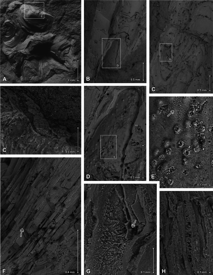
Figure 9. Scanning electron images of Palaenigma wrangeli (Schmidt, 1874).
(A, C) Specimen GIT 655-1, from Kükita 24 drillcore, Mustvee Parish, north-east Estonia, Vormsi Stage, showing fragment of a transversally ornamented periderm? near external surface of a pillar. (B, D–H) Specimen GIT 655-2, from Ellavere drillcore, Järva County, north-east Estonia, Vormsi? Stage. (B, D, E) Area with distinctive papillate surface and with wrinkles (arrow in E). (F–H) Details showing lamellate conch cross section with empty or filamentous interspaces (arrow in F). Note also the chimney like structures (arrow in G).

