Fig. 1.
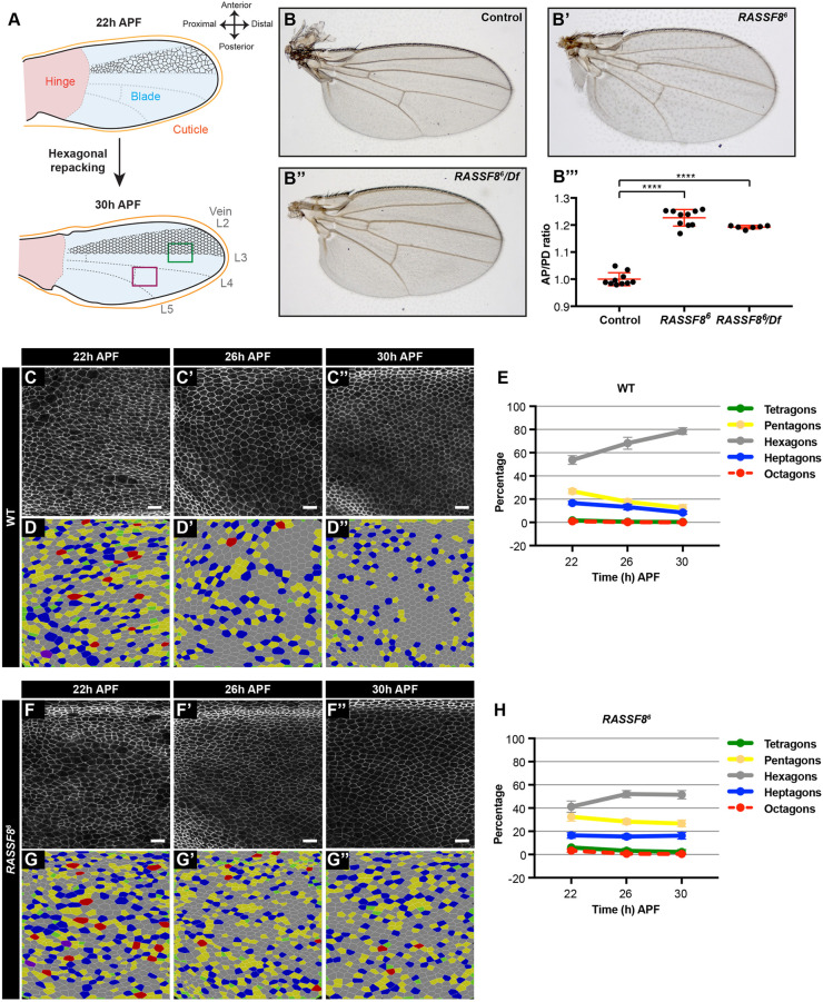
RASSF8 is required for pupal wing cell hexagonal packing. (A) Schematic diagram of pupal wing morphology, axes and development from 22 to 30 h after puparium formation (APF). Colour-coded rectangles indicate regions imaged for analyses. The region distal to the posterior crossvein is marked in purple and the region straddling the L3 vein is marked by a green rectangle. The positions of the longitudinal veins (L2-L5) and crossveins are indicated as dashed lines. As the wing hinge (shaded pink) contracts, the wing blade (shaded blue) extends in the PD axis and the wing epithelial cells (indicated in black between L2 and L3) reorder to form a hexagonal lattice. The wing images throughout this article are oriented as indicated in this diagram. (B) Wild-type wing, (B′) RASSF86 homozygous mutant wing and (B″) RASSF86/Df (3R)BSC321 deficiency heterozygous wing. (B‴) Quantification of relative wing roundness (ratio of AP to PD axis, normalised so that wild-type ratio=1). Data are mean±s.d. ANOVA (Tukey's correction): ****P<0.0001. As expected, because RASSF86 is a null mutant, the homozygous RASSF86 animals have a similar phenotype to the RASSF86/Df animals. (C-F) Hexagonal cell packing of wild-type and RASSF8 mutant wings at 22, 26 and 30 h APF. Images of Ecad::GFP-labelled wild-type (C-C″) and RASSF8 mutant (F-F″) wings at a region distal to the posterior crossvein (purple rectangle in A). Colour-coded images indicate the number of neighbours for each cell in wild-type (D-D″) and RASSF8 mutant (G-G″) wings, determined by using Tissue Analyzer (Aigouy et al., 2010). (E,H) Percentage of cells with four, five, six, seven and eight neighbours (colour coded as indicated) in wild type (D) and RASSF8 mutants (G). The red line (octagons) is dashed so the green line (tetragons) can be seen. Data are mean±s.d., n=1500-5000 cells from three to five individual wings. Scale bars: 10 μm. See Table S1 for raw data.

