



ABSTRACT
Nontyphoidal Salmonella (NTS) strains are associated with gastroenteritis worldwide but are also the leading cause of bacterial bloodstream infections in sub-Saharan Africa. The invasive NTS (iNTS) strains that cause bloodstream infections differ from standard gastroenteritis-causing strains by >700 single-nucleotide polymorphisms (SNPs). These SNPs are known to alter metabolic pathways and biofilm formation and to contribute to serum resistance and are thought to signify iNTS strains becoming human adapted, similar to typhoid fever-causing Salmonella strains. Identifying SNPs that contribute to invasion or increased virulence has been more elusive. In this study, we identified a SNP in the cache 1 signaling domain of diguanylate cyclase STM1987 in the invasive Salmonella enterica serovar Typhimurium type strain D23580. This SNP was conserved in 118 other iNTS strains analyzed and was comparatively absent in global S. Typhimurium isolates associated with gastroenteritis. STM1987 catalyzes the formation of bis-(3′,5′)-cyclic dimeric GMP (c-di-GMP) and is proposed to stimulate production of cellulose independent of the master biofilm regulator CsgD. We show that the amino acid change in STM1987 leads to a 10-fold drop in cellulose production and increased fitness in a mouse model of acute infection. Reduced cellulose production due to the SNP led to enhanced survival in both murine and human macrophage cell lines. In contrast, loss of CsgD-dependent cellulose production did not lead to any measurable change in in vivo fitness. We hypothesize that the SNP in stm1987 represents a pathoadaptive mutation for iNTS strains.
KEYWORDS: cellulose, diguanylate cyclase, STM1987, invasive Salmonella, CsgD independent, virulence, cellulose, cyclic-di-GMP, macrophage
INTRODUCTION
Salmonella enterica strains are important foodborne pathogens that cause a significant amount of morbidity and death worldwide (1). From a clinical perspective, the >2,500 serovars of S. enterica cause two main types of disease, namely, typhoid fever and gastroenteritis (2). Endemic in resource-poor settings, typhoid fever is a life-threatening systemic disease caused by typhoidal Salmonella (TS) serovars, primarily Typhi and Paratyphi (3). Gastroenteritis infections worldwide are caused by strains from nontyphoidal Salmonella (NTS) serovars, with serovars Typhimurium and Enteritidis being the most common (4). TS serovars are human restricted, while NTS serovars can cause disease in a wide range of hosts. Genomic changes such as gene acquisition, single-nucleotide polymorphisms (SNPs), and pseudogene accumulation have been associated with the restricted host range of TS serovars (5, 6). Over the past 3 decades, an invasive disease caused by NTS serovars has emerged as the third type of disease caused by S. enterica.
In sub-Saharan Africa, strains of the gastroenteritis-associated NTS serovars Typhimurium and Enteritidis are the leading cause of invasive bloodstream infections that are distinct from typhoid fever (7). These isolates are referred to as invasive NTS (iNTS). In 2017, iNTS serovars caused an estimated 535,000 cases of invasive bloodstream infections, resulting in 77,500 deaths (8). Bacteremia caused by iNTS serovars is common in children with severe anemia, HIV, malaria, or malnutrition and in immunocompromised adults. Symptoms include nonspecific fever, headaches, chills, malaise, and, in rare cases, diarrhea (9). Two closely related lineages of S. enterica serovar Typhimurium that belong to multilocus sequence typing sequence type 313 (ST313) are the most common causes of bacteremia caused by iNTS (10). Genome sequencing of S. Typhimurium D23580, a representative ST313 isolate that was isolated from a child in Malawi, identified >700 SNPs that were absent in non-ST313 isolates (10–12). These SNPs contribute to the ongoing genomic changes of ST313 isolates, which are thought to be evolving like human-adapted TS serovars (11).
Cellulose, composed of β-(1-4)-linked d-glucose units, is the most abundant polymer on earth. It is the major component of plant cell walls and is the starting material for numerous medical, industrial, and agricultural products (13). Cellulose is also one of the major components of the extracellular matrix of bacterial biofilms, and its production has been described in a wide variety of bacteria, such as Agrobacterium tumefaciens, Acetobacter xylinus, and Rhizobium spp. (14) in addition to clinically relevant S. enterica (15, 16) and E. coli strains (17, 18). In bacteria, cellulose is synthesized by a membrane-bound cellulose synthase complex composed of BcsA and BcsB subunits (19). BcsA is composed of four N-terminal and four C-terminal transmembrane helices separated by an extended intracellular loop that forms the glycosyltransferase (GT) domain. The eight transmembrane helices form a pore across the inner membrane, while the cytoplasmic GT domain contains the PilZ domain, which binds bis-(3′,5′)-cyclic dimeric GMP (c-di-GMP) (20). BcsB is in the periplasm and is anchored to one of the transmembrane helices of BcsA (19, 21). In the absence of c-di-GMP, BcsA is in an inactive state. Cyclic-di-GMP allosterically activates BcsA by binding to the PilZ domain (22).
Cyclic-di-GMP is an important bacterial secondary messenger involved in the regulation of diverse processes such as biofilm formation, motility, and virulence (23). Cyclic-di-GMP is synthesized from two molecules of GTP by diguanylate cyclase (DGC) enzymes and is degraded by specific phosphodiesterases (PDEs) (24). DGCs are typically composed of at least two domains; the highly conserved GGDEF motif required for c-di-GMP synthesis is present in the C-terminal domain (25), while the N-terminal domain typically consists of the sensory receptor that activates the GGDEF activity. The S. enterica genome encodes 12 GGDEF motif-containing proteins, many of which are constitutively expressed. Recombinant S. enterica strains lacking all 12 GGDEF motif-containing proteins are avirulent and impaired for biofilm formation (26, 27).
Biofilms are structured communities of bacterial cells encased in a self-produced extracellular polysaccharide matrix. For S. enterica, cellulose and curli fimbriae are the main components and have been shown to contribute to the long-term survival and persistence of cells in the environment (28). Transcription factor CsgD is the master regulator of biofilm formation in S. enterica; it positively regulates the biosynthesis of cellulose, curli fimbriae, and other biofilm components (29). Recently, S. Typhimurium was also shown to produce cellulose inside macrophages as an antivirulence factor (30). These diverse and opposing functions illustrate a complex role for cellulose in the Salmonella life cycle. As described above, the most important factor for cellulose biosynthesis is the production of c-di-GMP by DGCs, which respond to a variety of different environmental signals (14, 31, 32). The DGC AdrA induces cellulose production in lysogeny broth (LB) medium without salt at 28°C (biofilm-inducing conditions) in a CsgD-dependent manner, while STM1987 can induce cellulose production at 37°C in nutrient-deficient medium, independent of CsgD (29, 33). By treating the biofilms with cellulase, Solano et al. showed that biofilm obtained at 28°C in LB medium is composed of other extracellular matrix components besides cellulose, while biofilm obtained at 37°C in ATM medium is composed predominantly of cellulose (33, 34). Recently, Mills et al. showed that STM1987 can sense arginine, phytic acid, glucose, and GlcNAC at biologically relevant concentrations (35). Since arginine is thought to be a prominent signal inside macrophages (36–38), it was speculated that STM1987 might control cellulose production by S. enterica inside macrophages.
In this study, we identified a missense mutation in stm1987 in iNTS type strain S. Typhimurium D23580 that was conserved in all lineage I and II ST313 isolates from sub-Saharan Africa. Experiments showed that the presence of the altered STM1987 enzyme led to reduced cellulose production, increased rates of survival inside macrophages, and increased in vivo fitness. Our results indicate that CsgD-independent cellulose production plays a role in regulating S. Typhimurium virulence.
RESULTS
The presence of a SNP in stm1987 in S. Typhimurium strain D23580 results in an enzyme with reduced activity.
From a list of SNP differences between gastroenteritis-causing S. Typhimurium 14028 (ST19) and invasive S. Typhimurium strain D23580 (ST313) (Y. Wang, unpublished observations), we identified a C-to-G transversion SNP in S. Typhimurium D23580 located 566 bp downstream of the translational start site of stm1987. The mutation results in substitution of arginine for threonine at residue 189 in the cache 1 sensory domain (Fig. 1), a region that was shown to be critical for sensing external signals and regulating the overall enzyme activity of STM1987 (35).
FIG 1.

Schematic of stm1987 allele in S. Typhimurium 14028 and D23580. The 570-amino acid STM1987 protein is represented by the black rectangle and is shown to scale, with transmembrane (TM), cache 1 signaling, and GGDEF-DGC domains highlighted. The magnified region shows nucleotides between position 553 and position 579, with nucleotide 566 in bold (C in 14028 and G in D23580). Single-letter amino acids corresponding to each codon are shown above for S. Typhimurium 14028 and below for strain D23580. Amino acid residue 189 is shown in bold red (T for 14028 and R for D23580).
To determine whether the amino acid change in STM1987 would influence enzyme activity, the stm1987 alleles from S. Typhimurium strains 14028 and D23580 were cloned into pBR322 and used to complement a S. Typhimurium 14028 Δstm1987 mutant strain. Plasmid-based expression of stm1987 or other DGCs was previously demonstrated to result in the overproduction of cellulose (26, 33, 39). When macrocolonies of each strain were grown on medium containing calcofluor white, a fluorescent dye that stains cellulose (34), the strain complemented with the stm1987^ST14028 allele was noticeably brighter than the stm1987^D23580-complemented strain, and both were brighter than the pBR322 control (Fig. 2A). Cellulose production was also visualized by growing strains in liquid culture and quantifying the amount of biofilm material deposited at the air-liquid interface of the culture vessel (33, 34). After 24 h of growth at 37°C, the Δstm1987 strain complemented with the stm1987^ST14028 allele had a thick ring at the air-liquid interface, whereas the strain complemented with the stm1987^D23580 allele had only a faint ring and the pBR322 control strain had no visible ring (Fig. 2B). We confirmed that the curli biosynthesis operon (i.e., csgBAC) was not expressed under these conditions (see Fig. S1 in the supplemental material), indicating that cellulose was a primary component. Crystal violet (CV) staining showed that the stm1987^ST14028-complemented strain produced approximately 10 times more biomass attached to the glass than did the stm1987^D23580-complemented strain, which was slightly above background levels (Fig. 2C). We repeated the same experiment with a mutant strain unable to make cellulose (ΔbcsA mutant), and none of the plasmid-containing strains produced a visible ring at the air-liquid interface (Fig. S2). These experiments demonstrated that the single amino acid substitution in STM1987 in S. Typhimurium D23580 resulted in reduced cellulose production.
FIG 2.
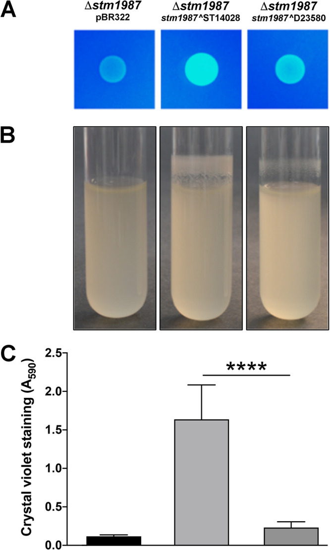
Phenotypic comparison of stm1987 alleles from S. Typhimurium 14028 and D23580. S. Typhimurium 14028 Δstm1987 was transformed with pBR322 (control) or pBR322 harboring the stm1987^ST14028 or stm1987^D23580 alleles. (A) Cells from overnight cultures were grown on tryptone agar supplemented with calcofluor white, and colonies were visualized under UV light after 12 h of growth at 28°C. (B) Strains were grown in LB for 24 h at 37°C, and tubes were examined for the presence of surface-attached biomass at the air-liquid interface. (C) The amount of attached biomass was quantified using CV staining, with the absorbance of the resulting solution being measured at 590 nm (A590). Columns represent the average values and error bars represent the standard deviations from five biological replicates. ****, P < 0.0001.
The SNP in stm1987 is conserved in invasive lineages of S. Typhimurium.
To determine whether the STM1987 mutation identified in S. Typhimurium D23580 was conserved, we examined the gene sequences in 118 additional ST313 S. Typhimurium strains. Whole-genome sequencing-based phylogeny has previously been used to cluster the ST313 strains from sub-Saharan Africa into closely related lineages I and II (10). We overlaid the stm1987 SNP information on a phylogenetic tree that was modeled after the work by Okoro et al. (10) (Fig. 3). All information pertaining to the strains in this tree, as well as additional isolates that were screened, is listed in Table S1 in the supplemental material. The stm1987 SNP was conserved in all lineage I isolates (44/44 shown in Fig. 3; 50 total) and lineage II isolates (57/57 shown in Fig. 3; 69 total). In contrast, the SNP was almost completely absent from a global clade of gastroenteritis-causing S. Typhimurium isolates of ST19, ST34, ST128, and ST568 (absent in 8/9 isolates shown in Fig. 3; 54/56 isolates total). S. Typhimurium strains DT24 (isolated from a patient in the United Kingdom) and 5634 (isolated from a patient in Kenya) were the only global isolates analyzed that possessed the SNP (Table S1). The conservation of the stm1987 SNP in invasive isolates from sub-Saharan Africa indicated that this mutation has been selectively maintained within this group of isolates.
FIG 3.

Conservation of stm1987 SNP in invasive S. Typhimurium lineages. The maximum likelihood phylogenetic tree was constructed from sequenced S. Typhimurium isolates (10) as reported previously (40). The isolates analyzed were divided into S. Typhimurium groups associated with gastroenteritis or invasive disease (lineages I and II) (10). The presence or absence of the STM1987 SNP is noted for each group; the red star shows the position of strain D23580 in the tree.
Reduced STM1987 activity is associated with increased survival rate of S. Typhimurium inside macrophages.
Production of cellulose was recently identified as an antivirulence factor for S. Typhimurium that was correlated with reduced survival inside macrophages. To test the role of STM1987 in this process, we compared the survival rates of S. Typhimurium D23580 and an engineered strain containing the stm1987^ST14028 allele inside human THP-1 and murine RAW macrophages. There was no difference in survival rates (Fig. 4A); however, we know that S. Typhimurium D23580 has other mutations that reduce overall cellulose production (i.e., bcsG truncation [41] and two csgD promoter mutations [40]). To test the effect of the stm1987 SNP in a strain that is able to make normal levels of cellulose, we introduced the stm1987^D23580 allele into S. Typhimurium 14028. The presence of the stm1987 SNP in S. Typhimurium 14028 led to significantly increased survival rates in both THP-1 and RAW macrophages (Fig. 4B). To compare the STM1987 enzymes directly to each other, we performed the same survival assays with plasmid-complemented S. Typhimurium 14028 Δstm1987 strains. Expression of stm1987^ST14028 correlated with the poorest survival, which was significantly reduced, compared to that of stm1987^D23580, and both enzymes caused decreased survival, compared to the vector-only control (Fig. 4C). To confirm that the survival effects were due to cellulose production, we performed the same experiment with a cellulose-negative (ΔbcsA mutant) strain. This time, there was no difference in survival rates between strains complemented with either enzyme or the plasmid alone (Fig. 4D).
FIG 4.

Analysis of the role of STM1987 and cellulose production in S. Typhimurium intramacrophage survival. Human THP-1 and murine RAW 264.7 macrophages were infected with S. Typhimurium D23580 or D23580 (stm1987^ST10428) (A), S. Typhimurium 14028 or 14028 (stm1987^D23580) (B), S. Typhimurium 14028 Δstm1987 strains transformed with pBR322, pBR322/stm1987^ST14028, or pBR322/stm1987^D23580 (C), or S. Typhimurium Δstm1987 ΔbcsA strains transformed with the same plasmids (D). Percent survival was determined as the proportion of intracellular CFU remaining at 18 h postinfection, compared to the initial CFU values measured after 30 min. Statistical significance between groups was noted. *, P < 0.05; **, P < 0.01; ***, P < 0.001; ****, P < 0.0001; ns, not significant, P > 0.05.
We performed additional survival experiments with an S. Typhimurium 14028 strain engineered to contain the bcsG mutation that is present in S. Typhimurium D23580. When this additional mutation was present, there was no significant difference in survival rates between strains expressing the STM1987 enzymes or the vector-only control (Fig. S3). This experiment demonstrated how a mutation in bcsG could mask the effects of stm1987.
Expression of stm1987 is independent of CsgD in S. Typhimurium.
In the context of Salmonella biofilm formation, cellulose production is primarily regulated by the activity of the DGC AdrA, which is positively regulated by the central biofilm regulator CsgD (16, 33, 42). In E. coli, a CsgD-independent pathway for cellulose production has been described; it is regulated by the DGC YedQ, which is a homolog of STM1987 (43). To investigate whether these same regulatory principles existed in S. Typhimurium, we monitored stm1987 and adrA expression using promoter-luciferase fusions designed to measure gene expression via light production. The expression profiles for stm1987 were the same in wild-type S. Typhimurium 14028 and a ΔcsgD mutant strain and were at similar levels in S. Typhimurium D23580 (Fig. 5A). In contrast, adrA expression was completely shut off in S. Typhimurium 14028 ΔcsgD and was greatly reduced in S. Typhimurium D23580 (Fig. 5B). The result in S. Typhimurium D23580 could be explained because csgD expression is significantly reduced in this strain due to the presence of two SNPs in the csgD promoter region (40). These experiments showed that stm1987 expression was not regulated by CsgD in S. Typhimurium, meaning that STM1987 likely participates in a CsgD-independent pathway for cellulose production, similar to YedQ in E. coli.
FIG 5.

Transcription of stm1987 is not CsgD dependent in S. Typhimurium. Expression curves for stm1987 (A) or adrA (B) promoter-lux fusions are shown for S. Typhimurium 14028 (black), 14028 ΔcsgD (red), or S. Typhimurium D23580 (gray) grown at 28°C in 1% tryptone medium. For each fusion, the luminescence values (counts per second [CPS]) were plotted as a function of time, with measurements recorded every 30 min. Each expression curve corresponds to an individual biological replicate culture.
The stm1987 SNP and CsgD-independent cellulose production contribute to S. Typhimurium virulence.
One of the major challenges in characterizing and understanding invasive S. enterica isolates from sub-Saharan Africa is the lack of an appropriate disease model. These iNTS isolates appear to be human adapted, with up to 1,000 SNPs that differentiate them from standard, gastroenteritis-causing NTS isolates. Due to the potential for multiple SNPs in the same pathways, it can be hard to pinpoint the effects of individual SNPs. In addition, broad antibiotic resistance profiles make genetic manipulations of these invasive isolates difficult.
Therefore, we performed pilot experiments with intraperitoneal (i.p.) injection of susceptible (Nramp−/−) C57BL/6 mice, which showed no difference in virulence between S. Typhimurium D23580 and the D23580 (stm1987^ST14028) strain (Fig. S4A and B). Based on the macrophage survival results, we reasoned that it was not possible to evaluate the impact of the stm1987 mutation in S. Typhimurium D23580 due to the presence of other mutations. Therefore, we evaluated the effect of the stm1987 SNP in the S. Typhimurium 14028 strain background.
C57BL/6 mice were challenged orally with 107 CFU of a 1:1 mixture of wild-type S. Typhimurium 14028 and S. Typhimurium 14028 (stm1987^D23580). At 4 to 7 days postinfection, mice were euthanized and the bacterial loads in the spleen, liver, cecum, and mesenteric lymph nodes (MLN) were determined. S. Typhimurium 14028 (stm1987^D23580) was recovered in greater proportions than the wild-type strain from the MLN and cecum, but there was no colonization difference measured in the liver and spleen (Fig. 6A). To bypass small intestinal colonization and to more directly assess systemic spread within the mouse, we performed additional competitive index (CI) experiments via i.p. injection. At 72 h postinfection, the S. Typhimurium 14028 (stm1987^D23580) strain was recovered in greater proportions than the wild-type 14028 strain in all organs analyzed (Fig. 6B). This gave a clear indication that the amino acid change in the cache 1 signaling domain of STM1987 could lead to increased in vivo fitness for S. Typhimurium. This also points to a role for CsgD-independent cellulose production inside macrophages as a virulence modifier.
FIG 6.

Role of the CsgD-independent and CsgD-dependent cellulose production pathways in S. Typhimurium virulence. CI infections were performed with S. Typhimurium 14028 and 14028 (stm1987^D23580) strains (A and B) or S. Typhimurium 14028 and 14028 (PcsgD^D7795) strains (C and D). C57BL/6 mice were challenged orally with 107 CFU (A and C) or i.p. with 104 CFU (B and D); each inoculum consisted of a 1:1 ratio of each pair of strains. At 4 to 7 days after oral infection or 72 h after i.p. infection, bacteria were enumerated from the spleen, liver, MLN, cecum, and kidney. CI values were calculated for each organ as follows: (CFU 14028mutant/14028wild-type)output/(CFU 14028mutant/14028wild-type)input. A CI value of 1, which represents a situation in which strains are equally virulent, is represented by the horizontal dotted line on each graph. Red circles represent CI values at which the 14028 strains containing the stm1987^D23580 or PcsgD^D7795 mutations were recovered in greater proportions than the wild-type strain. Statistical differences between groups of mice were noted. *, P < 0.05; **, P < 0.01; ***, P < 0.001; ns, not significant, P > 0.05.
The CsgD-dependent pathway for cellulose production has no apparent role in S. Typhimurium virulence.
As a control, we wanted to test the influence of CsgD-dependent cellulose production on the in vivo fitness of S. Typhimurium. We recently identified a csgD promoter SNP that abolished csgD transcription in S. Enteritidis D7795 and was conserved throughout the lineage of invasive S. Enteritidis strains (40). Correcting the csgD promoter SNP was enough to restore biofilm formation and cellulose production in this strain (40). As expected, there was no difference in virulence when SNP-containing or SNP-corrected invasive S. Enteritidis strains were tested directly (Fig. S4C). To more carefully evaluate the effects of this csgD promoter mutation, we compared the in vivo fitness of S. Typhimurium 14028 and a strain carrying the inactivating csgD promoter SNP, i.e., S. Typhimurium 14028 (PcsgD^D7795) (40), in both oral and i.p. challenge models. There was no significant difference in colonization between the strains 4 to 7 days after oral challenge (Fig. 6C) or 72 h after i.p. challenge (Fig. 6D). The overall conclusion from these experiments was that biofilm formation or CsgD-dependent cellulose production does not appear to alter the in vivo fitness of S. Typhimurium.
DISCUSSION
Invasive NTS serovars are thought to be evolving toward human adaptation through genomic degradation in the form of SNPs and the accumulation of pseudogenes, similar to TS serovars (11, 44, 45). The difference between gastroenteritis-associated and invasive S. Typhimurium strains reside in these genomic changes, some of which have been linked to the ability of iNTS serovars to cause systemic disease. Host immune susceptibility is also clearly a large factor, but the strains are different (9). In this study, we identified a conserved SNP present in invasive S. Typhimurium lineages that causes an amino acid substitution in the cache 1 signaling domain of DGC STM1987. This STM1987 mutation was correlated with reduced cellulose biosynthesis, increased survival in both human and murine macrophage cell lines, and increased in vivo fitness in murine infection. The stm1987 SNP was comparatively absent in global S. Typhimurium isolates that cause gastroenteritis.
There are at least two pathways for cellulose production in Salmonella, CsgD dependent and CsgD independent. Traditionally, cellulose production has been correlated with biofilm formation and persistence in nonhost environments (20). While initial studies indicated that cellulose had no role in virulence (18, 20, 28, 42, 46), it was recently shown that cellulose can be produced by Salmonella inside macrophages, serving as an antivirulence factor (30, 47). These opposing functions imply a complex role for cellulose in Salmonella biology. A CsgD-independent pathway for cellulose production was first identified in E. coli, involving the STM1987 homologue YedQ (43). All cellulose biosynthesis pathways converge at c-di-GMP-based activation of BcsA (22, 48–50), so it makes sense that the uncoupling of cellulose production from CsgD regulation could occur at the level of c-di-GMP biosynthesis. Our gene expression assays showed that stm1987 transcription was independent of CsgD in S. Typhimurium. Mills et al. showed that STM1987 stimulated cellulose production in response to putative host signals such as arginine and the cache 1 signaling domain was critical for this response (35, 51). Since arginine is thought to be a key metabolic signal inside macrophages (36–38), this led us to investigate the effect of the amino acid change in the cache 1 domain identified in S. Typhimurium D23580. When introduced into gastroenteritis-causing S. Typhimurium 14028, the missense mutation in STM1987 decreased cellulose production and led to increased in vivo fitness, whereas a SNP that shut off CsgD-dependent cellulose production did not appear to influence virulence. Regulating cellulose production via CsgD-dependent and CsgD-independent pathways that act through different DGCs might enable S. enterica strains to separate cellulose production inside the macrophage and under biofilm-inducing conditions.
Several genomic changes in invasive S. Typhimurium isolates (11, 12, 44) have been associated with clear virulence or disease phenotypes. Hammarlöf et al. identified a SNP in the promoter region of pgtE that increased the expression of this outer membrane protease, which promoted survival and dissemination of ST313 isolates during infection (52). Carden et al. showed that pseudogenization of effector sseI, part of the Salmonella pathogenicity island-2 (SPI-2) type 3 secretion system, contributes to the hyperdissemination of D23580 from the gut to systemic organs (53). Ramachandran et al. showed that ST313 isolates survive better inside murine and human macrophages, compared to ST19 strains, due to reduced flagellin production, although the specific genomic changes responsible were not identified (54). In our experiments with S. Typhimurium D23580, the effect of reduced STM1987 activity on virulence was likely masked by the presence of a premature stop codon in bcsG (40, 41), which encodes a component of cellulose synthase that modifies the growing β-d-1,4-glucan chain with phosphoethanolamine (55). BcsG is required for cellulose production in Salmonella and E. coli (55, 56). Based on the results of our intramacrophage survival assays, we hypothesize that the existing mutation in bcsG masks any colonization advantage provided by the SNP in stm1987 in S. Typhimurium D23580. The stm1987 mutation is present in both lineage I and lineage II ST313 isolates and in the newly described lineage III group of isolates (57), whereas the bcsG mutation is found only in lineage II (40, 41). This indicates that the SNP in stm1987 is a more ancient mutation, and it also suggests that adaptation is an ongoing process, whereby the amount of cellulose produced by S. Typhimurium D23580 would be further reduced due to the presence of a bcsG mutation. The stepwise evolutionary process for these ST313 isolates has recently been described in great detail (57). A real-world example of adaptive evolution of iNTS strains was shown by Klemm et al., who used comparative genomics to track changes over time in a Salmonella serovar Enteritidis strain causing recurrent infections in an immunocompromised, interleukin-12 β1-receptor-deficient patient (45).
Several groups have attempted to establish animal models to evaluate the virulence of human-adapted ST313 isolates. Following oral inoculation, ST313 isolates are able to colonize organs systemically in susceptible mice (11, 58, 59). Carden et al. used the streptomycin-treated mouse model to specifically analyze the rate of dissemination to systemic sites, compared to S. Typhimurium ST19 strain SL1344 (53). Parsons et al. showed that, following oral inoculation, ST313 isolates D23580 and Q456 caused more invasive infections in chickens (i.e., spleen and liver), compared to ST19 isolates F98 and 4/74 (60). However, their result was not reproduced by Lacharme-Lora et al., who showed that ST313 strain D23580 was less virulent than ST19 strain 4/74 in both inbred and outbred chickens (61). S. Typhimurium D23580 has also been shown to colonize chick embryos and Galleria mellonella larvae (61). Rhesus macaques infected via the intragastric route developed only mild diarrhea, with no significant pathology in the liver, ileum, colon, and MLN (58). Although these animal infection models demonstrate that ST313 strains can infect nonhuman hosts, it is not well known how each disease model relates to iNTS infection in humans, which is distinct from gastroenteritis and typhoid fever (7–9). To evaluate the effect of the stm1987 SNP on virulence, we infected susceptible C57BL/6 mice via the i.p. route. In this model, iNTS strains were able to rapidly colonize the organs systemically as well as to disseminate to distant tissues via the bloodstream, confirming the results of other laboratories (11, 62).
In light of the difficulties with animal models and the presence of other mutations in iNTS strains, our results suggest that moving individual mutations into standard, more well-defined NTS strains is a valid way to test their role in virulence. In addition, because iNTS strains are typically resistant to multiple antibiotics (10, 44, 63), the use of sensitive non-ST313 strains allows easier genomic modifications. In our case, we were able to show that the stm1987 SNP conferred increased in vivo fitness, compared to a wild-type strain, with both oral and i.p. routes of infection. We cannot explain why no difference was observed in the spleen and liver after oral challenge. However, following oral ingestion it takes about 72 h for Salmonella to reach the liver and spleen, when mice start to exhibit signs of infection (64). It is possible that at later stages of infection the balance between virulence and host resistance to infection may favor Salmonella (65), and hence replication may be largely unrestricted in a diseased mouse, making it more difficult to differentiate between strains. Intraperitoneal injection bypasses the oral invasion steps and is predicted to speed access to macrophages and internal organs. For this reason, we concluded that the increased fitness of S. Typhimurium 14028 (stm1987^D23580) in all organs tested following i.p. injection was correlated with increased rates of survival inside macrophages. Two possibilities for how reduced cellulose production could aid in intramacrophage survival are increased growth rate and increased efficiency of SPI-2 function at the interface between the S. Typhimurium outer membrane and the vacuolar membrane (66).
The importance of c-di-GMP in the life cycle of pathogenic bacterial species is highlighted by our results. Cyclic-di-GMP has been implicated in diverse processes such as biofilm formation, motility, cell cycle progression, and virulence (31, 67, 68). The domains for DGC activity (i.e., GGDEF) and PDE activity (i.e., EAL or HD-GYP) can be found separately or together in hybrid proteins that contain both domains; most S. Typhimurium strains encode five GGDEF, seven GGDEF-EAL, and 10 EAL domain proteins (69). In general, low levels of c-di-GMP enhanced by the actions of PDEs promote Salmonella virulence in mice and increased survival inside macrophages (70), while high c-di-GMP levels enhanced by DGCs promote biofilm formation and environmental persistence. The activities of most DGCs and PDEs are controlled by the sensory domains present in the N-terminal regions of the proteins (71). Independent of transcription and translation, the sensory domains allow bacteria to respond to changes in environmental conditions, such as oxygen, redox state, temperature, light, nutritional signals, and the presence of quorum-sensing molecules. These sensory domains can be localized to the cytoplasm, such as PAS (Per-Arnt-Sim), GAF, and globin domains, associated with membranes, such as MASE and MHYT domains, or in the periplasm, such as CHASE, Cache 1, and CSS domains (71). Sarenko et al. showed that the DGC and PDE proteins in E. coli fall into two categories, i.e., (i) DGCs and PDEs that form a tightly interconnected network and regulate a particular phenotype, such as biofilm formation, or (ii) distinct enzymes that are not connected to other proteins but contribute to controlling the overall cellular pool of c-di-GMP under specific conditions (72). The specificity of the signaling domain may be the main indicator of the conditions under which distinct DGCs and PDEs are induced. For example, the cache 1 domain of STM1987 appears to have a critical role in responding to intracellular signals inside macrophages, hypothesized to be arginine (35), but it is not known whether that is its only function. It was surprising that a single amino acid change in the cache 1 domain affected the overall enzymatic activity, independent of the GGDEF domain. Although we did not test whether the two proteins were equally stable, our results suggest that mutations in signaling domains can alter the activity of c-di-GMP-associated enzymes. For sensory domains like PAS, which can detect a broad diversity of environmental signals, such as molecular oxygen, light, small metabolites, and temperature (71), it is possible that mutations could increase or limit the types of signals detected. Such mutations or SNPs in signaling domains could contribute to bacterial evolution in terms of adaption to new ecological niches and/or hosts.
Our results indicate that the CsgD-independent pathway for cellulose production, acting through the activity of STM1987, regulates S. enterica survival and virulence inside macrophages. We recently showed that CsgD-dependent biofilms are formed during acute infections in mice, with curli fimbriae being detected in the distal gastrointestinal tract (80). Since curli fimbriae are a pathogen-associated molecular pattern (PAMP) that activates Toll-like receptors (TLRs) (TLR-1 and TLR-2) and intracellular NOD-like receptors (73, 74), it is tempting to speculate that cellulose could also play a role in this. How the CsgD-dependent and -independent pathways fit within the fine-tuned pathogenesis of S. enterica strains is still up for debate and requires further investigation.
MATERIALS and METHODS
Bacterial strains, media, and growth conditions.
The bacterial strains used in this study are listed in Table S2 in the supplemental material. For standard growth, strains were inoculated from frozen stocks onto LB agar (LB, 1% NaCl, 1.5% agar) supplemented with an appropriate antibiotic (50 μg ml−1 kanamycin [Kan], 34 μg ml−1 chloramphenicol [Cam], or 5 μg ml−1 tetracycline [Tet]) and were grown overnight at 37°C. Isolated colonies were used to inoculate 5 ml LB, and the culture was incubated for 18 h at 37°C with agitation at 200 rpm. To visualize cellulose production, 2-μl aliquots of overnight culture were spotted on 1% tryptone agar supplemented with calcofluor white (fluorescent brightener 28; Sigma-Aldrich) (200 μg ml−1) (34). To evaluate cellulose production by CV staining, 5 × 10⁶ cells were inoculated into 5 ml LB and incubated for 18 h at 37°C with agitation at 200 rpm. For macrophage survival assays, cells from overnight cultures were diluted 1:100 into fresh LB and grown at 37°C until the cultures reached an optical density at 600 nm of 0.7. For murine infection experiments, overnight cultures grown in LB were diluted to the desired CFU concentration in 100 mM HEPES (pH 8) and used to infect mice.
Generation of S. Typhimurium 14028 mutant strains.
The S. Typhimurium 14028 Δstm1987 and S. Typhimurium 14028 Δstm1987 ΔbcsA strains were generated using the lambda red recombinase knockout procedure (75). Primers STM14_2608-ko_sense and STM14_2608-ko_antisense or bcsA-ko_sense and bcsA-ko_antisense, containing 50-nucleotide sequences on either side of the stm1987 or bcsA gene (Table S2), were used to amplify the cat gene from pKD3 using Phusion high-fidelity DNA polymerase (New England BioLabs). The PCR products were purified and electroporated into S. Typhimurium 14028 cells containing pKD46. Mutant strains were selected by growth at 37°C on LB agar supplemented with 7 μg ml−1 Cam before restreaking on LB agar containing 34 μg ml−1 Cam. The mutations were moved into a clean strain background by transduction with P22 phage (76). The cat gene was resolved from the chromosome using pCP20 (75). Sanger DNA sequencing of PCR products amplified from the chromosome of the Δstm1987 strain using primers STM1987detect1 and STM1987detect2 confirmed the loss of the stm1987 open reading frame, while primers bcsAdetect1 and bcsAdetect2 were used to confirm the loss of the bcsA open reading frame.
Cellulose quantification using CV staining.
In vitro production of cellulose by S. Typhimurium was visible as a ring of adherent cells at the air-liquid interface. After 24 h of growth, the culture broth was discarded, and the culture tubes were washed twice with water to remove any nonadherent cells before air drying for 10 min. Eight milliliters of CV solution (0.1% [wt/vol]) was added to each tube to completely cover the ring of adherent cells, and tubes were incubated for 10 min at room temperature. The CV solution was removed, and tubes were washed twice with water before air drying for 10 min. To dissolve bound CV, 8 ml of 95% ethanol was added to each tube, and the tubes were incubated at room temperature for 15 min. The optical density of the resulting solution was measured at 590 nm.
Reference genome sequences and identification of polymorphisms in S. Typhimurium strains.
Whole-genome sequences were obtained from the National Centre for Biotechnology Information (NCBI) via the following accession numbers: NC_016856 (S. Typhimurium 14028) and NC_016854 (S. Typhimurium D23580). Genome assemblies of S. Typhimurium isolates from sub-Saharan Africa and different parts of the world were investigated for a SNP in stm1987 through in silico PCR. Names, accession numbers, and metadata pertaining to all strains that were screened are listed in Table S1.
Generation of bacterial luciferase reporters and pBR322-stm1987 plasmid vectors.
To generate a luciferase reporter to measure stm1987 expression, the promoter-containing DNA region was PCR amplified from S. Typhimurium 14028 using primers STM14_2408for1 and STM14_2408rev2 (Table S3) and Phusion high-fidelity DNA polymerase (New England BioLabs), with reaction conditions as outlined by the manufacturer. The resulting PCR product was purified, sequentially digested with XhoI and BamHI (New England BioLabs), and ligated using T4 DNA ligase (New England BioLabs) into the pCS26 vector cut with XhoI and BamHI. Primers pZE05 and pZE06 were used to verify the successful fusion of the stm1987 promoter region to luxCDABE. The generation of csgB and adrA luciferase fusions was described previously (15, 28).
To generate pBR322/stm1987 plasmid vectors, DNA fragments containing stm1987 with native promoters were PCR amplified from genomic DNA of S. Typhimurium 14028 or S. Typhimurium D23580 using primers STM1987forEco and STM1987revAatII (Table S3). PCR products were purified, digested with EcoRI and AatII, and ligated into digested pBR322 using T4 DNA ligase.
Luciferase reporter assays.
For bioluminescence assays, overnight cultures of reporter strains of S. Typhimurium were diluted 1:600 in LB without salt (i.e., LBns) or 1% tryptone supplemented with 50 μg ml−1 Kan, to a final volume of 150 μl per well in 96-well clear-bottom black plates (Costar number 9520; Corning Inc.). To minimize evaporation of the medium during the assay, cultures were overlaid with 50 μl of mineral oil. Cultures were assayed for absorbance at 600 nm (0.1 s) and luminescence (1 s) every 30 min during growth at 28°C or 37°C, with agitation, in a Victor X3 multilabel plate reader (Perkin-Elmer).
Genome engineering in Salmonella Typhimurium 14028 and D23580.
The I-SceI suicide plasmid system (77) was used for genome engineering either to introduce or to correct the stm1987 SNP in S. Typhimurium strains. The stm1987 gene was PCR amplified from S. Typhimurium 14028 and D23580 using primers STM1987forEco and STM1987revHind (Table S3) and Phusion high-fidelity DNA polymerase (New England BioLabs). Purified PCR products were digested with BamHI and PstI (New England Biolabs) and ligated into BamHI/PstI-digested pSEVA212. Two clones were selected in E. coli S17-1 (λpir) and used for all subsequent experiments. Purified pSEVA212/stm1987 constructs were transformed into S. Typhimurium 14028 or D23580 by mating, with selection for growth on M9 minimal agar supplemented with 1 mM MgSO4, 0.2% glucose, and 100 μg ml−1 Kan (M9-Glc-Kan100). Merodiploid strains with pSEVA212/stm1987 plasmid inserted into the genome were confirmed by restreaking onto M9-Glc-Kan100 agar. Purified pSEVA628S (250 ng) (78) was transformed into merodiploid strains by electroporation, with selection on LB agar supplemented with 1 mM m-toluate and 20 μg ml−1 gentamicin. Resulting colonies were restreaked on LB agar supplemented with 20 μg ml−1 gentamicin, streaked on LB agar supplemented with 50 μg ml−1 Kan to confirm the loss of the pSEVA212 plasmid, and streaked on TCR plates (1% tryptone, 1.5% agar, 40 μg ml−1 Congo red) to check the biofilm phenotype. Colonies were selected from TCR plates and grown at 37°C for two overnight growth steps without gentamicin to generate cells lacking pSEVA628S. Final colonies were streaked onto (i) LB agar, (ii) LB agar with 50 μg ml−1 Kan, (iii) LB agar with 20 μg ml−1 gentamicin, and (iv) TCR plates to select the desired phenotypes. At this stage, two colonies were chosen for further analysis. Primers STM1987checkFOR and STM1987checkREV (Table S3) were used to confirm the presence or absence of the SNP in stm1987 at nucleotide 566. The S. Typhimurium 14028 and S. Enteritidis D7795 strains containing the inactivating csgD promoter SNP were described previously (40).
Murine infection experiments incorporating randomization and blinding.
Six- to 8-week-old female C57BL/6 mice were purchased from the Jackson Laboratory (Bar Harbor, ME) and assigned to cage groups using a randomization table prepared in Microsoft Excel; individual mice were marked with ear notches.
For CI experiments, two groups of six mice were challenged with a mixed inoculum consisting of a 1:1 ratio of S. Typhimurium 14028 and 14028 (stm1987^D23580) or 14028 and 14028 (PcsgD^D7795) either by oral gavage (107 CFU) or i.p. (104 CFU). To apply blinding to the experiments, each strain was marked with either Kan resistance or Cam resistance sig70_16 luxCDABE constructs (79), and two different challenges were prepared (i.e., challenge 1, wild-type-Kan/mutant-Cam; challenge 2, wild-type-Cam/mutant-Kan). Vaccine and Infectious Disease Organization animal care technicians then gave challenge 1 and challenge 2 to different groups of mice, and the cage assignments were revealed to members of the White laboratory only after the final results were analyzed.
To determine the optimal time point for evaluation of iNTS colonization, two groups of six mice were challenged i.p. with 104 CFU of S. Typhimurium D23580 or S. Enteritidis D7795 strains containing the Kan resistance sig70_16-luxCDABE construct (79). At 24 h, 48 h, and 72 h postinfection, two mice from each group were humanely euthanized. In subsequent infections, groups of six mice were challenged i.p. with 104 CFU of S. Typhimurium D23580 (with or without the stm1987 SNP) or S. Enteritidis D7795 (with or without the PcsgD SNP), each containing the Kan-resistant, sig70_16-luxCDABE construct (79). Blinding was not possible because the strains are broadly antibiotic resistant and we were not able to use the Cam marker.
For all infection trials, mice were weighed daily and monitored for clinical signs of infection. Mice that had a >20% drop in weight were humanely euthanized. Mice infected via oral gavage were euthanized 5 to 7 days postinfection, and spleen, liver, MLN, and cecum were collected. Mice infected i.p. were humanely euthanized 72 h postinfection, and spleen, liver, MLN, kidney, and blood samples were collected. For all experiments, collected organs were placed in a 2-ml Eppendorf Safe-Lock tube containing 1 ml of phosphate-buffered saline and a 5-mm steel bead (Qiagen number 69989) and were homogenized for 5 min at 30 Hz using a mixer mill (Retsch number MM400). To determine the number of CFU in the organ homogenates, serial dilutions were plated on LB agar supplemented with 50 μg ml−1 Kan or 20 μg ml−1 Cam. The same procedure was performed for initial challenge doses to ensure the proper inoculation size.
Ethics statement.
All animals were cared for and used in accordance with the guidelines of the Canadian Council on Animal Care and the regulations of the University of Saskatchewan Committee on Animal Care and Supply. All animal experiments were performed under animal use protocol 20190071, which was approved by the University of Saskatchewan Animal Research Ethics Board.
Macrophage growth conditions.
THP-1 monocytes were maintained in RPMI 1640 complete medium (Fisher Scientific number 11875085) supplemented with 10% heat-denatured fetal bovine serum (FBS) (SAFC Biosciences number 12103C). The monocytes were differentiated with phorbol-12-myristate-14-acetate ester (PMA) and seeded at 1.25 × 10⁵ cells per 0.5 ml per well in 48-well plates 3 days prior to infection. Upon PMA treatment, THP-1 monocytes differentiate into adherent macrophage-like cells and stop dividing. One day before S. Typhimurium infection, after the third day of treatment with PMA, the medium was replaced with fresh RPMI 1640 medium.
RAW 264.7 macrophages were grown in Dulbecco’s modified Eagle’s medium (DMEM) (Millipore Sigma number D5796) supplemented with 10% heat-denatured FBS, 1 mM sodium pyruvate, and 50 μg ml−1 gentamicin. Prior to seeding of cells into multiwell plates, the cells were washed with Versene, centrifuged at 1,200 rpm, and resuspended in DMEM with 10% FBS (without antibiotics). RAW 264.7 cells were seeded at 1.25 × 10⁵ S. Typhimurium cells per 0.5 ml per well in a 48-well plate. The cells were allowed to adhere for at least 3 h before infection and continued growing for the duration of the assay.
Macrophage survival assay.
A total of 1.25 × 10⁵ macrophages were infected with S. Typhimurium at a multiplicity of infection (MOI) of 100 for RAW 264.7 and a MOI of 10 for THP-1 macrophages. Infected macrophages were centrifuged at 1,000 rpm for 1 min and incubated at 37°C for 30 min. Following S. Typhimurium internalization, macrophages were washed twice with warm PBS, and medium containing 20 μg ml−1 gentamicin was added to kill extracellular bacteria. Macrophages were lysed with 0.1% Triton X-100 at 30 min and 18 h postinfection to enumerate intracellular S. Typhimurium. To determine the number of intramacrophage bacteria, lysed cell mixtures were serially diluted and plated on LB agar or LB agar supplemented with 5 μg ml−1 Tet. Percent macrophage survival was calculated as follows: [(CFU of bacteria recovered at 18 h)/(CFU of bacteria recovered at 30 min) × 100].
Statistical analysis.
Statistical analysis was performed using GraphPad Prism version 8.0. The normality of each data set was first assessed using the Shapiro-Wilk normality test. For CV assays, the mean difference between groups was compared using one-way analysis of variance with post hoc analysis via the Holm-Sidak multiple-comparison test. For murine infections, if data sets were normally distributed, then comparisons were performed using unpaired t tests with Welch’s corrections. If any data sets were not normally distributed, then comparisons were performed using the Mann-Whitney test. For CI experiments, if the data were not normally distributed, then the Wilcoxon signed-rank test was used to determine whether the median CI was significantly different from 1.0. If the data were normally distributed, then a one-sample t test was used to determine whether the mean was significantly different from 1.0. For macrophage survival assays, if the data were normally distributed, then comparisons of the percent survival from multiple experiments were performed using unpaired t tests. If the data were not normally distributed, then comparisons were performed using the Mann-Whitney test.
ACKNOWLEDGMENTS
We are grateful to Colette Wheler, Sherri Tetland, Jan Erickson, and the Vaccine and Infectious Disease Organization animal care staff for professional help with the animal experiments and to Patrick Musicha and Nicholas A. Feasey at the Liverpool School of Tropical Medicine for final phylogenetic tree building.
This research was supported by a Discovery Grant from the Natural Sciences and Engineering Research Council of Canada (NSERC) (grant 2017-05737) and the Jarislowsky Chair in Biotechnology to A.P.W. A.S.S. was supported by a graduate research fellowship and College of Medicine devolved scholarship from the University of Saskatchewan. K.D.M. was supported by a Canada Graduate Scholarship from NSERC and a fellowship from the Saskatchewan Health Research Foundation. L.R.B. was supported by an NSERC undergraduate award and a Biomedical Research Award from the University of Saskatchewan. The funders had no role in study design, data collection and analysis, decision to publish, or preparation of the manuscript. Published as VIDO manuscript series no. 905.
Author contributions were as follows: conceptualization: A.S.S., K.D.M., and A.P.W.; methodology: A.P.W.; formal analysis: A.S.S. and A.P.W.; investigation: A.S.S., L.R.B., Y.W., M.B.P., B.C., N.J.H., and M.C.C.; resources: J.M.C. and A.P.W.; data curation: A.S.S. and A.P.W.; writing, original draft preparation: A.S.S. and A.P.W.; writing, review and editing: A.P.W. and A.S.S.; supervision: J.M.C. and A.P.W.; project administration: A.P.W.; funding acquisition: A.P.W. All authors have read and agreed to the published version of the manuscript.
Footnotes
Supplemental material is available online only.
Contributor Information
Aaron P. White, Email: aaron.white@usask.ca.
Manuela Raffatellu, University of California San Diego School of Medicine.
REFERENCES
- 1.Kirk MD, Pires SM, Black RE, Caipo M, Crump JA, Devleesschauwer B, Döpfer D, Fazil A, Fischer-Walker CL, Hald T, Hall AJ, Keddy KH, Lake RJ, Lanata CF, Torgerson PR, Havelaar AH, Angulo FJ. 2015. World Health Organization estimates of the global and regional disease burden of 22 foodborne bacterial, protozoal, and viral diseases, 2010: a data synthesis. PLoS Med 12:e1001921. doi: 10.1371/journal.pmed.1001921. [DOI] [PMC free article] [PubMed] [Google Scholar]
- 2.Gal-Mor O, Boyle EC, Grassl GA. 2014. Same species, different diseases: how and why typhoidal and non-typhoidal Salmonella enterica serovars differ. Front Microbiol 5:391. doi: 10.3389/fmicb.2014.00391. [DOI] [PMC free article] [PubMed] [Google Scholar]
- 3.Buckle GC, Walker CLF, Black RE. 2012. Typhoid fever and paratyphoid fever: systematic review to estimate global morbidity and mortality for 2010. J Glob Health 2:e010401. doi: 10.7189/jogh.01.010401. [DOI] [PMC free article] [PubMed] [Google Scholar]
- 4.Majowicz SE, Musto J, Scallan E, Angulo FJ, Kirk M, O'Brien SJ, Jones TF, Fazil A, Hoekstra RM. 2010. The global burden of nontyphoidal Salmonella gastroenteritis. Clin Infect Dis 50:882–889. doi: 10.1086/650733. [DOI] [PubMed] [Google Scholar]
- 5.Sabbagh SC, Forest CG, Lepage C, Leclerc J-M, Daigle F. 2010. So similar, yet so different: uncovering distinctive features in the genomes of Salmonella enterica serovars Typhimurium and Typhi. FEMS Microbiol Lett 305:1–13. doi: 10.1111/j.1574-6968.2010.01904.x. [DOI] [PubMed] [Google Scholar]
- 6.Kingsley RA, Baumler AJ. 2000. Host adaptation and the emergence of infectious disease: the Salmonella paradigm. Mol Microbiol 36:1006–1014. doi: 10.1046/j.1365-2958.2000.01907.x. [DOI] [PubMed] [Google Scholar]
- 7.Ao TT, Feasey NA, Gordon MA, Keddy KH, Angulo FJ, Crump JA. 2015. Global burden of invasive nontyphoidal Salmonella disease, 2010. Emerg Infect Dis 21:941–949. doi: 10.3201/eid2106.140999. [DOI] [PMC free article] [PubMed] [Google Scholar]
- 8.GBD. 2017. Non-Typhoidal Salmonella Invasive Disease Collaborators. 2019. The global burden of non-typhoidal Salmonella invasive disease: a systematic analysis for the Global Burden of Disease Study 2017. Lancet Infect Dis 19:1312–1324. doi: 10.1016/S1473-3099(19)30418-9. [DOI] [PMC free article] [PubMed] [Google Scholar]
- 9.Feasey NA, Dougan G, Kingsley RA, Heyderman RS, Gordon MA. 2012. Invasive non-typhoidal Salmonella disease: an emerging and neglected tropical disease in Africa. Lancet 379:2489–2499. doi: 10.1016/S0140-6736(11)61752-2. [DOI] [PMC free article] [PubMed] [Google Scholar]
- 10.Okoro CK, Kingsley RA, Connor TR, Harris SR, Parry CM, Al-Mashhadani MN, Kariuki S, Msefula CL, Gordon MA, de Pinna E, Wain J, Heyderman RS, Obaro S, Alonso PL, Mandomando I, MacLennan CA, Tapia MD, Levine MM, Tennant SM, Parkhill J, Dougan G. 2012. Intracontinental spread of human invasive Salmonella Typhimurium pathovariants in sub-Saharan Africa. Nat Genet 44:1215–1221. doi: 10.1038/ng.2423. [DOI] [PMC free article] [PubMed] [Google Scholar]
- 11.Okoro CK, Barquist L, Connor TR, Harris SR, Clare S, Stevens MP, Arends MJ, Hale C, Kane L, Pickard DJ, Hill J, Harcourt K, Parkhill J, Dougan G, Kingsley RA. 2015. Signatures of adaptation in human invasive Salmonella Typhimurium ST313 populations from sub-Saharan Africa. PLoS Negl Trop Dis 9:e0003611. doi: 10.1371/journal.pntd.0003611. [DOI] [PMC free article] [PubMed] [Google Scholar]
- 12.Canals R, Hammarlöf DL, Kröger C, Owen SV, Fong WY, Lacharme-Lora L, Zhu X, Wenner N, Carden SE, Honeycutt J, Monack DM, Kingsley RA, Brownridge P, Chaudhuri RR, Rowe WPM, Predeus AV, Hokamp K, Gordon MA, Hinton JCD. 2019. Adding function to the genome of African Salmonella Typhimurium ST313 strain D23580. PLoS Biol 17:e3000059. doi: 10.1371/journal.pbio.3000059. [DOI] [PMC free article] [PubMed] [Google Scholar]
- 13.Gorgieva S, Trček J. 2019. Bacterial cellulose: production, modification and perspectives in biomedical applications. Nanomaterials (Basel) 9:1352. doi: 10.3390/nano9101352. [DOI] [PMC free article] [PubMed] [Google Scholar]
- 14.Ross P, Mayer R, Benziman M. 1991. Cellulose biosynthesis and function in bacteria. Microbiol Rev 55:35–58. doi: 10.1128/MR.55.1.35-58.1991. [DOI] [PMC free article] [PubMed] [Google Scholar]
- 15.White AP, Surette MG. 2006. Comparative genetics of the rdar morphotype in Salmonella. J Bacteriol 188:8395–8406. doi: 10.1128/JB.00798-06. [DOI] [PMC free article] [PubMed] [Google Scholar]
- 16.Römling U, Bokranz W, Rabsch W, Zogaj X, Nimtz M, Tschäpe H. 2003. Occurrence and regulation of the multicellular morphotype in Salmonella serovars important in human disease. Int J Med Microbiol 293:273–285. doi: 10.1078/1438-4221-00268. [DOI] [PubMed] [Google Scholar]
- 17.Bokranz W, Wang X, Tschäpe H, Römling U. 2005. Expression of cellulose and curli fimbriae by Escherichia coli isolated from the gastrointestinal tract. J Med Microbiol 54:1171–1182. doi: 10.1099/jmm.0.46064-0. [DOI] [PubMed] [Google Scholar]
- 18.Zogaj X, Nimtz M, Rohde M, Bokranz W, Romling U. 2001. The multicellular morphotypes of Salmonella typhimurium and Escherichia coli produce cellulose as the second component of the extracellular matrix. Mol Microbiol 39:1452–1463. doi: 10.1046/j.1365-2958.2001.02337.x. [DOI] [PubMed] [Google Scholar]
- 19.Omadjela O, Narahari A, Strumillo J, Mélida H, Mazur O, Bulone V, Zimmer J. 2013. BcsA and BcsB form the catalytically active core of bacterial cellulose synthase sufficient for in vitro cellulose synthesis. Proc Natl Acad Sci U S A 110:17856–17861. doi: 10.1073/pnas.1314063110. [DOI] [PMC free article] [PubMed] [Google Scholar]
- 20.Römling U, Galperin MY. 2015. Bacterial cellulose biosynthesis: diversity of operons, subunits, products, and functions. Trends Microbiol 23:545–557. doi: 10.1016/j.tim.2015.05.005. [DOI] [PMC free article] [PubMed] [Google Scholar]
- 21.Krasteva PV, Bernal-Bayard J, Travier L, Martin FA, Kaminski P-A, Karimova G, Fronzes R, Ghigo J-M. 2017. Insights into the structure and assembly of a bacterial cellulose secretion system. Nat Commun 8:2065. doi: 10.1038/s41467-017-01523-2. [DOI] [PMC free article] [PubMed] [Google Scholar]
- 22.Morgan JLW, McNamara JT, Zimmer J. 2014. Mechanism of activation of bacterial cellulose synthase by cyclic di-GMP. Nat Struct Mol Biol 21:489–496. doi: 10.1038/nsmb.2803. [DOI] [PMC free article] [PubMed] [Google Scholar]
- 23.Lamprokostopoulou A, Monteiro C, Rhen M, Römling U. 2010. Cyclic di-GMP signalling controls virulence properties of Salmonella enterica serovar Typhimurium at the mucosal lining. Environ Microbiol 12:40–53. doi: 10.1111/j.1462-2920.2009.02032.x. [DOI] [PubMed] [Google Scholar]
- 24.Kader A, Simm R, Gerstel U, Morr M, Romling U. 2006. Hierarchical involvement of various GGDEF domain proteins in rdar morphotype development of Salmonella enterica serovar Typhimurium. Mol Microbiol 60:602–616. doi: 10.1111/j.1365-2958.2006.05123.x. [DOI] [PubMed] [Google Scholar]
- 25.Galperin MY, Nikolskaya AN, Koonin EV. 2001. Novel domains of the prokaryotic two-component signal transduction systems. FEMS Microbiol Lett 203:11–21. doi: 10.1111/j.1574-6968.2001.tb10814.x. [DOI] [PubMed] [Google Scholar]
- 26.Solano C, García B, Latasa C, Toledo-Arana A, Zorraquino V, Valle J, Casals J, Pedroso E, Lasa I. 2009. Genetic reductionist approach for dissecting individual roles of GGDEF proteins within the c-di-GMP signaling network in Salmonella. Proc Natl Acad Sci U S A 106:7997–8002. doi: 10.1073/pnas.0812573106. [DOI] [PMC free article] [PubMed] [Google Scholar]
- 27.Latasa C, Echeverz M, García B, Gil C, García-Ona E, Burgui S, Casares N, Hervás-Stubbs S, Lasarte JJ, Lasa I, Solano C. 2016. Evaluation of a Salmonella strain lacking the secondary messenger c-di-GMP and RpoS as a live oral vaccine. PLoS One 11:e0161216. doi: 10.1371/journal.pone.0161216. [DOI] [PMC free article] [PubMed] [Google Scholar]
- 28.White AP, Gibson DL, Kim W, Kay WW, Surette MG. 2006. Thin aggregative fimbriae and cellulose enhance long-term survival and persistence of Salmonella. J Bacteriol 188:3219–3227. doi: 10.1128/JB.188.9.3219-3227.2006. [DOI] [PMC free article] [PubMed] [Google Scholar]
- 29.Steenackers H, Hermans K, Vanderleyden J, Keersmaecker SCJD. 2012. Salmonella biofilms: an overview on occurrence, structure, regulation and eradication. Food Res Int 45:502–531. doi: 10.1016/j.foodres.2011.01.038. [DOI] [Google Scholar]
- 30.Pontes MH, Lee E-J, Choi J, Groisman EA. 2015. Salmonella promotes virulence by repressing cellulose production. Proc Natl Acad Sci U S A 112:5183–5188. doi: 10.1073/pnas.1500989112. [DOI] [PMC free article] [PubMed] [Google Scholar]
- 31.Römling U, Galperin MY, Gomelsky M. 2013. Cyclic di-GMP: the first 25 years of a universal bacterial second messenger. Microbiol Mol Biol Rev 77:1–52. doi: 10.1128/MMBR.00043-12. [DOI] [PMC free article] [PubMed] [Google Scholar]
- 32.Hengge R. 2009. Principles of c-di-GMP signalling in bacteria. Nat Rev Microbiol 7:263–273. doi: 10.1038/nrmicro2109. [DOI] [PubMed] [Google Scholar]
- 33.García B, Latasa C, Solano C, Portillo FG, Gamazo C, Lasa I. 2004. Role of the GGDEF protein family in Salmonella cellulose biosynthesis and biofilm formation. Mol Microbiol 54:264–277. doi: 10.1111/j.1365-2958.2004.04269.x. [DOI] [PubMed] [Google Scholar]
- 34.Solano C, García B, Valle J, Berasain C, Ghigo J-M, Gamazo C, Lasa I. 2002. Genetic analysis of Salmonella enteritidis biofilm formation: critical role of cellulose. Mol Microbiol 43:793–808. doi: 10.1046/j.1365-2958.2002.02802.x. [DOI] [PubMed] [Google Scholar]
- 35.Mills E, Petersen E, Kulasekara BR, Miller SI. 2015. A direct screen for c-di-GMP modulators reveals a Salmonella Typhimurium periplasmic l-arginine–sensing pathway. Sci Signal 8:ra57. doi: 10.1126/scisignal.aaa1796. [DOI] [PubMed] [Google Scholar]
- 36.Gogoi M, Datey A, Wilson KT, Chakravortty D. 2016. Dual role of arginine metabolism in establishing pathogenesis. Curr Opin Microbiol 29:43–48. doi: 10.1016/j.mib.2015.10.005. [DOI] [PMC free article] [PubMed] [Google Scholar]
- 37.Das P, Lahiri A, Lahiri A, Chakravortty D. 2010. Modulation of the arginase pathway in the context of microbial pathogenesis: a metabolic enzyme moonlighting as an immune modulator. PLoS Pathog 6:e1000899. doi: 10.1371/journal.ppat.1000899. [DOI] [PMC free article] [PubMed] [Google Scholar]
- 38.Bronte V, Zanovello P. 2005. Regulation of immune responses by l-arginine metabolism. Nat Rev Immunol 5:641–654. doi: 10.1038/nri1668. [DOI] [PubMed] [Google Scholar]
- 39.Simm R, Morr M, Kader A, Nimtz M, Römling U. 2004. GGDEF and EAL domains inversely regulate cyclic di-GMP levels and transition from sessility to motility. Mol Microbiol 53:1123–1134. doi: 10.1111/j.1365-2958.2004.04206.x. [DOI] [PubMed] [Google Scholar]
- 40.MacKenzie KD, Wang Y, Musicha P, Hansen EG, Palmer MB, Herman DJ, Feasey NA, White AP. 2019. Parallel evolution leading to impaired biofilm formation in invasive Salmonella strains. PLoS Genet 15:e1008233. doi: 10.1371/journal.pgen.1008233. [DOI] [PMC free article] [PubMed] [Google Scholar]
- 41.Singletary LA, Karlinsey JE, Libby SJ, Mooney JP, Lokken KL, Tsolis RM, Byndloss MX, Hirao LA, Gaulke CA, Crawford RW, Dandekar S, Kingsley RA, Msefula CL, Heyderman RS, Fang FC. 2016. Loss of multicellular behavior in epidemic African nontyphoidal Salmonella enterica serovar Typhimurium ST313 strain D23580. mBio 7:e02265-15. doi: 10.1128/mBio.02265-15. [DOI] [PMC free article] [PubMed] [Google Scholar]
- 42.Romling U, Rohde M, Olsen A, Normark S, Reinkoster J. 2000. AgfD, the checkpoint of multicellular and aggregative behaviour in Salmonella typhimurium regulates at least two independent pathways. Mol Microbiol 36:10–23. doi: 10.1046/j.1365-2958.2000.01822.x. [DOI] [PubMed] [Google Scholar]
- 43.Da Re S, Ghigo J-M. 2006. A CsgD-independent pathway for cellulose production and biofilm formation in Escherichia coli. J Bacteriol 188:3073–3087. doi: 10.1128/JB.188.8.3073-3087.2006. [DOI] [PMC free article] [PubMed] [Google Scholar]
- 44.Kingsley RA, Msefula CL, Thomson NR, Kariuki S, Holt KE, Gordon MA, Harris D, Clarke L, Whitehead S, Sangal V, Marsh K, Achtman M, Molyneux ME, Cormican M, Parkhill J, MacLennan CA, Heyderman RS, Dougan G. 2009. Epidemic multiple drug resistant Salmonella Typhimurium causing invasive disease in sub-Saharan Africa have a distinct genotype. Genome Res 19:2279–2287. doi: 10.1101/gr.091017.109. [DOI] [PMC free article] [PubMed] [Google Scholar]
- 45.Klemm EJ, Gkrania-Klotsas E, Hadfield J, Forbester JL, Harris SR, Hale C, Heath JN, Wileman T, Clare S, Kane L, Goulding D, Otto TD, Kay S, Doffinger R, Cooke FJ, Carmichael A, Lever AML, Parkhill J, MacLennan CA, Kumararatne D, Dougan G, Kingsley RA. 2016. Emergence of host-adapted Salmonella Enteritidis through rapid evolution in an immunocompromised host. Nat Microbiol 1:15023. doi: 10.1038/nmicrobiol.2015.23. [DOI] [PubMed] [Google Scholar]
- 46.Serra DO, Richter AM, Hengge R. 2013. Cellulose as an architectural element in spatially structured Escherichia coli biofilms. J Bacteriol 195:5540–5554. doi: 10.1128/JB.00946-13. [DOI] [PMC free article] [PubMed] [Google Scholar]
- 47.Ahmad I, Rouf SF, Sun L, Cimdins A, Shafeeq S, Guyon SL, Schottkowski M, Rhen M, Römling U. 2016. BcsZ inhibits biofilm phenotypes and promotes virulence by blocking cellulose production in Salmonella enterica serovar Typhimurium. Microb Cell Fact 15:177. doi: 10.1186/s12934-016-0576-6. [DOI] [PMC free article] [PubMed] [Google Scholar]
- 48.Sokaribo AS, Hansen EG, McCarthy M, Desin TS, Waldner LL, MacKenzie KD, Mutwiri G, Herman NJ, Herman DJ, Wang Y, White AP. 2020. Metabolic activation of CsgD in the regulation of Salmonella biofilms. Microorganisms 8:964. doi: 10.3390/microorganisms8070964. [DOI] [PMC free article] [PubMed] [Google Scholar]
- 49.Cowles KN, Willis DK, Engel TN, Jones JB, Barak JD. 2016. Diguanylate cyclases AdrA and STM1987 regulate Salmonella enterica exopolysacchar- ide production during plant colonization in an environment-dependent manner. Appl Environ Microbiol 82:1237–1248. doi: 10.1128/AEM.03475-15. [DOI] [PMC free article] [PubMed] [Google Scholar]
- 50.Bhowmick PP, Devegowda D, Ruwandeepika HAD, Fuchs TM, Srikumar S, Karunasagar I, Karunasagar I. 2011. gcpA (stm1987) is critical for cellulose production and biofilm formation on polystyrene surfaces by Salmonella enterica serovar Weltevreden in both high and low nutrient medium. Microb Pathog 50:114–122. doi: 10.1016/j.micpath.2010.12.002. [DOI] [PubMed] [Google Scholar]
- 51.Römling U. 2015. Small molecules with big effects: cyclic di-GMP–mediated stimulation of cellulose production by the amino acid l-arginine. Sci Signal 8:fs12. doi: 10.1126/scisignal.aac4734. [DOI] [PubMed] [Google Scholar]
- 52.Hammarlöf DL, Kröger C, Owen SV, Canals R, Lacharme-Lora L, Wenner N, Schager AE, Wells TJ, Henderson IR, Wigley P, Hokamp K, Feasey NA, Gordon MA, Hinton JCD. 2018. Role of a single noncoding nucleotide in the evolution of an epidemic African clade of Salmonella. Proc Natl Acad Sci U S A 115:E2614–E2623. doi: 10.1073/pnas.1714718115. [DOI] [PMC free article] [PubMed] [Google Scholar]
- 53.Carden SE, Walker GT, Honeycutt J, Lugo K, Pham T, Jacobson A, Bouley D, Idoyaga J, Tsolis RM, Monack D. 2017. Pseudogenization of the secreted effector gene sseI confers rapid systemic dissemination of S. Typhimurium ST313 within migratory dendritic cells. Cell Host Microbe 21:182–194. doi: 10.1016/j.chom.2017.01.009. [DOI] [PMC free article] [PubMed] [Google Scholar]
- 54.Ramachandran G, Perkins DJ, Schmidlein PJ, Tulapurkar ME, Tennant SM. 2015. Invasive Salmonella Typhimurium ST313 with naturally attenuated flagellin elicits reduced inflammation and replicates within macrophages. PLoS Negl Trop Dis 9:e3394. doi: 10.1371/journal.pntd.0003394. [DOI] [PMC free article] [PubMed] [Google Scholar]
- 55.Thongsomboon W, Serra DO, Possling A, Hadjineophytou C, Hengge R, Cegelski L. 2018. Phosphoethanolamine cellulose: a naturally produced chemically modified cellulose. Science 359:334–338. doi: 10.1126/science.aao4096. [DOI] [PubMed] [Google Scholar]
- 56.Sun L, Vella P, Schnell R, Polyakova A, Bourenkov G, Li F, Cimdins A, Schneider TR, Lindqvist Y, Galperin MY, Schneider G, Römling U. 2018. Structural and functional characterization of the BcsG subunit of the cellulose synthase in Salmonella typhimurium. J Mol Biol 430:3170–3189. doi: 10.1016/j.jmb.2018.07.008. [DOI] [PMC free article] [PubMed] [Google Scholar]
- 57.Pulford CV, Perez-Sepulveda BM, Canals R, Bevington JA, Bengtsson RJ, Wenner N, Rodwell EV, Kumwenda B, Zhu X, Bennett RJ, Stenhouse GE, Silva PMD, Webster HJ, Bengoechea JA, Dumigan A, Tran-Dien A, Prakash R, Banda HC, Alufandika L, Mautanga MP, Bowers-Barnard A, Beliavskaia AY, Predeus AV, Rowe WPM, Darby AC, Hall N, Weill F-X, Gordon MA, Feasey NA, Baker KS, Hinton JCD. 2020. Stepwise evolution of Salmonella Typhimurium ST313 causing bloodstream infection in Africa. Nat Microbiol doi: 10.1038/s41564-020-00836-1. [DOI] [PMC free article] [PubMed] [Google Scholar]
- 58.Ramachandran G, Panda A, Higginson EE, Ateh E, Lipsky MM, Sen S, Matson CA, Permala-Booth J, DeTolla LJ, Tennant SM. 2017. Virulence of invasive Salmonella Typhimurium ST313 in animal models of infection. PLoS Negl Trop Dis 11:e0005697. doi: 10.1371/journal.pntd.0005697. [DOI] [PMC free article] [PubMed] [Google Scholar]
- 59.Yang J, Barrila J, Roland KL, Kilbourne J, Ott CM, Forsyth RJ, Nickerson CA. 2015. Characterization of the invasive, multidrug resistant non-typhoidal Salmonella strain D23580 in a murine model of infection. PLoS Negl Trop Dis 9:e0003839. doi: 10.1371/journal.pntd.0003839. [DOI] [PMC free article] [PubMed] [Google Scholar]
- 60.Parsons BN, Humphrey S, Salisbury AM, Mikoleit J, Hinton JCD, Gordon MA, Wigley P. 2013. Invasive non-typhoidal Salmonella typhimurium ST313 are not host-restricted and have an invasive phenotype in experimentally infected chickens. PLoS Negl Trop Dis 7:e2487. doi: 10.1371/journal.pntd.0002487. [DOI] [PMC free article] [PubMed] [Google Scholar]
- 61.Lacharme-Lora L, Owen SV, Blundell R, Canals R, Wenner N, Perez-Sepulveda B, Fong WY, Gilroy R, Wigley P, Hinton JCD. 2019. The use of chicken and insect infection models to assess the virulence of African Salmonella Typhimurium ST313. PLoS Negl Trop Dis 13:e0007540. doi: 10.1371/journal.pntd.0007540. [DOI] [PMC free article] [PubMed] [Google Scholar]
- 62.Micoli F, Rondini S, Alfini R, Lanzilao L, Necchi F, Negrea A, Rossi O, Brandt C, Clare S, Mastroeni P, Rappuoli R, Saul A, MacLennan CA. 2018. Comparative immunogenicity and efficacy of equivalent outer membrane vesicle and glycoconjugate vaccines against nontyphoidal Salmonella. Proc Natl Acad Sci U S A 115:10428–10433. doi: 10.1073/pnas.1807655115. [DOI] [PMC free article] [PubMed] [Google Scholar]
- 63.Feasey NA, Hadfield J, Keddy KH, Dallman TJ, Jacobs J, Deng X, Wigley P, Barquist LB, Langridge GC, Feltwell T, Harris SR, Mather AE, Fookes M, Aslett M, Msefula C, Kariuki S, Maclennan CA, Onsare RS, Weill F-X, Hello SL, Smith AM, McClelland M, Desai P, Parry CM, Cheesbrough J, French N, Campos J, Chabalgoity JA, Betancor L, Hopkins KL, Nair S, Humphrey TJ, Lunguya O, Cogan TA, Tapia MD, Sow SO, Tennant SM, Bornstein K, Levine MM, Lacharme-Lora L, Everett DB, Kingsley RA, Parkhill J, Heyderman RS, Dougan G, Gordon MA, Thomson NR. 2016. Distinct Salmonella Enteritidis lineages associated with enterocolitis in high-income settings and invasive disease in low-income settings. Nat Genet 48:1211–1217. doi: 10.1038/ng.3644. [DOI] [PMC free article] [PubMed] [Google Scholar]
- 64.Nilsson OR, Kari L, Steele-Mortimer O. 2019. Foodborne infection of mice with Salmonella Typhimurium. PLoS One 14:e0215190. doi: 10.1371/journal.pone.0215190. [DOI] [PMC free article] [PubMed] [Google Scholar]
- 65.Grant AJ, Restif O, McKinley TJ, Sheppard M, Maskell DJ, Mastroeni P. 2008. Modelling within-host spatiotemporal dynamics of invasive bacterial disease. PLoS Biol 6:e74. doi: 10.1371/journal.pbio.0060074. [DOI] [PMC free article] [PubMed] [Google Scholar]
- 66.Cornelis GR. 2006. The type III secretion injectisome. Nat Rev Microbiol 4:811–825. doi: 10.1038/nrmicro1526. [DOI] [PubMed] [Google Scholar]
- 67.Hall CL, Lee VT. 2018. Cyclic-di-GMP regulation of virulence in bacterial pathogens. Wiley Interdiscip Rev RNA 9:e1454. doi: 10.1002/wrna.1454. [DOI] [PMC free article] [PubMed] [Google Scholar]
- 68.Ahmad I, Lamprokostopoulou A, Guyon SL, Streck E, Barthel M, Peters V, Hardt W-D, Römling U. 2011. Complex c-di-GMP signaling networks mediate transition between virulence properties and biofilm formation in Salmonella enterica serovar Typhimurium. PLoS One 6:e28351. doi: 10.1371/journal.pone.0028351. [DOI] [PMC free article] [PubMed] [Google Scholar]
- 69.Römling U. 2020. Chapter 24, Cyclic di-GMP signaling in Salmonella enterica serovar Typhimurium, p 395–425. In Chou SH, Guiliani N, Lee VT, Römling U (ed), Microbial cyclic di-nucleotide signaling. Springer, Cham, Switzerland. [Google Scholar]
- 70.Petersen E, Mills E, Miller SI. 2019. Cyclic-di-GMP regulation promotes survival of a slow-replicating subpopulation of intracellular Salmonella Typhimurium. Proc Natl Acad Sci U S A 116:6335–6340. doi: 10.1073/pnas.1901051116. [DOI] [PMC free article] [PubMed] [Google Scholar]
- 71.Dayton H, Smiley MK, Forouhar F, Harrison JJ, Price-Whelan A, Dietrich LEP. 2020. Chapter 9, Sensory domains that control cyclic di-GMP-modulating proteins: a critical frontier in bacterial signal transduction, p 137–158. In Chou SH, Guiliani N, Lee VT, Römling U (ed), Microbial cyclic di-nucleotide signaling. Springer, Cham, Switzerland. [Google Scholar]
- 72.Sarenko O, Klauck G, Wilke FM, Pfiffer V, Richter AM, Herbst S, Kaever V, Hengge R. 2017. More than enzymes that make or break cyclic di-GMP: local signaling in the interactome of GGDEF/EAL domain proteins of Escherichia coli. mBio 8:e01639-17. doi: 10.1128/mBio.01639-17. [DOI] [PMC free article] [PubMed] [Google Scholar]
- 73.Tükel Ç, Raffatellu M, Humphries AD, Wilson RP, Andrews-Polymenis HL, Gull T, Figueiredo JF, Wong MH, Michelsen KS, Akçelik M, Adams LG, Bäumler AJ. 2005. CsgA is a pathogen-associated molecular pattern of Salmonella enterica serotype Typhimurium that is recognized by Toll-like receptor 2: CsgA is a PAMP recognized by TLR2. Mol Microbiol 58:289–304. doi: 10.1111/j.1365-2958.2005.04825.x. [DOI] [PubMed] [Google Scholar]
- 74.Rapsinski GJ, Wynosky-Dolfi MA, Oppong GO, Tursi SA, Wilson RP, Brodsky IE, Tükel Ç. 2015. Toll-like receptor 2 and NLRP3 cooperate to recognize a functional bacterial amyloid, curli. Infect Immun 83:693–701. doi: 10.1128/IAI.02370-14. [DOI] [PMC free article] [PubMed] [Google Scholar]
- 75.Datsenko KA, Wanner BL. 2000. One-step inactivation of chromosomal genes in Escherichia coli K-12 using PCR products. Proc Natl Acad Sci U S A 97:6640–6645. doi: 10.1073/pnas.120163297. [DOI] [PMC free article] [PubMed] [Google Scholar]
- 76.Maloy SR, Stewart VJ, Taylor RK. 1996. Genetic analysis of pathogenic bacteria: a laboratory manual. Cold Spring Harbor Laboratory Press, Plainview, NY. [Google Scholar]
- 77.Martínez-García E, de Lorenzo V. 2011. Engineering multiple genomic deletions in Gram-negative bacteria: analysis of the multi-resistant antibiotic profile of Pseudomonas putida KT2440. Environ Microbiol 13:2702–2716. doi: 10.1111/j.1462-2920.2011.02538.x. [DOI] [PubMed] [Google Scholar]
- 78.Silva-Rocha R, Martínez-García E, Calles B, Chavarría M, Arce-Rodríguez A, de Heras AL, Páez-Espino AD, Durante-Rodríguez G, Kim J, Nikel PI, Platero R, de Lorenzo V. 2013. The Standard European Vector Architecture (SEVA): a coherent platform for the analysis and deployment of complex prokaryotic phenotypes. Nucleic Acids Res 41:D666–D675. doi: 10.1093/nar/gks1119. [DOI] [PMC free article] [PubMed] [Google Scholar]
- 79.Shivak DJ, MacKenzie KD, Watson NL, Pasternak JA, Jones BD, Wang Y, DeVinney R, Wilson HL, Surette MG, White AP. 2016. A modular, Tn7-based system for making bioluminescent or fluorescent Salmonella and Escherichia coli strains. Appl Environ Microbiol 82:4931–4943. doi: 10.1128/AEM.01346-16. [DOI] [PMC free article] [PubMed] [Google Scholar]
- 80.Miller AL, Pasternak JA, Medeiros NJ, Nicastro LK, Tursi SA, Hansen EG, et al. 2020. In vivo synthesis of bacterial amyloid curli contributes to joint inflammation during S. Typhimurium infection. PLoS Pathog 16(7):e1008591. doi: 10.1371/journal.ppat.1008591. [DOI] [PMC free article] [PubMed] [Google Scholar]