Abstract
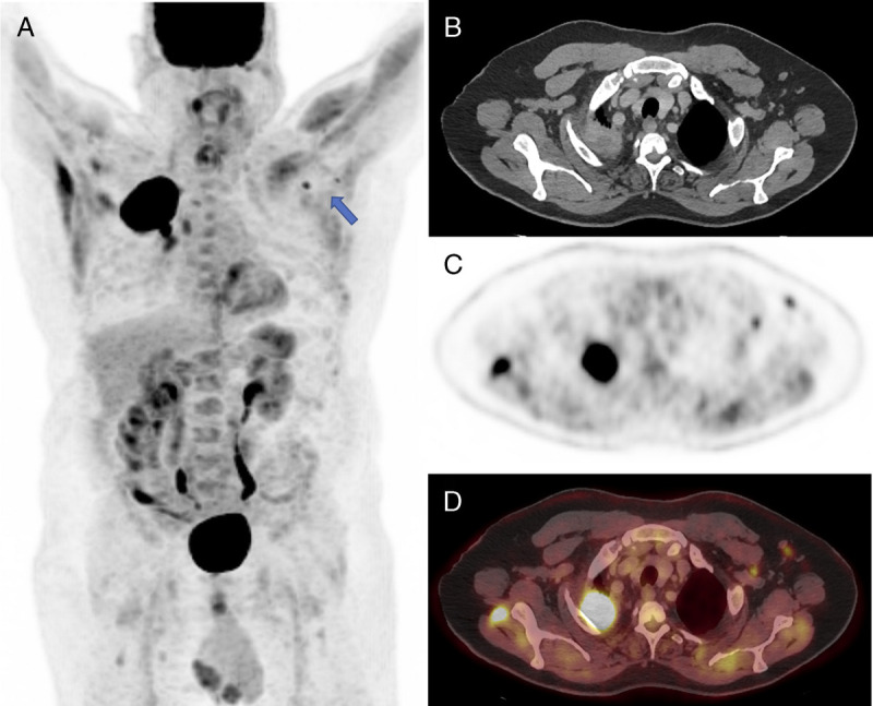
World-wide mass COVID-19 vaccination has been deployed starting with those most vulnerable, including the elderly and cancer patients. A 70-year-old man with right lung cancer underwent staging FDG PET/CT, which demonstrated an avid right lung mass with avid hilar and mediastinal nodes. Avid left axillary nodes of benign configuration were also noted. The patient had the Oxford-AstraZeneca COVID-19 vaccination in the left arm a week earlier. On reflection, the axillary nodes were concluded to be reactive related to this. This is a potential COVID-19 vaccination associated pitfall on PET/CT that should be considered when interpreting FDG PET/CT images.
Key Words: COVID-19 vaccination, 18F-FDG PET/CT, pitfall, axillary lymph nodes, inflammatory/reactive
FIGURE 1.

A 70-year-old man with right lung cancer. A half-body (skull base to mid-thigh) staging FDG PET/CT scan to assess disease extent; MIP (A) demonstrated a markedly avid right lung apical mass and avid ipsilateral hilar and mediastinal nodes. Contralateral axillary nodes of a similar avidity were also noted (arrow). These are clearly demonstrated on the axial CT, PET, and fused PET/CT (B–D). This raised the suspicion of nodal involvement. On CT correlation, the left axillary nodes had a benign configuration. It was noted that the tracer was administered via the right antecubital fossa and hence unlikely to be related.1 The patient, however, gave history of Oxford-AstraZeneca COVID-19 vaccination in the left upper arm 1 week earlier as part of the current mass vaccination program. FDG PET/CT can identify disease within nonenlarged lymph nodes; however, nonmalignant causes of avid nodes are also identified, such as reactive nodes in response to infection or inflammation.2 Axillary lymphadenopathy has been found associated with various vaccinations on FDG PET/CT, such as vaccinations for H1N1, human papilloma virus, and influenza.3–6 On reviewing, the CT component and correlating with the clinical history, it was concluded that the left axillary nodes were reactive, in response to recent ipsilateral vaccination. Similar findings were recently described associated with COVID-19 mRNA vaccines (Pfizer-BioNTech).7,8 This highlights a new pitfall related to the current mass COVID-19 vaccination that PET/CT reporters should be aware of. If it is not considered, this may result in inadvertent upstaging and overtreatment; in this case, PET/CT staging could have been potentially escalated from M0 to M1b.9
Footnotes
Conflicts of interest and sources of funding: none declared.
Contributor Information
Julie Searle, Email: Julie.searle1@nhs.net.
Richard Hopkins, Email: richard.hopkins1@nhs.net.
Iain Douglas Lyburn, Email: iain.lyburn@nhs.net.
REFERENCES
- 1.Long NM, Smith CS. Causes and imaging features of false positives and false negatives on F-PET/CT in oncologic imaging. Insights Imaging. 2011;2:679–998. [DOI] [PMC free article] [PubMed] [Google Scholar]
- 2.Shreve PD, Anai Y, Wahl RL. Pitfalls in oncologic diagnosis with FDG PET imaging: physiologic and benign variants. Radiographics. 1999;19:61–77. [DOI] [PubMed] [Google Scholar]
- 3.Burger IA Husmann L Hany TF, et al. Incidence and intensity of F-18 FDG uptake after vaccination with H1N1 vaccine. Clin Nucl Med. 2011;36:848–853. [DOI] [PubMed] [Google Scholar]
- 4.Williams G, Joyce RM, Parker JA. False-positive axillary lymph node on FDG-PET/CT scan resulting from immunization. Clin Nucl Med. 2006;31:731–732. [DOI] [PubMed] [Google Scholar]
- 5.Coates EE Costner PJ Nason MC, et al. Lymph node activation by PET/CT following vaccination with licensed vaccines for human papillomaviruses. Clin Nucl Med. 2017;42:329–334. [DOI] [PMC free article] [PubMed] [Google Scholar]
- 6.Shirone N Shinkai T Yamane T, et al. Axillary lymph node accumulation on FDG-PET/CT after influenza vaccination. Ann Nucl Med. 2012;26:248–252. [DOI] [PubMed] [Google Scholar]
- 7.Eifer M, Eshet Y. Imaging of COVID-19 vaccination at FDG PET/CT. Radiology. 2021;210030. [DOI] [PMC free article] [PubMed] [Google Scholar]
- 8.Xu G, Lu Y. COVID-19 mRNA vaccination-induced lymphadenopathy mimics lymphoma progression on FDG PET/CT. Clin Nucl Med. 2021;46:353–354. [DOI] [PubMed] [Google Scholar]
- 9.Feng SH, Yang ST. The new 8th TNM staging system of lung cancer and its potential imaging interpretation pitfalls and limitations with CT image demonstrations. Diagn Interv Radiol. 2019;25:270–279. [DOI] [PMC free article] [PubMed] [Google Scholar]


