Abstract
This article comments on:
Klaus Lunau, Daniela Scaccabarozzi, Larissa Willing and Kingsley Dixon, A bee’s eye view of remarkable floral colour patterns in the Southwest Australian biodiversity hotspot revealed by false colour photography’, Annals of Botany, Volume 128, Issue 7, 2 December 2021, Pages 821–824 https://doi.org/10.1093/aob/mcab088
Keywords: Flower signalling, false colour photography, floral colour patterns
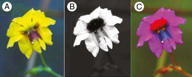
One of the joys of botanists, gardeners, photographers and the general public is that flower displays can present a dizzying array of patterned colours. Whilst such displays are often enthralling to human observers with our blue-, green- and red-sensitive trichromatic vision (Fig. 1A), humans are not the biological agent that promoted the evolution of such colour complexity (Dyer et al., 2021), and thus flowers may have very different colours and patterns when considering pollinator vision (Fig. 1B, C). In the past two decades, botanists have thus shifted from describing floral colours based on human visual comparisons of samples using reference colour charts, to embrace the use of spectrometers to obtain an observer-independent spectral signature describing colour information (van der Kooi et al., 2019). These techniques have shed light on the complexities of plant community assembly by considering competition or facilitation to attract animal visitors as they perceive the world (Kemp et al., 2019; Garcia et al., 2020). Whilst spectrometers remain the gold standard for the precise quantitative appraisal of flower colours, this method remains limited to point measurements of small areas. This limitation makes it challenging to obtain a holistic view of the complex flower patterns that an animal observer might perceive when foraging.
Fig. 1.
Digital photographic images showing the various spectral components of the pattern displayed by the flowers of Velleia trinervis (typical collora length 9–13 mm) using the method given by Lunau et al. (2021). (A) Digital, red, green and blue (RGB) image as perceived by human colour vision. (B) UV reflectance component of the same flower when imaged through a specialized UV-transmitting filter. (C) False colour representation integrating the UV-, BLUE- and GREEN-component channels of images B and A, respectively.
In this issue of Annals of Botany, Lunau et al. (2021) embraced this challenge by employing digital false-colour imaging to map flower colours from Western Australia. Australia is an important comparative study site for understanding flower evolution due to its geological isolation as an island continent, and well-documented evidence that flower colours are signals that have evolved due to biotic pollination (Dyer et al., 2012; Dalrymple et al., 2020). Furthermore, its native plants frequently reflect UV patterns to enhance signalling to bees (Dyer, 1996). In their new study, Lunau et al. (2021) employed a specially modified UV-sensitive digital camera fitted with a fused-quartz lens to capture two-dimensional information reflected from the entire flower surface. They then used digital false-colour imaging techniques to translate the spectral signals as might be seen by a pollinator into a biologically useful visual representation that we can interpret. This method employed waveband-specific light filtering and enabled the translation of the UV-, BLUE- and GREEN-component camera channels that allow bee vision to be displayed as a mixture of blue, green and red colours as perceived by typical human trichromatic vision.
Lunau et al. (2021) surveyed 55 species from four plant communities and 16 families, frequently observing complex floral signals in terms of colour patterns, floral guides, stamens, and pollen- and stamen-mimicking structures. The striking images show that colour information is often far more complex than the story told by just considering the primary petal colour of a flower, or a typical colour photograph taken in the field. One of the best known UV floral patterns is a bullseye effect in which the outer petals reflect significant amounts of UV whilst the inner flower parts absorb UV and create a salient target for landing bees (fig. 1A–C in Lunau et al., 2021). Several but not all yellow flowers in the Lunau et al. (2021) survey had such bullseye patterns, showing that a human colour such as ‘yellow’ might actually appear as completely different colours to biologically relevant pollinators.
In their paper, Lunau et al. (2021) show the amazing floral guides of Labichea lanceolata (their fig. 2D), Lotus subiflorus (fig. 3A), or the floral patterning of bird-pollinated Kennedia rubicunda (fig. 5D) and K. prostrata (fig. 5F). The false colour photographs produced by Lunau et al. (2021) reveal much additional information not available in conventional photographs, showing us that floral colour patterning is often more salient for pollinators. This is important as many insect pollinators such as bees are known to be attracted to colourful edges, and floral nectar guides are a key feature in interpreting how bees interact with flowers (Lunau, 1992). The technique for surveying flowers with false colour imaging thus has potential value for both taxonomic classification, as well as informing functional and behavioural studies of how various flower traits may have evolved as potential signals.
Perhaps due to the difficulty of objectively quantifying the spatial component of flower patterns from point samples, the role of patterning in visual communication between plants and animals remains understudied. Birds with high-resolution vision are likely to be capable of discriminating complex flower patterns made up of different elements (Caves et al., 2018), information that they can potentially use to drive decisions in natural environments (Spottiswoode and Stevens, 2010). The presence of markings on the petals of bird-pollinated flowers in Lunau et al. (2021) such as K. prostrata (their fig. 5F) suggests that these pattern elements may play a role in visual communication. Likewise, by improving our understanding of flower patterns using false-colour imaging, it may be possible to reformulate and test new hypotheses regarding the effect of the multiple spatial elements of flowers on the behaviour of pollinators. An additional advantage of the use of photographic images is the potential to obtain two-dimensional arrays of calibrated spectral information (Garcia et al., 2013) and assess how understudied factors such as flower pigment density influence colour signals and pollinator behaviour in complex environments (Garcia et al., 2014, 2020; van der Kooi et al., 2019). Exploring the world as experienced by pollinators via new photographic techniques thus promises to open up new avenues of research to improve our understanding of flower pollination, and the success of angiosperms.
LITERATURE CITED
- Caves EM, Brandley NC, Johnsen S. 2018. Visual acuity and the evolution of signals. Trends in Ecology & Evolution 33: 358–372. [DOI] [PubMed] [Google Scholar]
- Dalrymple RL, Kemp DJ, Flores-Moreno H, et al. 2020. Macroecological patterns in flower colour are shaped by both biotic and abiotic factors. The New Phytologist 228: 1972–1985. [DOI] [PubMed] [Google Scholar]
- Dyer AG. 1996. Reflection of near-ultraviolet radiation from flowers of Australian native plants. Australian Journal of Botany 44: 473–488. [Google Scholar]
- Dyer AG, Boyd-Gerny S, McLoughlin S, Rosa MG, Simonov V, Wong BB. 2012. Parallel evolution of angiosperm colour signals: common evolutionary pressures linked to hymenopteran vision. Proceedings. Biological Sciences 279: 3606–3615. [DOI] [PMC free article] [PubMed] [Google Scholar]
- Dyer AG, Jentsch A, Burd M, et al. 2021. Fragmentary blue: resolving the rarity paradox in flower colors. Frontiers in Plant Science 11: 618203. [DOI] [PMC free article] [PubMed] [Google Scholar]
- Garcia JE, Dyer AG, Greentree AD, Spring G, Wilksch PA. 2013. Linearisation of RGB camera responses for quantitative image analysis of visible and UV photography: a comparison of two techniques. PLoS ONE 8: e79534. [DOI] [PMC free article] [PubMed] [Google Scholar]
- Garcia JE, Greentree AD, Shrestha M, Dorin A, Dyer AG. 2014. Flower colours through the lens: quantitative measurement with visible and ultraviolet digital photography. PLoS ONE 9: e96646. [DOI] [PMC free article] [PubMed] [Google Scholar]
- Garcia JE, Phillips RD, Peter CI, Dyer AG. 2020. Changing how biologists view flowers—color as a perception not a trait. Frontiers in Plant Science 11: 1–15. [DOI] [PMC free article] [PubMed] [Google Scholar]
- Kemp JE, Bergh NG, Soares M, Ellis AG. 2019. Dominant pollinators drive non-random community assembly and shared flower colour patterns in daisy communities. Annals of Botany 123: 277–288. [DOI] [PMC free article] [PubMed] [Google Scholar]
- van der Kooi CJ, Dyer AG, Kevan PG, Lunau K. 2019. Functional significance of the optical properties of flowers for visual signalling. Annals of Botany 123: 263–276. [DOI] [PMC free article] [PubMed] [Google Scholar]
- Lunau K. 1992. A new interpretation of flower guide colouration: absorption of ultraviolet light enhances colour saturation. Plant Systematics and Evolution 183: 51–65. [Google Scholar]
- Lunau K, Scaccabarozzi D, Willing L, Dixon KW. 2021. A bee’s eye view of remarkable floral colour patterns in the Southwest Australian biodiversity hotspot revealed by false colour photography. Annals of Botany, 128: 821–834. [DOI] [PMC free article] [PubMed] [Google Scholar]
- Spottiswoode CN, Stevens M. 2010. Visual modeling shows that avian host parents use multiple visual cues in rejecting parasitic eggs. Proceedings of the National Academy of Sciences of the United States of America 107: 8672–8676. [DOI] [PMC free article] [PubMed] [Google Scholar]


