Abstract
The foot-and-mouth disease virus (FMDV) causes a highly infectious disease of all cloven-footed animals. The RNA genome of the virus continuously evolves, leading to the generation of new strains; this necessitates the selection of new vaccine strains to ensure complete protection. Infection with one FMDV serotype does not provide cross-protection against the other FMDV serotypes. Many of the recovered animals may become carriers of the FMDV, but they still remain susceptible to the other serotypes. Coinfection with multiple FMDV serotypes has been reported and studied to understand the virus evolution. Isolation and characterization of all the involved serotypes in the mixed infection case is essential to understand the molecular evolution of the virus. In this study, two cases of coinfection were studied by selective isolation of each of the FMDV serotypes under the cross-serotype-specific immune pressure. It was estimated that the virus present in a minimum of 10−0.92 TCID50 could be isolated from the mixed population containing other serotypes in infective doses of 100.25 TCID50 or less. All involved serotypes present in the mixed infection cases were isolated, without any cross-contamination. Virus characterization revealed that genotype 2 was of serotype A virus from a sample collected in 1995, which was last reported in 1986, indicating a possible subdued prevalence of the genetic group even after vanishing from the field.
Keywords: Coinfection, Foot-and-mouth disease virus, Virus isolation, TaqMan assay
Introduction
Foot-and-mouth disease (FMD) is a highly contagious and infectious disease of cloven-hoofed animals. It is caused by seven immunologically distinct serotypes of FMD virus (FMDV) belonging to the genus Aphthovirus of the family Picornaviridae [1]. More than one FMDV serotype and many of their genotypes are circulating in most of the FMD-endemic countries [2]. Three (serotypes O, A, and Asia1) of the seven serotypes, and many of their genotypes/lineages, are simultaneously circulating in India [3, 4]. Serotype O has historically been dominant in India, whereas serotype C has not been recorded since 1995 [4]. On the other hand, serotypes A and Asia1 are not widespread, but they continue to prevail, with sporadic incidences. The spatial distribution of FMD in the last 15 years reveals that any one of the three serotypes is endemic in most of the areas of the country, whereas other serotypes have never been reported. However, the simultaneous circulation of two or more serotypes could also be observed in a few pockets of the country (unpublished data). The plausible reason(s) behind the differential distribution is(are) yet to be understood, but high animal density, significant animal movements, and improper vaccination have been considered some of the factors behind the simultaneous circulation of multiple serotypes in some geographical areas. Recovery from infection of one serotype does not offer protection against strains of the other serotypes [2]. The emergence of new genotypes/lineages has been linked to the evolution of the fittest virus under pressure and to the pre-existing immunity to the genotype in circulation [5]. In contrast, re-emergence of the previously existing genotypes/lineages suggests that the virus genetic group latently survived and re-appeared [2, 6]. Infected animals (bovine, caprine, and ovine) may carry FMDV without showing characteristic clinical symptoms for a long period of time [7]. The average extinction time for the carrier status in FMD-infected Indian farms has been calculated as 13.1 ± 0.2 months [8]. However, carrier animals remain susceptible to infection to other serotypes. All these factors provide suitable conditions for coinfection with two or more serotypes of FMDV [9]. Occasional inter-serotypic recombination, found in the genome of the FMDV, has been linked to coinfection [8, 10, 11]. The detection of coinfection using molecular methods has been reported from many parts of the world, including India, but isolation of both the serotypes in cell culture is essential to completely characterize the virus. The objective of this study was therefore to investigate and characterize FMDV coinfection in cattle. Selective isolation of both the serotypes in cell culture was attained by the application of cross-neutralizing antibodies. The isolated virus serotypes were then genetically and antigenically characterized for genetic recombination.
Materials and methods
FMDV isolates and serotype-specific sera
Two cell culture–adapted materials obtained from coinfected animals, IND393/1988 (contain serotype O and Asia1) and IND53/1995 (documented to contain serotype O and A), were obtained from the National FMD virus repository maintained at the Project Directorate on Foot and Mouth Disease (PDFMD), Mukteswar, and freshened up in a BHK-21 cell monolayer. Three vaccine strains, O (IND R2/1975), A (IND 40/2000), and Asia1 (IND 63/1972), were also revived in BHK-21 cells for virus quantification. The details of the virus used in the study are presented in Table 1. Revived viruses were quantified by end-point dilution as described by Reed and Muench [12]. Serotype-specific hyperimmune sera against three FMD vaccine strains raised in healthy rabbits were obtained from the FMD serum repository maintained at PDFMD, Mukteswar.
Table 1.
Details of FMD virus isolates and clinical samples used in the study
| S no | Virus number | Type of materials | FMDV serotype | Place of collection | Year of collection |
|---|---|---|---|---|---|
| 1 | INDR2/1975 | Cell culture isolate | O | Ranipet, Tamilnadu | 1975 |
| 2 | IND63/1972 | Cell culture isolate | Asia1 | Maharashtra | 1972 |
| 3 | IND40/2000 | Cell culture isolate | A | Banneraghatta, Anekal Bangalore, Karnataka | 2000 |
| 4 | IND393/1988 | Cell culture isolate | O and Asia1 | Hessarghatta, Bangalore, Karnataka | 1988 |
| 5 | IND53/1995 | Cell culture isolate | O and A | Hyderabad Andhra Pradesh | 1995 |
| 6 | PD195/2013 | Tongue material from FMD-infected animal | A | Ranchi Jharkhand | 2013 |
| 7 | PD335/13 | Tongue material from FMD-infected animal | Asia1 | Bilaspur Chattisgarh | 2013 |
| 8 | PD44/2015 | Tongue material from FMD-infected animal | O | BK Palya Bangalore Karnataka | 2015 |
| 9 | PD1321/14 | Tongue material from FMD-infected animal | O | Narsinghpur Madhya Pradesh | 2014 |
Serotype-specific TaqMan assay
The TaqMan chemistry-based real-time (qRT-PCR) assay for the quantification of serotypes O, A, and Asia1 was used for serotype detection and growth curve analysis. The primers and probes specific for serotypes O and A were designed in this study (Table 2). Primers and probes specific for serotype Asia1 have been designed previously [13]. Total RNA was extracted using the RNeasy mini kit (Qiagen, Germany) in an automated genome extractor (Qiacube, Qiagen, Germany). The one-step RT-PCR kit (Qiagen, Germany) was used for assembling the serotype-specific TaqMan qRT-PCR. In brief, 5 μl of 5 × RT-PCR buffer, 0.5 μl of 10 mM each of the dNTP set, 0.5 μl each of 10 pmol forward and reverse primers, 0.5 μl of ROX reference dye (Applied Biosystems, USA), 0.5 μl of RT enzyme mix, 0.25 μl of serotype-specific probe (10 mM), and 2 μl of the viral RNA were mixed together in thin-walled PCR tubes with clear caps, and the volume was adjusted to 25 μl. The qRT-PCR was performed in the 7500 cycler (Applied Biosystems, USA) under the following conditions: initial holding at 50 °C for 30 min for reverse transcription, 95 °C for 15 min for initial denaturation, followed by 40 cycles of denaturation at 95 °C for 15 s and annealing at 60 °C. The assay was evaluated for the specific detection of each serotype by testing RNA extracted from serially tenfold-diluted vaccine strains of virus serotypes.
Table 2.
Details of primers and probes used in the study
| S no | Name of the primer/probe | Primer/probe sequence | Serotype specificity | Reference |
|---|---|---|---|---|
| 1 | O primer F | YGCTGCCYACYTCCTTCA | O | In this study |
| 2 | O primer R | GAGGCCGRGGRCAGTAYGTTTC | O | In this study |
| 3 | A primer F | ATGAAGCGYGCCGARCTCTACTG | A | In this study |
| 4 | Asia1 F | TTY AAY TAC GGY GCT GTR AAG GCT G | Asia1 | [13] |
| 5 | A probe | 6FAM-TCTCGTCGCARGACAGACRC-TAMRA | A | In this study |
| 6 | Asia1 probe | 6FAM-ACA CCA CYC ARG ACC GCC GTA A-TAMRA | Asia1 | [13] |
| 7 | NK 61 | GACATGTCCTCCTGCATCTG | Universal | [12] |
Selective isolation of FMDV serotypes
The virus neutralization assay was performed to determine the amounts of homologous and heterologous antibodies required to completely neutralize the vaccine strains of O, A, and Asia1 [12]. Briefly, 100 μl of the cell culture supernatant of IND392/1988 was mixed with 2% FBS-DMEM medium containing serotype O–specific hyperimmune sera in 1:1000 dilutions in tube 1 and serotype Asia1–specific hyperimmune sera in 1:1200 dilutions in tube 2 for selective virus isolation. Similarly, 100 μl of the cell culture supernatant of IND53/1995 was mixed in 2 ml of 2% FBS-DMEM containing serotype O–specific hyperimmune sera in 1:1000 dilutions in tube 3 and serotype A–specific hyperimmune sera in 1:1000 dilutions in tube 4. All virus/serum mixtures were incubated at 37 °C for 1 h, and BHK-21 cells with 70–80% confluency in cell culture tubes (Nunc, Thermo) were washed with 2% FBS-DMEM medium, followed by transferring the virus/serum mixture, and the BHK-21 tubes were incubated at 37 °C. Characteristic cytopathic effects (CPE) were observed and passaged blindly until the obvious CPE appeared. The RNA was extracted from each passaged virus, and virus serotypes were estimated by qRT-PCR as described in the previous section.
Antigenic characterization of the viruses isolated from the mixed population
The antigenic relationship of the isolated viruses from the mixed population with that of the vaccine virus was estimated by performing the 2-dimensional microneutralization test (2D-MNT) as described previously [12]. Phenotypic characterization of the isolated virus from the mixed virus was performed by plaque assay as described previously [12].
Genetic characterization of the isolated viruses through deep sequencing
Complete P1-coding regions of the isolated viruses were amplified using NK61 reverse and L463 forward primers as described elsewhere [14]. The amplified and gel-purified P1 region PCR amplicon was used for library preparation for deep sequencing through Ion Torrent (Applied Biosystems), applying the Ion PI Library preparation kit as prescribed by the manufacturer. Library quality was assured using the Bioanalyzer (Agilent) before sequencing. The sequence contigs were aligned against the reference sequence of the P1 region of R2/1975 FMDV in the Ion Torrent server, and the aligned sequence was visualized using the IGV tools (available at https://software.broadinstitute.org/software/igv/igvtools). Phylogenetic analysis and sequence comparisons were performed using the MEGA 6.06 software [15]. Recombination analysis was carried out using the Recombination Detection Program (RDP) 4 [16] and the SimPlot [17] software.
Results
Real-time PCR assay
The primers and probe used in this study showed serotype specificity, without any cross-amplification of other serotypes when tested at various concentrations (data not shown). As the threshold cycle (Ct) values showed near perfect linear regression with the TCID50 [13, 18], the Ct values were extrapolated into TCID50 from the standard curve to monitor the growth kinetics.
Selective FMDV serotype isolation
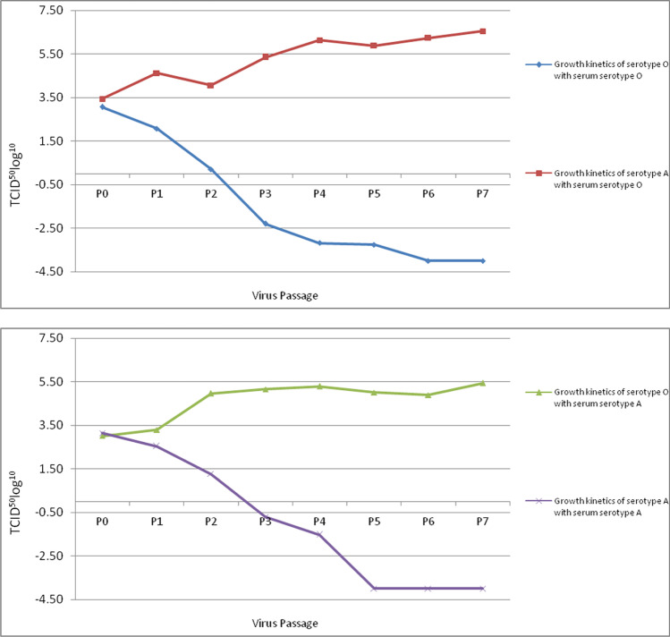
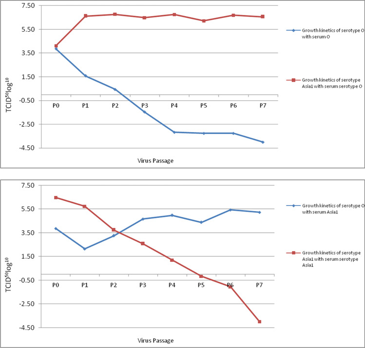
Various combinations of virus and serum concentrations were evaluated to attain complete inhibition of target virus serotype replication in the BHK-21 cells (data not presented). The serotype-specific hyperimmune serum in < 1:256 dilution completely inhibited FMDV serotype replication, with a concentration ≤ 107.32 TCID50 in BHK-21 cells. The FMDV serotypes with < 10−0.53 TCID50 concentration were non-specifically inhibited with hyperimmune serum in < 1:64 dilutions but not with higher dilutions. Individual serotypes O (O/IND53/1995) and A (A/IND53/1995) were successfully isolated from the coinfected cell culture suspension material of IND53/1995 after P-5 (Fig. 1) without cross-contamination from other serotypes. Similarly, serotypes O (O/IND393/1988) and Asia1 (Asia1/IND393/1988) were successfully isolated from the coinfected cell culture suspension of IND393/1988 without any cross-contamination after P-7 (Fig. 2). No traces of cross-contamination of other serotypes were evident by qRT-PCR.
Fig. 1.
Growth kinetics of serotypes O and A from the coinfected field isolate of IND53/1995 virus. Individual virus serotypes O and A could be isolated upon application of cross-serotype-specific antibody pressure and no traces of other serotype could be observed after passage 6. In this figure, 4 TCID50/50 μl indicates no detectable RNA in qRTPCR
Fig. 2.
Growth kinetics of serotypes O and Asia1 from the coinfected field isolate of IND393/1988 virus. Individual virus serotypes O and Asia1 could be isolated upon application of cross-serotype-specific antibody pressure, and no traces of other serotype could be observed after passage 6. In this figure, 4 TCID50/50 μl indicates no detectable RNA in qRTPCR
Antigenic characterization of the isolated virus
The virus neutralization assay of the isolated viruses confirmed the absence of any cross-contamination. The effect of cross-serotype immune pressure on the targeted virus serotype was evaluated by the 2D-MNT assay. The virus serotypes A/IND53/1995, O/IND53/1995, O/IND393/1988, and Asia1/IND393/1988 showed r-values of 0.45, 0.77, 0.59, and 0.69, respectively, in comparison to the respective vaccine strains. The r-values were above the accepted cutoff value of 0.3, which indicates sufficient protection coverage offered by the vaccine strain to these strains. Phenotypic characterization of the isolated virus from the mixed virus population demonstrated no significant changes in plaque morphology when compared to the original virus.
Genetic characterization
The P1-coding regions of about 2.2 Kb of the isolated viruses were amplified using previously described primers and subjected to deep sequencing on the Ion Torrent platform. The P1 nucleotide sequences were annotated and submitted to GenBank.
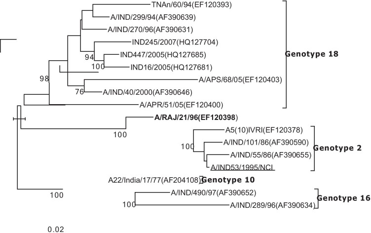
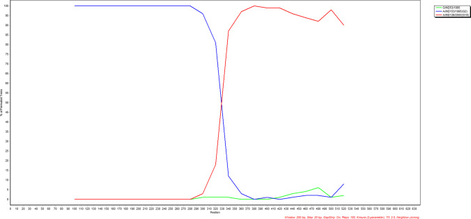
Phylogenetic analysis revealed that Asia1/IND393/1988 (accession number #MW524747) belonged to sub-lineage CI. Although O/IND393/1988 (accession number #MW524748) did not group with any of the previously identified major lineages [4], it clustered within the ME-SA topotype, closely with lineage branch B. Both the sequences obtained from coinfected sample did not reveal any recombination events. The O/IND53/1995 (accession number #MW524745) belonged to the lineage O/ME-SA/branch CII, whereas A/IND53/1995 (accession number #MW524746) belonged to the lineage genotype 2. Surprisingly, the genotype 2 serotype A virus was recovered from the sample collected in 1995, which has not been recorded in India since 1986 (Fig. 3). From 1986 to 2001, only genotypes 16 and 18 were co-circulating in the field [5]. Recombination analysis of the P1-coding region of A/IND53/1995 revealed a clear mosaic pattern; the 5′end of VP1 (~ 320 bp) was characteristic of genotype 2 and the ~ 230-bp end at 3′ was characteristic of genotype 18 (Fig. 4).
Fig. 3.
Maximum likelihood tree depicting the phylogenetic relationship of type A viruses. The virus recovered from the coinfected sample is underlined and the recombinant virus collected in 1996 is in bold face
Fig. 4.
Boot scan evidence for recombination between genotype 2 (A/IND/153/1995) and genotype 18 (A/IND/26/2000)
Discussion
Three of seven FMDV serotypes (serotypes O, A, and Asai1) and many of their genotypes/lineages are simultaneously circulating in India. Asymmetrical appearance and disappearance of different serotypes and their genotypes suggest a highly complex landscape epidemiology of FMDV in endemic settings. The FMDV genotypes/lineages continue to evolve in a particular area and occasionally spread to the larger areas, for example, the rapid dissemination of the Ind2001d strain in the year 2013 in India. Outbreaks may occur as waves of infections upon the introduction of different serotypes in the population, infect susceptible animals, and then move on to reappear a few years later when a new susceptible population is established [19]. Alternatively, one serotype may persist in a region and rarely appear clinically, producing only mild infections in the young stock when they lose their maternal antibodies. Clinical disease may thus only be seen when a new serotype is introduced [2]. The precise role of carrier and coinfected animals still needs to be completely understood in the epidemiology and evolution of FMDV. Similar speculations have been made in the East Africa region, where serotype O is the most prevalent one, followed by A, strongly suggesting the possibility of coinfection with both or even more serotypes within some animals [20]. Klein et al. [8] demonstrated that most sub-clinical infections in Asian Buffaloes are caused by the A/IRN/2005 sub-lineage, and there is a high probability of a subsequent infection of the same animal with another FMDV serotype, for example, Asia1, increasing the likelihood of inter-serotype recombination. Similarly, Li et al. [11] demonstrated that the Asia1/HNK/CHA/05 virus might have resulted from inter-serotypic recombination of the two parental viruses O/Tibet/CHA/99 and Asia1/IND/491/97.
Hence, it is essential to detect and characterize the coinfection cases of FMD with multiple serotypes to improve our understandings about the landscape epidemiology, virus evolution, genetic recombinations, and the carrier status of the animals, for which the isolation of serotypes present in the coinfected samples is essential. The detection of coinfection with different serotypes was made possible with the latest molecular techniques, such as real-time PCR. Genetic characterization of all involved serotypes in a mixed infection is now performed with deep-sequencing techniques. However, each of the involved serotypes in the coinfected samples should be isolated in the cell culture system for complete antigenic and genetic characterization. Nevertheless, generally, any one serotype which dominates cell culture isolation is isolated. It has been demonstrated that during infection of the cell culture with different RNA, virus strains can promote Red Queen dynamics (RQD) [21]. Under RQD, both strains improve their fitness relative to their ancestors, but with no relative gain between them. They thus coexist along their evolution in cell cultures, but this coexistence ends eventually as a consequence of competitive exclusion, and only one virus serotype dominates in the end [22]. Hence, in most of the clinical materials obtained from the coinfected animals, only one virus serotype is isolated, whereas the other serotype(s) gets eliminated in subsequent passages. In rare instances, the isolated virus in the cell culture contained both the virus serotype populations [13]. Two of such original cell culture suspension materials (IND53/1995 contained serotypes O and Asia1; IND393/1988 contained serotypes O and A) were examined in this study. The cross-serology approach for virus isolation from mixed infection was optimized by experimentally mixing FMDV vaccine serotypes of O, A, and Asia1 in various known concentrations, where one serotype was inhibited with serotype-specific serum to allow the growth of other serotypes. Cross-serology approaches have previously been evaluated for the isolation of desired viruses from mixed virus population, for example the isolation of the avian influenza virus from cloacal samples containing avian influenza and Newcastle disease viruses [23]. Serotype-specific antibodies were carefully applied for the selective isolation of both serotypes involved in the coinfection. It was observed that high concentrations (< 1:64 dilution) of serotype-specific hyperimmune serum could non-specifically inhibit the other serotypes if present in concentrations less than 10−0.53 TCID50. Hence, the serum concentration has to be selected carefully, considering the tentative initial load in the sample. Based on the experimental mixing of FMDV vaccine strains, it was estimated that the targeted virus serotype present in as low as 10−0.92 TCID50 could be isolated from the mixed population containing another serotype in an infective dose of 100.25 TCID50 or less. The copy number of targeted virus serotypes continued to increase after P-1, whereas that of other serotypes continued to decrease after P-2 during the passage on BHK-21 under cross-serotype-specific immune pressure (Figs. 1 and 2). The virus serotype, inhibited by the serum in the mixed population, could not be detected by the qRT-PCR after P-5. Each of the involved serotypes O (O/IND53/1995) and serotype A (A/IND53/1995) was isolated individually. Similarly, viruses of serotype O (O/IND393/1988 and serotype Asia1/INDI393/1988) were isolated individually without any remnants of cross-contamination. All four isolated viruses showed a relationship value (r-value) > 0.3 against their respective vaccine strains, indicating that the antibodies induced by vaccine strains offer sufficient protection to these viruses. The P1-coding regions of isolated viruses, encoding the four structural proteins (VP1, VP2, VP3, and VP4), were amplified and subjected to deep sequencing. The reads were aligned and analyzed for phylogenetic and genetic recombinations.
Asia1/IND393/1988 was found to belong to the sub-lineage CI of Asia1. In contrast, O/IND393/1988 did not group with any of the previously identified major lineages [4], but it clustered within the ME-SA topotype close to lineage branch B. The virus O/IND53/1955 belonged to the lineage O/ME-SA/branch CII, whereas A/IND53/1955 belonged to the lineage genotype 2. Genotype 2 of FMDV serotype A has not been recorded since 1986 [5], but it could be identified in the sample collected in the year 1995. Recombination analysis did not reveal any recombination in the isolated virus, but the P1-coding region of A/IND53/1995 revealed a clear mosaic pattern: the 5′end of VP1 (~ 320 bp) was characteristics of genotype 2 and the ~ 230-bp end at 3′ was characteristic of genotype 18 (Fig. 3). Interestingly, a recombinant serotype A virus between genotypes 2 and 18 was isolated in 1996 from the state of Rajasthan, India, and the recombination breakpoint was identified in the VP1 region [24]. This finding further highlights the hypothesis that extinct genetic groups, such as genotype 2, that are not observed in any field outbreaks are still able to maintain a limited subdued prevalence and may contribute to the recombination and evolution of viruses.
The starting clinical material used in this study was a cell-adapted coinfected sample with two serotypes. To assess the application of cross-serology for selective isolation from clinical material, two tongue epithelial materials (O/PD332/2014 and A/PD53/2014) were experimentally mixed and subjected to selective isolation as described above. Each of the target serotypes was successfully isolated in the cell culture without any remnants of other serotypes (data not presented). More detection and selective isolation from coinfected samples will lead to a further understanding of the FMD epidemiology and evolution. In our laboratory, several samples have been found positive for more than one serotype during the last decade (data not presented), but all involved virus serotypes could not be isolated from those samples. The present method of selective isolation should help to further improve our understanding of this issue.
It may also be speculated that although the virus serotypes isolated from the clinical materials were of one particular lineage, the initial clinical materials, especially oro-pharyngeal fluid, might contain two or more lineages of the same serotype, and one lineage was selected upon serial passage through the cell culture system. Hence, the cross-serology approach for virus isolation could be extended further for the characterization of specific lineages from mixed infected samples. The detection of two or more lineages in the clinical materials is possible through molecular techniques such as lineage-differentiating PCR [25, 26]. The demonstration and isolation of FMDV of different lineages from the same animals will further add to the understanding of the FMD landscape epidemiology.
Funding
This study was funded by Indian Council of Agricultural Research (Project Code: IXX11548).
Data availability
All the raw data are available with the corresponding authors and can be shared upon reasonable request.
Declarations
Ethics approval
This article does not contain any studies with animals performed by any of the authors.
Conflict of interest
The authors declare no competing interests.
Footnotes
Responsible Editor: Fernando R. Spilki
Publisher's note
Springer Nature remains neutral with regard to jurisdictional claims in published maps and institutional affiliations.
Contributor Information
Sonalika Mahajan, Email: sonalikam1@gmail.com.
Gaurav Kumar Sharma, Email: gaurvet@gmail.com.
References
- 1.Racaniello VR (2001) Picornaviridae: the viruses and their replication. In: Knipe D.M. and P.M. Howley (eds) Fields Virology, 4th edn. Lippincott Williams and Wilkins, Philadelphia. Vol. 1, pp 685–722.
- 2.Kitching RP. Future research on foot and mouth disease. Rev Sci Tech. 2002;21:885–889. doi: 10.20506/rst.21.3.1379. [DOI] [PubMed] [Google Scholar]
- 3.Pattnaik B, Subramaniam S, Sanyal A, Mohapatra JK, Dash BB, Ranjan R, Rout M. Foot-and-mouth disease: global status and future road map for control and prevention in India. Agric Res. 2012;1:132–147. doi: 10.1007/s40003-012-0012-z. [DOI] [Google Scholar]
- 4.Subramaniam S, Sanyal A, Mohapatra JK, Sharma GK, Biswal JK, Ranjan R, Rout M, Das B, Bisht P, Mathapati BS, Dash BB, Pattnaik B. Emergence of a novel lineage genetically divergent from the predominant Ind 2001 lineage of serotype O foot-and-mouth disease virus in India. Infect Genet Evol. 2013;18:1–7. doi: 10.1016/j.meegid.2013.04.027. [DOI] [PubMed] [Google Scholar]
- 5.Mohapatra JK, Subramaniam S, Pandey LK, Pawar SS, De A, Das B, Sanyal A, Pattnaik B. Phylogenetic structure of serotype A foot-and-mouth disease virus: global diversity and the Indian perspective. J Gen Virol. 2011;92:873–879. doi: 10.1099/vir.0.028555-0. [DOI] [PubMed] [Google Scholar]
- 6.Sanyal A, Subramaniam S, Mohapatra JK, Tamilselvan RP, Singh NK, Hemadri D, Pattnaik B. Phylogenetic analysis of Indian serotype Asia1 foot-and-mouth-disease virus isolates revealed emergence and reemergence of different genetic lineages. Vet Microbiol. 2010;144:198–202. doi: 10.1016/j.vetmic.2009.12.034. [DOI] [PubMed] [Google Scholar]
- 7.Alexandersen S, Zhang Z, Donaldson AI. Aspects of the persistence of foot-and-mouth disease virus in animals–the carrier problem. Microbes Infect. 2002;4:1099–1110. doi: 10.1016/S1286-4579(02)01634-9. [DOI] [PubMed] [Google Scholar]
- 8.Klein J, Hussain M, Ahmad M, Normann P, Afzal M, Alexandersen S. Genetic characterisation of the recent foot-and-mouth disease virus subtype A/IRN/2005. Virol J. 2007;15(4):122. doi: 10.1186/1743-422X-4-122. [DOI] [PMC free article] [PubMed] [Google Scholar]
- 9.Knowles NJ, Samuel AR (1995) Polymerase chain reaction amplification and cycle sequencing of the 1D (VP1) gene of foot-and mouth disease viruses. Report of the Session of the Standing Technical Committee of the European Commission for the Control of Foot-and-Mouth disease held at Vienna, Austria, September 19–22, 1994. FAO, Rome, pp 45–53
- 10.Jackson T, Ellard FM, Ghazaleh RA, Brookes SM, Blakemore WE, Corteyn AH, Stuart DI, Newman JW, King AM. Efficient infection of cells in culture by type O foot-and-mouth disease virus requires binding to cell surface heparan sulfate. J Virol. 1996;70:5282–5287. doi: 10.1128/jvi.70.8.5282-5287.1996. [DOI] [PMC free article] [PubMed] [Google Scholar]
- 11.Li D, Shang YJ, Liu ZX, Liu XT, Cai XP. Molecular relationships between type Asia1 new strain from China and type O Panasia strains of foot-and-mouth-disease virus. Virus Genes. 2007;35:273–279. doi: 10.1007/s11262-006-0073-9. [DOI] [PubMed] [Google Scholar]
- 12.Sarangi LN, Mohapatra JK, Subramaniam S, Sanyal A, Pattnaik B. Antigenic site variation in foot-and-mouth disease virus serotype O grown under vaccinal serum antibodies in vitro. Virus Res. 2013;176:273–279. doi: 10.1016/j.virusres.2013.07.003. [DOI] [PubMed] [Google Scholar]
- 13.Selvan T (2010) Analysis of replication dynamics of mixed foot-and-mouth disease virus populations using serotype differentiating multiplex QPCR. Thesis submitted to Deemed University IVRI, Izatnagar Bareilly (UP) India
- 14.Pandey LK, Mohapatra JK, Subramaniam S, Sanyal A, Pande V, Pattnaik B. Evolution of serotype A foot-and-mouth disease virus capsid under neutralizing antibody pressure in vitro. Virus Res. 2014;181:72–76. doi: 10.1016/j.virusres.2014.01.009. [DOI] [PubMed] [Google Scholar]
- 15.Tamura K, Stecher G, Peterson D, Filipski A, Kumar S. MEGA6: Molecular Evolutionary Genetics Analysis version 6.0. Mol Biol Evol. 2013;30:2725–2729. doi: 10.1093/molbev/mst197. [DOI] [PMC free article] [PubMed] [Google Scholar]
- 16.Martin DP, Murrell B, Golden M, Khoosal A, Muhire B (2015) RDP4: Detection and analysis of recombination patterns in virus genomes. Virus Evolution 1: vev003 [DOI] [PMC free article] [PubMed]
- 17.Lole KS, Bollinger RC, Paranjape RS, Gadkari D, Kulkarni SS, Novak NG, Ingersoll R, Sheppard HW, Ray SC. Full-length human immunodeficiency virus type 1 genomes from subtype C-infected seroconverters in India, with evidence of intersubtype recombination. J Virol. 1999;73(1):152–60. doi: 10.1128/JVI.73.1.152-160.1999. [DOI] [PMC free article] [PubMed] [Google Scholar]
- 18.Sachs LA, Schnurr D, Yagi S, Lachowicz-Scroggins ME, Widdicombe JH. Quantitative real-time PCR for rhinovirus, and its use in determining the relationship between TCID50 and the number of viral particles. J Virol Methods. 2011;171:212–218. doi: 10.1016/j.jviromet.2010.10.027. [DOI] [PubMed] [Google Scholar]
- 19.Kitching RP. The role of the World Reference Laboratories for Foot-and-Mouth Disease and for Rinderpest. Ann N Y Acad Sci. 2000;916:139–146. doi: 10.1111/j.1749-6632.2000.tb05284.x. [DOI] [PubMed] [Google Scholar]
- 20.Balinda SN, Siegismund HR, Muwanika VB, Sangula AK, Masembe C, Ayebazibwe C, Normann P, Belsham GJ. Phylogenetic analyses of the polyprotein coding sequences of serotype O foot-and-mouth disease viruses in East Africa: evidence for interserotypic recombination. Virol J. 2010;7:199. doi: 10.1186/1743-422X-7-199. [DOI] [PMC free article] [PubMed] [Google Scholar]
- 21.Van Valen L. A new evolutionary law. Evol Theory. 1973;1:1–30. [Google Scholar]
- 22.Saldaña J, Elena SF, Solé RV. Coinfection and superinfection in RNA virus populations: a selection-mutation model. Math Biosci. 2003;183:135–160. doi: 10.1016/S0025-5564(03)00038-5. [DOI] [PubMed] [Google Scholar]
- 23.El Zowalaty ME, Chander Y, Redig PT, Abd El Latif HK, El Sayed MA, Goyal SM. Selective isolation of Avian influenza virus (AIV) from cloacal samples containing AIV and Newcastle disease virus. J Vet Diagn Invest. 2011;23:330–332. doi: 10.1177/104063871102300222. [DOI] [PubMed] [Google Scholar]
- 24.Nagendrakumar SB, Madhanmohan M, Rangarajan PN, Srinivasan VA. Genetic analysis of foot-and-mouth disease virus serotype A of Indian origin and detection of positive selection and recombination in leader protease-and capsid-coding regions. J Biosci. 2009;34:85–101. doi: 10.1007/s12038-009-0011-9. [DOI] [PMC free article] [PubMed] [Google Scholar]
- 25.Mohapatra JK, Sanyal A, Hemadri D, Tosh C, Rasool TJ, Bandyopadhyay SK. A novel genetic lineage differentiating RT-PCR as a useful tool in molecular epidemiology of foot-and-mouth disease in India. Arch Virol. 2006;151:803–809. doi: 10.1007/s00705-005-0673-z. [DOI] [PubMed] [Google Scholar]
- 26.Mohapatra JK, Subramaniam S, Tosh C, Hemadri D, Sanyal A, Periyasamy TR, Rasool TJ. Genotype differentiating RT-PCR and sandwich ELISA: handy tools in epidemiological investigation of foot and mouth disease. J Virol Methods. 2007;143:117–121. doi: 10.1016/j.jviromet.2007.02.008. [DOI] [PubMed] [Google Scholar]
Associated Data
This section collects any data citations, data availability statements, or supplementary materials included in this article.
Data Availability Statement
All the raw data are available with the corresponding authors and can be shared upon reasonable request.


