



Abstract
Although medical therapy is the preferred first-line treatment for patients with chronic coronary syndrome (CCS), revascularization remains an important consideration. We present a review that identifies the three diagnostic technologies most important to guiding the decision to revascularize patients with CCS: (1) cardiac computed tomography, (2) intracoronary imaging, and (3) lesion-specific physiological guidance.
Keywords: chronic coronary syndrome, medical therapy, revascularization, intravascular ultrasound, optical coherence tomography, cardiac computed tomography, fractional flow reserve, instantaneous wave-free ratio
Atherosclerosis remains the leading cause of death worldwide. 1 Globally, the rate of major adverse cardiac events (MACE), defined as the composite of nonfatal stroke, nonfatal myocardial infarction (MI), and cardiovascular death, continues to increase. The United States, in contrast, has seen a 70% decrease in the incidence of MACE over the last five decades. 2 This decline has been attributed primarily to a greater understanding of disease pathophysiology and advancements in medical therapy (MT), as well as improvements in revascularization technology. 3
For the revascularization of patients with acute coronary syndrome (ACS), application of each new technology, such as drug-eluting stents, intravascular ultrasound (IVUS), and fractional flow reserve (FFR), has resulted in an incremental reduction in MACE. 4 5 6 7 8 Revascularization in patients with chronic coronary syndrome (CCS), while effective at alleviating symptoms, has not consistently translated into reduced MACE. The major CCS trials, which are well summarized in recent statistically sophisticated meta-analyses, 9 10 tell a story of equivalence, at the very least with regard to all-cause mortality, of revascularization as compared with MT.
The difference in revascularization outcomes for ACS and CCS is not surprising, given the fact that these syndromes represent pathophysiologically distinct manifestations of the remitting and relapsing process of atherosclerosis. In ACS, disruption of an unstable atherosclerotic plaque, or plaque erosion, leads to acute thrombosis and partial or total arterial occlusion, and subsequent MI. In contrast, CCS most often manifests as a stenosis that has slowly evolved over time to physiologically restrict blood flow. 11 12
Indications and end-point goals for revascularization are also different between the two syndromes. For ACS, treatment reduces or prevents MI from acute thrombosis and results in an immediate reduction in the risk of death. For CCS, the goals of revascularization are twofold (1) to ameliorate symptoms of angina and dyspnea so that the CCS patient can exercise and thus achieve a healthier lifestyle, and (2) to stabilize lesions and in turn prevent future episodes of ACS. Perhaps because of the immediacy of results post-therapy, it has been easier to design trials showing the efficacy of revascularization for ACS than for CCS.
The 2019 European Society Cardiology guidelines for the diagnosis and management of CCS, informed by trials like COURAGE and FAME-2, established MT as the consensus first-line therapy for CCS. 13 Revascularization was recommended as an initial therapeutic option only for patients with complex disease, such as those with left main lesions and low ejection fraction. 14 The ISCHEMIA trial, published in April 2020, provided the best evidence yet in support of MT as an initial strategy for the treatment of CCS. 15 Nevertheless, researchers continue to seek to identify a subcohort of CCS patients who can potentially benefit from revascularization, mindful that the key to their identification and management is innovation in lesion visualization and other diagnostic technologies. The purpose of this review is to identify and discuss the most important of these emerging modalities.
Methods
A search of PubMed was performed with the following terms: (“revascularization” AND “coronary artery”) NOT (atrial OR cerebrovascular OR pulmonary OR mitral OR aortic). Results were then filtered to include only English language clinical trials and meta-analyses published between January 1, 2010 and May 15, 2021. Two of the authors, (WF and MW), screened the publications to identify those focused on diagnostic technologies. Those records remaining were then discussed by the entire writing group.
Results
The initial search identified 2,496 records. Of these, 2,402 were excluded during screening for one or more of the following reasons: (1) focus was on ACS rather than CCS, (2) authors reported conflicting or equivocal results, or (3) discussion was wholly unrelated to diagnostic technological innovation. This left 94 publications for review and discussion by the entire writing group. The writing group met several times to discuss their review of the literature and selected, by consensus, the 10 most interesting clinical trials of technologies that guide revascularization of CCS, reported since January 2010, to include as topics of this review. The ten trials are as follows: SCOT-HEART, ISCHEMIA, RESCUE, IVUS-XPL, ULTIMATE, OPINION, ILUMIEN III, FAME 2, IFR SWEDEHEART, and DEFINE-FLAIR. Additional literature review was performed as necessary to support the integrated discussion of these trials presented below.
Discussion
Cardiac CT
Of the noninvasive imaging modalities reviewed in this publication, cardiac computed tomography (CT) has emerged as the most important. Unlike coronary angiography, which produces projection cineradiographs of the contrast-enhanced coronary artery lumen, cardiac CT creates four dimensional isometric images that characterize the entirety of the heart, including the coronary artery lumen, wall, plaque, and periarterial fat.
Multiple trials demonstrate that cardiac CT, particularly when coupled with CT-FFR (an ersatz FFR that is computationally derived from cardiac CT data, Fig. 1 ), is superior to other functional imaging modalities, such as stress echocardiography and nuclear stress testing, in diagnostic performance, convenience of use, or both. For example, in the SCOT-HEART trial, patients who underwent cardiac CT had revascularization earlier but not at a greater overall rate, and thus were treated more promptly when clinically indicated. In addition, patients who had a cardiac CT suffered from significantly less fatal and nonfatal MI (odds ratio = 0.59, p = 0.004), though they had similar rates of all-cause mortality compared with the control group. Lastly, patients from the cardiac CT arm were more likely to receive and stay on MT. 16 17
Fig. 1.
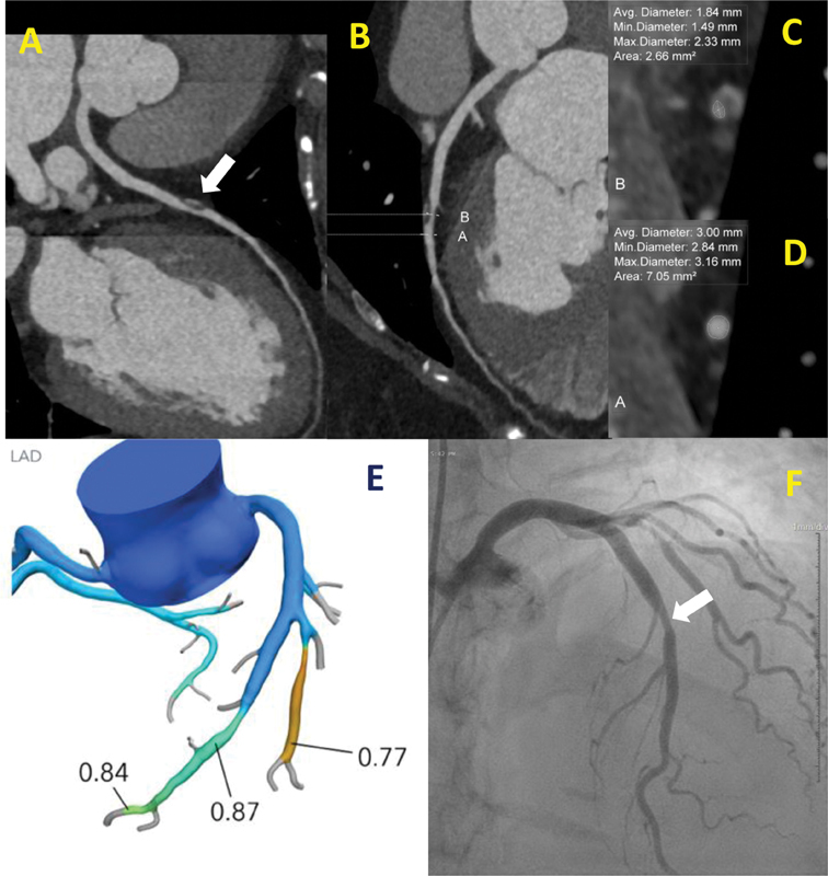
Cardiac computed tomography (CT) of a mild-to-moderate coronary artery stenosis with computed tomography fractional flow reserve (CT-FFR) and angiographic correlation. ( A, B ) Curved planar reformats of the left anterior descending artery showing a large focal plaque. ( C, D ) Closeup of double oblique multiplanar reformatted images with lumen measurements at the site of greatest stenosis and at the distal normal lumen. ( E ) CT-FFR with a value distal to the left anterior descending lesion of 0.87. ( F ) Angiographic image. White arrow = lesion.
Ironically, it is the ISCHEMIA trial, rather than SCOT-HEART, which established the superiority of cardiac CT over functional imaging. The trial, which enrolled 5179 patients, was designed to determine whether patients with positive functional imaging tests would benefit from an initial strategy of invasive coronary angiography (ICA) and any consequent revascularization. The composite primary outcome chosen was a combination of the following: (1) death from cardiovascular causes, (2) MI, (3) cardiovascular hospitalization (i.e., due to unstable angina or heart failure) or (4) resuscitated cardiac arrest. Patients were randomized to either ICA or MT alone as an initial treatment strategy (an important caveat is that patients with significant left main disease were excluded, a point which will be further discussed below). Initial revascularization rates in the ICA group were high (78%), which was in line with general expectations. Eventually, 23% of patients in the MT arm went on to undergo revascularization. However, in rigorous and lengthy statistical analysis, the results of the trial were clear, with equivalence in the rate of the combined primary outcome between the ICA and MT groups. 15
The implications of the ISCHEMIA result are threefold. First, ISCHEMIA confirmed MT as a first-line therapy for CCS. Second, it established that there is little or no benefit to functional testing for CCS once patients with left main disease were excluded. Finally, because by design ISCHEMIA used cardiac CT to exclude patients with left main disease, cardiac CT was demonstrated to be of great utility in the initial triage of CCS patients. As a significant aside, the ISCHEMIA investigators determined during post-hoc analysis that 20% of the patients enrolled were found to have no flow limiting lesions on cardiac CT 18 ; all the studies performed for ISCHEMIA patients were rigorously reviewed in core laboratories. In light of the well-established negative predictive value of cardiac CT, 19 this finding further emphasizes the value of cardiac CT over functional imaging modalities.
Although somewhat underpowered (1050 enrolled rather than 4300 as planned) and weakened by suboptimal patient compliance to MT, the results of the 2020 RESCUE trial are nonetheless a significant and confirmatory afternote to the ISCHEMIA trial. RESCUE was designed to test the noninferiority of cardiac CT to single photon emission computed tomography (SPECT) myocardial perfusion in the setting of CCS and in essence, allowed for a point-by-point comparison of the two modalities. 20 For patients triaged initially to MT, the primary measured outcome was MACE or revascularization, and for those triaged initially to revascularization, the primary measured outcome was MACE alone. Both arms had a similarly low rate of MACE or revascularization (hazard ratio [HR]: 1.03; p = 0.19). However, CCTA emerged as the better predictor of MACE and revascularization ( p = 0.02) when compared SPECT, reaffirming the utility of cardiac CT over functional imaging.
Large trials, including SCOT-HEART and RESCUE, have to date not incorporated CT-FFR into comparisons of cardiac CT to functional imaging. In recent years, CT-FFR has become an important technological improvement to cardiac CT, with more recent trials demonstrating the diagnostic benefits of adding physiologic data to traditional CT imaging. Of these studies, the DISCOVER-FLOW trial is particularly noteworthy as it offers a direct comparison of cardiac CT alone to cardiac CT with CT-FFR, using invasive FFR as the gold standard. Although powered at a per vessel, rather than per patient, level, the study demonstrated that the addition of CT-FFR improved the average area under the receiver operator characteristics (ROC) curve from 0.75 to 0.90. 21 To improve the power and expand on the findings of the DISCOVER-FLOW trial, the NXT trial, which was similar in design and reported shortly thereafter, demonstrated an improvement in the area under the ROC curve from 0.68 to 0.81 at the per patient level. 22
Intracoronary Imaging
Technical challenges and radiation safety concerns prevent integration of cardiac CT as an intraprocedural modality. However, two intraprocedural cross-sectional imaging modalities, IVUS and optical coherence tomography (OCT), have been employed to complement conventional angiographic guidance for some time ( Figs. 2 3 4 ). As both involve the use of intravascular probes, they are referred to collectively as intracoronary imaging (ICI). As their names suggest, these modalities use either sound (IVUS) or visible light (OCT) to characterize the three-dimensional morphology and pathology of diseased coronary artery segments and to locate the ostia of nearby branches. The goal of ICI technology is to improve outcomes of percutaneous coronary intervention (PCI) through more precise utilization, stent sizing, and placement. 23
Fig. 2.
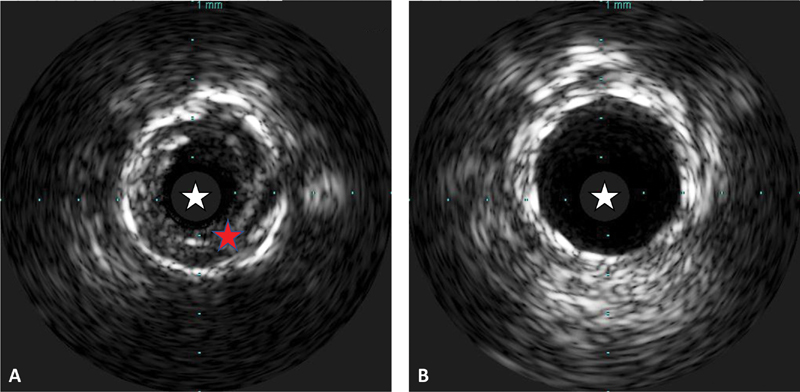
Intravascular ultrasound (IVUS) of pre- and post-percutaneous coronary intervention. ( A ) An IVUS image of a diseased coronary artery. ( B ) The same coronary artery segment after percutaneous coronary intervention. After intervention, the lumen area is much larger compared with preintervention. The white star indicates the IVUS catheter lumen. The red star indicates the atherosclerotic plaque.
Fig. 3.
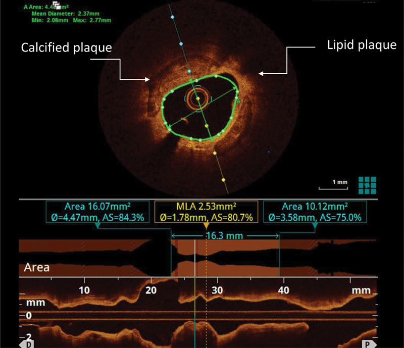
Optical coherence tomography image of a culprit lesion. The image at the top is a cross-sectional view of the culprit lesion in a coronary artery. A calcified plaque and a lipid plaque can be identified. The image at the bottom is a longitudinal view of the same coronary artery segment. This longitudinal view gives an accurate assessment of the length of the lesion, which can be used to optimize percutaneous coronary intervention. Image courtesy of Dr. Mark Rabbat M.D. from Loyola University Medical Center.
Fig. 4.

Optical coherence tomography illustrating intracoronary and stent thrombus. ( A ) De novo intracoronary thrombus. ( B ) Stent thrombosis. The white star indicates the thrombi. The intracoronary and stent lumen is outlined by the dotted line in both images.
Several strong meta-analyses support the use of IVUS guidance for PCI over angiographic guidance alone. 24 25 26 27 The most recent, rigorous, and important of these studies, published in the Journal of the American Heart Association by Darmoch et al, compared data from nearly 30 thousand patients, including ∼11,500 in the IVUS cohort. 27 The investigators found IVUS significantly reduced the risk of both cardiovascular mortality and MI during PCI compared with angiography alone. Furthermore, although the meta-analyses included both ACS (43% in the IVUS group and 34% in the angiography group) and CCS patients, meta-regression analysis demonstrated that the risk reduction of IVUS was independent of whether an ACS event had prompted revascularization. The implication of this finding is that IVUS improves the outcome of PCI for both ACS and CCS patients.
Among clinical trials of ICI technology, IVUS-XPL stands out, not only because its size (1400 patients) and length of follow-up (5 years), but also because of its exclusive focus on CCS patients. 28 29 30 IVUS-XPL enrolled patients with typical angina or positive functional test results; patients with ACS were excluded. Randomization occurred if initial diagnostic angiography revealed a long (>28 mm) lesion deemed appropriate for revascularization. Patients were assigned either to IVUS-guided or angiography-guided groups for stent placement. Five-year follow-up data, completed for 85% of patients, showed a significant reduction in MACE for the IVUS group (HR: 0.50; 95% confidence interval [CI]: 0.34–0.75; p = 0.001). This result reflected a markedly reduced risk for risk for target lesion revascularization. The risk of cardiac death also trended lower in the IVUS group ( p = 0.07).
Although touted as an “all-comer trial,” nearly 80% of the similarly sized ULTIMATE trial patients were treated for ACS, calling into question the applicability of its results to CCS patients. The trial also suffered because interventionalists adhered to “IVUS-defined optimal criteria” that in retrospect may have been too difficult to reasonably achieve in most cases. 31 Nevertheless, when 3-year follow-up results of ULTIMATE were reported in February 2021, IVUS guidance for stent placement proved significantly superior to angiographic guidance in preventing the composite primary outcome of target vessel failure (while this endpoint included cardiac death and target-vessel MI, only target-vessel revascularization actually showed a significant HR reduction). 32
The beautiful imagery of OCT, the newest of the technologies discussed herein, has captured the attention of many investigators ( Figs. 3 and 4 ). The well-organized OPINION trial set out to establish the noninferiority of OCT compared with IVUS to guide revascularization in CCS patients and did just that. 33 34 35 ILUMIEN III, a pilot study comparing OCT, IVUS, and angiographic guidance, was too underpowered to arrive at any conclusion. 35 However, it inspired the now ongoing ILUMIEN IV trial, which will enroll at least 2,500 patients undergoing PCI and randomize them, in a 1:1 ratio, to either OCT or conventional angiographic guidance. The inclusion criteria for this trial are written in such a way as to capture those ACS and CCS patients with complex disease that are both likely to suffer from MACE and most likely to benefit from ICI guidance. 36
Lesion-Specific Physiological Guidance
Unlike the imaging modalities discussed above, lesion-specific physiological guidance, which includes FFR and the newer instantaneous wave-free ratio (iFR), uses pressure wire-based indices to quantify the physiologic significance of a coronary stenosis and its potential to induce myocardial ischemia ( Figs. 5 and 6 ). FFR is an established and well-tested technology supported by over a decade of evidence from randomized controlled trials. Although iFR is the newer of the two techniques, and more sophisticated in conception, is simpler and easier to perform. Another advantage of iFR is that the technique, unlike FFR, does not require the administration of a vasodilator.
Fig. 5.
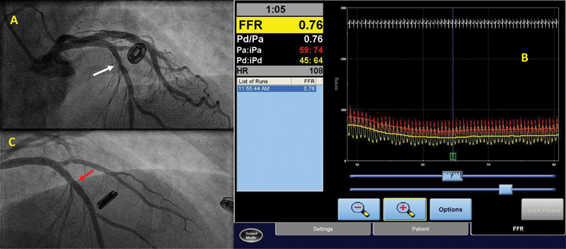
Fractional flow reserve (FFR) in an angiographically moderate coronary artery stenosis. ( A ) A coronary angiogram of the left coronary system in a right anterior oblique cranial projection. The mid left anterior descending coronary artery has an angiographically moderate stenosis of 50 to 60% (white arrow). ( B ) The FFR assessment of this stenosis, which is positive with a value of 0.76 (<0.80 is considered positive). ( C ) The same segment after percutaneous coronary intervention with placement of a drug-eluting stent (red arrow).
Fig. 6.

Instantaneous wave-free ratio (iFR) in an angiographically moderate coronary artery stenosis. ( A ) A coronary angiogram of the left coronary system in an anteroposterior caudal projection. The obtuse marginal-1 vessel has a long 50 to 70% stenosis (white arrow). An iFR wire can be seen in the obtuse marginal-1 branch (red arrow). ( B ) The iFR pressure tracings with a final value of 0.97. As seen in the image, multiple runs were performed, and all iFR values were negative (>0.89).
The seminal FAME study randomized 1,005 patients to either FFR guidance or angiographic guidance for stent placement. Two-year follow-up, reported in 2010, confirmed a durable reduction in MACE for FFR-guided PCI compared with angiography-guided PCI in patients with CCS. 8 37 38 The subsequent FAME 2 trial compared FFR-guided PCI with MT to MT alone, with a composite primary outcome of death, MI, or urgent revascularization, in patients with CCS 39 40 The trial was halted prematurely, with much fanfare, after enrolling 1,220 patients, due to early emergence of an 8% risk reduction in the primary endpoint for the FFR-guided PCI group, primarily owing to reduction in urgent revascularizations. At 5-year follow-up, the rate of the composite primary end point remained lower in the FFR-guided PCI group (13.9 vs. 27.0%; HR: 0.46; 95% CI: 0.34–0.63; p < 0.001), again due primarily to reduced urgent revascularizations, an impressive result. 41 A post-hoc analysis of pooled data from FAME and FAME 2 revealed that measuring the FFR values after PCI can also provide very useful clinical information. After examining data from 639 patients with a total of 837 lesions, the analysis found greater symptomatic relief (odds ratio: 1.33; p = 0.02) and lower event rates (HR: 2.01; p = 0.04) when there was a larger improvement in post-PCI FFR. 42
The iFR-SWEDEHEART trial was designed to demonstrate the noninferiority of iFR compared with FFR and succeeded in its purpose. Two-thousand thirty-seven patients were randomized to FFR- or iFR-guided PCI with a primary outcome of death, nonfatal MI, or unplanned revascularization. At 12 months, the groups had similar clinical event rates (6.7 vs. 6.1%; HR: 1.12, p = 0.53). 43 44 The similarly designed DEFINE-FLAIR trial (with results published the same day as the iFR-SWEDEHEART trial) also demonstrated noninferiority of iFR compared with FFR. 45 In this study of 2,492 patients, iFR was found to be noninferior to FFR with regard to risk of MACEs at 12 months (7.0% in FFR group vs. 6.8% in iFR group; HR: 0.95; p = 0.78; p < 0.001 for noninferiority). In both trials, FFR patients were far more likely to experience chest pain during revascularization than IFR patients, which was expected given that chest pain is a well-known side effect of adenosine. Importantly, DEFINE-FLAIR found iFR to be associated with shorter procedural time (median time of 40.5 minutes in the iFR group vs. 45 minutes in the FFR group; p = 0.001).
Conclusion
Publication of the Scandinavian Simvastatin Survival Study in 1994 revolutionized MT for CCS. 46 MT and revascularization are both complimentary and competing therapies for CCS, as they seek to improve symptoms and prevent future events. Both types of therapy have evolved over time, leading to more options for patients with CCS. It has taken technological advances in diagnosis, patient selection, procedural guidance, stent technology, and procedural technique more than a quarter of a century to redefine and recapture a role for revascularization in CCS patients.
Drug eluting stents and the global shift toward radial access have improved the tolerability and safety of PCI for all comers and need to be acknowledged as important advances in the treatment of CCS; as the tolerability and safety of revascularization improve, its attractiveness as an option to reduce symptoms in patients with CCS grows. 47 48 However, in light of excellent MT for CCS that will no doubt continue to improve, the keys to incorporation of revascularization into the treatment of CCS are patient selection and lesion selection. The three technologies discussed herein, cardiac CT, ICI, and lesion-specific physiological guidance, have over the past decade proven to be the best diagnostic tools in our armamentarium. Like MT, these diagnostic modalities continue to improve. CT-FFR is an improvement over conventional cardiac CT. OCT may eventually prove to be superior to IVUS, not just aesthetically, but also in its accuracy. iFR has already begun to supplant FFR because of its simplicity, efficiency, and freedom from side effects.
In patients with CCS, optimal MT is the standard of care. Revascularization can be considered for patients with angina that persists despite maximal tolerated antianginal regimen. Other CCS patients who may benefit from revascularization include those often excluded from clinical trials: those with left ventricular dysfunction, heart failure, left main disease, and severe multivessel disease. All CCS patients, the majority of whom will receive MT, benefit from improvements in diagnostic technology.
Acknowledgments
On the submission site, there is a five-author limit. If our manuscript is selected for publication, in lieu of including the following individuals as authors, we would like to acknowledge them: Dr.'s Wael Aljaroudi, William Bates, Mark Rabbat, Sean Javaheri, Raymond Mai, and Homeyar Dinshaw.
Funding Statement
Funding Dr. Winkler receives research support from Terarecon, Ziosoft, Algomedica, and Teleflex. Dr. Arora receives research support from Terarecon. Dr. Weintraub is funded by grants from the National Institutes of Health (HL142097, HL134354, R56AG064895, and AR070029).
Conflict of Interest None declared.
Authors' Contributions
MW substantially contributed to the concept and design, drafting of the manuscript, literature review, summary and qualitative synthesis of publications related to noninvasive cardiovascular imaging and intracoronary imaging, and selection and annotation of figures.
RP was involved in literature review, some drafting, qualitative synthesis of publications related to lesion specific physiological guidance, and selection and annotation of figures.
WF was involved in literature review, some drafting, editing, and manuscript preparation.
VA was involved in concept and design, editing, and minor drafting.
NW was involved in concept and design, editing, final draft, integrity of the manuscript.
References
- 1.The top Ten Causes of Death. World Health Organization Website https://www.who.int/news-room/fact-sheets/detail/the-top-10-causes-of-death. Updated 9 December 2020, Accessed July 29, 2021
- 2.Ma J, Ward E M, Siegel R L, Jemal A.Temporal trends in mortality in the United States, 1969-2013 JAMA 2015314161731–1739. 10.1001/jama.2015.12319Erratum in: JAMA. 2015 Dec 8;314(22):2415. PMID: 26505597 [DOI] [PubMed] [Google Scholar]
- 3.Mensah G A, Wei G S, Sorlie P D. Decline in cardiovascular mortality: possible causes and implications. Circ Res. 2017;120(02):366–380. doi: 10.1161/CIRCRESAHA.116.309115. [DOI] [PMC free article] [PubMed] [Google Scholar]
- 4.Agostoni P, Biondi-Zoccai G G, Gasparini G L. Is bare-metal stenting superior to balloon angioplasty for small vessel coronary artery disease? Evidence from a meta-analysis of randomized trials. Eur Heart J. 2005;26(09):881–889. doi: 10.1093/eurheartj/ehi116. [DOI] [PubMed] [Google Scholar]
- 5.Sabaté M, Brugaletta S, Cequier A.Clinical outcomes in patients with ST-segment elevation myocardial infarction treated with everolimus-eluting stents versus bare-metal stents (EXAMINATION): 5-year results of a randomised trial Lancet 2016387(10016):357–366. [DOI] [PubMed] [Google Scholar]
- 6.NORSTENT Investigators . Bønaa K H, Mannsverk J, Wiseth R. Drug-eluting or bare-metal stents for coronary artery disease. N Engl J Med. 2016;375(13):1242–1252. doi: 10.1056/NEJMoa1607991. [DOI] [PubMed] [Google Scholar]
- 7.Buccheri S, Franchina G, Romano S. Clinical outcomes following intravascular imaging-guided versus coronary angiography-guided percutaneous coronary intervention with stent implantation: a systematic review and Bayesian network meta-analysis of 31 studies and 17,882 patients. JACC Cardiovasc Interv. 2017;10(24):2488–2498. doi: 10.1016/j.jcin.2017.08.051. [DOI] [PubMed] [Google Scholar]
- 8.FAME Study Investigators . Tonino P A, De Bruyne B, Pijls N H. Fractional flow reserve versus angiography for guiding percutaneous coronary intervention. N Engl J Med. 2009;360(03):213–224. doi: 10.1056/NEJMoa0807611. [DOI] [PubMed] [Google Scholar]
- 9.Vij A, Kassab K, Chawla H.Invasive therapy versus conservative therapy for patients with stable coronary artery disease: an updated meta-analysis Clin Cardiol 20214405675–682.; Epub ahead of print 10.1002/clc.23592 [DOI] [PMC free article] [PubMed] [Google Scholar]
- 10.Bangalore S, Maron D J, Stone G W, Hochman J S. Routine revascularization versus initial medical therapy for stable ischemic heart disease: a systematic review and meta-analysis of randomized trials. Circulation. 2020;142(09):841–857. doi: 10.1161/CIRCULATIONAHA.120.048194. [DOI] [PubMed] [Google Scholar]
- 11.Libby P, Theroux P. Pathophysiology of coronary artery disease. Circulation. 2005;111(25):3481–3488. doi: 10.1161/CIRCULATIONAHA.105.537878. [DOI] [PubMed] [Google Scholar]
- 12.Meikle P J, Wong G, Tsorotes D. Plasma lipidomic analysis of stable and unstable coronary artery disease. Arterioscler Thromb Vasc Biol. 2011;31(11):2723–2732. doi: 10.1161/ATVBAHA.111.234096. [DOI] [PubMed] [Google Scholar]
- 13.ESC Scientific Document Group Knuuti J, Wijns W, Saraste A.2019 ESC Guidelines for the diagnosis and management of chronic coronary syndromes Eur Heart J 20204103407–477. 10.1093/eurheartj/ehz425Erratum in: Eur Heart J. 2020 Nov 21;41(44):4242. PMID: 31504439 [DOI] [PubMed] [Google Scholar]
- 14.Wolff G, Dimitroulis D, Andreotti F. Survival benefits of invasive versus conservative strategies in heart failure in patients with reduced ejection fraction and coronary artery disease: a meta-analysis. Circ Heart Fail. 2017;10(01):e003255. doi: 10.1161/CIRCHEARTFAILURE.116.003255. [DOI] [PubMed] [Google Scholar]
- 15.ISCHEMIA Research Group . Maron D J, Hochman J S, Reynolds H R. Initial invasive or conservative strategy for stable coronary disease. N Engl J Med. 2020;382(15):1395–1407. doi: 10.1056/NEJMoa1915922. [DOI] [PMC free article] [PubMed] [Google Scholar]
- 16.SCOT-HEART Investigators . Newby D E, Adamson P D, Berry C. Coronary CT angiography and 5-year risk of myocardial infarction. N Engl J Med. 2018;379(10):924–933. doi: 10.1056/NEJMoa1805971. [DOI] [PubMed] [Google Scholar]
- 17.SCOT-HEART Investigators . Adamson P D, Williams M C, Dweck M R. Guiding therapy by coronary CT angiography improves outcomes in patients with stable chest pain. J Am Coll Cardiol. 2019;74(16):2058–2070. doi: 10.1016/j.jacc.2019.07.085. [DOI] [PMC free article] [PubMed] [Google Scholar]
- 18.COME-CCT Consortium . Haase R, Schlattmann P, Gueret P. Diagnosis of obstructive coronary artery disease using computed tomography angiography in patients with stable chest pain depending on clinical probability and in clinically important subgroups: meta-analysis of individual patient data. BMJ. 2019;365:l1945:1–15. doi: 10.1136/bmj.l1945. [DOI] [PMC free article] [PubMed] [Google Scholar]
- 19.Mancini G BJ, Leipsic J, Budoff M J. CT angiography followed by invasive angiography in patients with moderate or severe ischemia-insights from the ISCHEMIA Trial. JACC Cardiovasc Imaging. 2021;14(07):1384–1393. doi: 10.1016/j.jcmg.2020.11.012. [DOI] [PMC free article] [PubMed] [Google Scholar]
- 20.RESCUE investigators * . Stillman A E, Gatsonis C, Lima J AC. Coronary computed tomography angiography compared with single photon emission computed tomography myocardial perfusion imaging as a guide to optimal medical therapy in patients presenting with stable angina: the rescue trial. J Am Heart Assoc. 2020;9(24):e017993. doi: 10.1161/JAHA.120.017993. [DOI] [PMC free article] [PubMed] [Google Scholar]
- 21.Koo B K, Erglis A, Doh J H. Diagnosis of ischemia-causing coronary stenoses by noninvasive fractional flow reserve computed from coronary computed tomographic angiograms. Results from the prospective multicenter DISCOVER-FLOW (Diagnosis of Ischemia-Causing Stenoses Obtained Via Noninvasive Fractional Flow Reserve) study. J Am Coll Cardiol. 2011;58(19):1989–1997. doi: 10.1016/j.jacc.2011.06.066. [DOI] [PubMed] [Google Scholar]
- 22.Min J K, Leipsic J, Pencina M J. Diagnostic accuracy of fractional flow reserve from anatomic CT angiography. JAMA. 2012;308(12):1237–1245. doi: 10.1001/2012.jama.11274. [DOI] [PMC free article] [PubMed] [Google Scholar]
- 23.Mintz G S. Clinical utility of intravascular imaging and physiology in coronary artery disease. J Am Coll Cardiol. 2014;64(02):207–222. doi: 10.1016/j.jacc.2014.01.015. [DOI] [PubMed] [Google Scholar]
- 24.Parise H, Maehara A, Stone G W, Leon M B, Mintz G S. Meta-analysis of randomized studies comparing intravascular ultrasound versus angiographic guidance of percutaneous coronary intervention in pre-drug-eluting stent era. Am J Cardiol. 2011;107(03):374–382. doi: 10.1016/j.amjcard.2010.09.030. [DOI] [PubMed] [Google Scholar]
- 25.Elgendy I Y, Mahmoud A N, Elgendy A Y, Bavry A A. Outcomes with intravascular ultrasound-guided stent implantation: a meta-analysis of randomized trials in the era of drug-eluting stents. Circ Cardiovasc Interv. 2016;9(04):e003700. doi: 10.1161/CIRCINTERVENTIONS.116.003700. [DOI] [PubMed] [Google Scholar]
- 26.Bavishi C, Sardar P, Chatterjee S. Intravascular ultrasound-guided vs angiography-guided drug-eluting stent implantation in complex coronary lesions: meta-analysis of randomized trials. Am Heart J. 2017;185:26–34. doi: 10.1016/j.ahj.2016.10.008. [DOI] [PubMed] [Google Scholar]
- 27.Darmoch F, Alraies M C, Al-Khadra Y, Moussa Pacha H, Pinto D S, Osborn E A. Intravascular ultrasound imaging-guided versus coronary angiography-guided percutaneous coronary intervention: a systematic review and meta-analysis. J Am Heart Assoc. 2020;9(05):e013678. doi: 10.1161/JAHA.119.013678. [DOI] [PMC free article] [PubMed] [Google Scholar]
- 28.Cho S, Shin D H, Kim J S. Rationale and design: impact of intravascular ultrasound guidance on long-term clinical outcomes of everolimus-eluting stents in long coronary lesions. Contemp Clin Trials. 2015;40:90–94. doi: 10.1016/j.cct.2014.11.012. [DOI] [PubMed] [Google Scholar]
- 29.IVUS-XPL Investigators Hong S J, Kim B K, Shin D H.Effect of intravascular ultrasound-guided vs angiography-guided everolimus-eluting stent implantation: the IVUS-XPL randomized clinical trial JAMA 2015314202155–2163. 10.1001/jama.2015.15454Erratum in: JAMA. 2016 Feb 2;315(5):518. Kim, Yonghoon [corrected to Kim, Yong Hoon]. PMID: 26556051 [DOI] [PubMed] [Google Scholar]
- 30.IVUS-XPL Investigators . Hong S J, Mintz G S, Ahn C M. Effect of intravascular ultrasound-guided drug-eluting stent implantation: 5-year follow-up of the IVUS-XPL randomized trial. JACC Cardiovasc Interv. 2020;13(01):62–71. doi: 10.1016/j.jcin.2019.09.033. [DOI] [PubMed] [Google Scholar]
- 31.Zhang J, Gao X, Kan J. Intravascular ultrasound versus angiography-guided drug-eluting stent implantation: the ULTIMATE trial. J Am Coll Cardiol. 2018;72(24):3126–3137. doi: 10.1016/j.jacc.2018.09.013. [DOI] [PubMed] [Google Scholar]
- 32.ULTIMATE Investigators . Gao X F, Ge Z, Kong X Q. 3-year outcomes of the ULTIMATE trial comparing intravascular ultrasound versus angiography-guided drug-eluting stent implantation. JACC Cardiovasc Interv. 2021;14(03):247–257. doi: 10.1016/j.jcin.2020.10.001. [DOI] [PubMed] [Google Scholar]
- 33.Kubo T, Shinke T, Okamura T. Optical frequency domain imaging vs. intravascular ultrasound in percutaneous coronary intervention (OPINION trial): study protocol for a randomized controlled trial. J Cardiol. 2016;68(05):455–460. doi: 10.1016/j.jjcc.2015.11.007. [DOI] [PubMed] [Google Scholar]
- 34.OPINION Investigators . Kubo T, Shinke T, Okamura T. Optical frequency domain imaging vs. intravascular ultrasound in percutaneous coronary intervention (OPINION trial): one-year angiographic and clinical results. Eur Heart J. 2017;38(42):3139–3147. doi: 10.1093/eurheartj/ehx351. [DOI] [PMC free article] [PubMed] [Google Scholar]
- 35.Ali Z A, Karimi Galougahi K, Maehara A. Outcomes of optical coherence tomography compared with intravascular ultrasound and with angiography to guide coronary stent implantation: one-year results from the ILUMIEN III: OPTIMIZE PCI trial. EuroIntervention. 2021;16(13):1085–1091. doi: 10.4244/EIJ-D-20-00498. [DOI] [PMC free article] [PubMed] [Google Scholar]
- 36.Ali Z, Landmesser U, Karimi Galougahi K. Optical coherence tomography-guided coronary stent implantation compared to angiography: a multicentre randomised trial in PCI - design and rationale of ILUMIEN IV: OPTIMAL PCI. EuroIntervention. 2021;16(13):1092–1099. doi: 10.4244/EIJ-D-20-00501. [DOI] [PMC free article] [PubMed] [Google Scholar]
- 37.FAME Study Investigators Fearon W F, Tonino P A, De Bruyne B, Siebert U, Pijls N H.Rationale and design of the fractional flow reserve versus angiography for multivessel evaluation (FAME) study Am Heart J 200715404632–636. 10.1016/j.ahj.2007.06.012Erratum in: Am Heart J. 2007 Dec;154(6):1243. PMID: 17892983 [DOI] [PubMed] [Google Scholar]
- 38.FAME Study Investigators . Pijls N H, Fearon W F, Tonino P A. Fractional flow reserve versus angiography for guiding percutaneous coronary intervention in patients with multivessel coronary artery disease: 2-year follow-up of the FAME (Fractional Flow Reserve Versus Angiography for Multivessel Evaluation) study. J Am Coll Cardiol. 2010;56(03):177–184. doi: 10.1016/j.jacc.2010.04.012. [DOI] [PubMed] [Google Scholar]
- 39.FAME 2 Trial Investigators De Bruyne B, Pijls N H, Kalesan B.Fractional flow reserve-guided PCI versus medical therapy in stable coronary disease N Engl J Med 201236711991–1001. 10.1056/NEJMoa1205361Erratum in: N Engl J Med. 2012 Nov;367(18):1768. Mobius-Winckler, Sven [corrected to Möbius-Winkler, Sven]. PMID: 22924638 [DOI] [PubMed] [Google Scholar]
- 40.FAME 2 Trial Investigators De Bruyne B, Fearon W F, Pijls N H.Fractional flow reserve-guided PCI for stable coronary artery disease N Engl J Med 2014371131208–1217. 10.1056/NEJMoa1408758Erratum in: N Engl J Med. 2014 Oct 9;371(15):1465. PMID: 25176289 [DOI] [PubMed] [Google Scholar]
- 41.FAME 2 Investigators . Xaplanteris P, Fournier S, Pijls N HJ. Five-year outcomes with PCI guided by fractional flow reserve. N Engl J Med. 2018;379(03):250–259. doi: 10.1056/NEJMoa1803538. [DOI] [PubMed] [Google Scholar]
- 42.Fournier S, Ciccarelli G, Toth G G. Association of improvement in fractional flow reserve with outcomes, including symptomatic relief, after percutaneous coronary intervention. JAMA Cardiol. 2019;4(04):370–374. doi: 10.1001/jamacardio.2019.0175. [DOI] [PMC free article] [PubMed] [Google Scholar]
- 43.Götberg M, Christiansen E H, Gudmundsdottir I. Instantaneous Wave-Free Ratio versus Fractional Flow Reserve guided intervention (iFR-SWEDEHEART): rationale and design of a multicenter, prospective, registry-based randomized clinical trial. Am Heart J. 2015;170(05):945–950. doi: 10.1016/j.ahj.2015.07.031. [DOI] [PubMed] [Google Scholar]
- 44.iFR-SWEDEHEART Investigators . Götberg M, Christiansen E H, Gudmundsdottir I J. Instantaneous wave-free ratio versus fractional flow reserve to guide PCI. N Engl J Med. 2017;376(19):1813–1823. doi: 10.1056/NEJMoa1616540. [DOI] [PubMed] [Google Scholar]
- 45.Davies J E, Sen S, Dehbi H M. Use of the instantaneous wave-free ratio or fractional flow reserve in PCI. N Engl J Med. 2017;376(19):1824–1834. doi: 10.1056/NEJMoa1700445. [DOI] [PubMed] [Google Scholar]
- 46.Pedersen T R, Kjekshus J, Berg K.Randomised trial of cholesterol lowering in 4444 patients with coronary heart disease: the Scandinavian Simvastatin Survival Study (4S) Lancet 1994344(8934):1383–1389. [PubMed] [Google Scholar]
- 47.Marlevi D, Edelman E R. Vascular lesion-specific drug delivery systems: JACC state-of-the-art review. J Am Coll Cardiol. 2021;77(19):2413–2431. doi: 10.1016/j.jacc.2021.03.307. [DOI] [PMC free article] [PubMed] [Google Scholar]
- 48.MATRIX Investigators Valgimigli M, Frigoli E, Leonardi S.Radial versus femoral access and bivalirudin versus unfractionated heparin in invasively managed patients with acute coronary syndrome (MATRIX): final 1-year results of a multicentre, randomised controlled trial Lancet 2018392(10150):835–848. [DOI] [PubMed] [Google Scholar]