Abstract
Although melatonin is an astonishing molecule, it is possible that chemistry will help in the discovery of new compounds derived from it that may exceed our expectations regarding antioxidant protection and perhaps even neuroprotection. This review briefly summarizes the significant amount of data gathered to date regarding the multiple health benefits of melatonin and related compounds. This review also highlights some of the most recent directions in the discovery of multifunctional pharmaceuticals intended to act as one-molecule multiple-target drugs with potential use in multifactorial diseases, including neurodegenerative disorders. Herein, we discuss the beneficial activities of melatonin derivatives reported to date, in addition to computational strategies to rationally design new derivatives by functionalization of the melatonin molecular framework. It is hoped that this review will promote more investigations on the subject from both experimental and theoretical perspectives.
Keywords: multifactorial diseases, multifunctional drugs, antioxidant, neuroprotection, free radical scavengers, metal chelators, oxidative stress, DNA repair
1. Introduction
Chemistry is a science for which every detail matters. Thus, every structural modification, regardless how small, will have consequences on the behavior of chemicals. The rational design of new molecules with potential pharmacological benefits is based on this idea. In this context, a highly challenging task is to find single chemical entities with multiple biological activities, because they are expected to have better success when used in the treatment of complex (multifactorial) diseases. Such chemicals are known as multifunctional drugs (MFD), and are also referred to as magic bullets or master keys due to their improved therapeutic effects and fewer side effects. They are also referred to as dual-mechanism, dual-ligand, bifunctional, multimechanistic, multimodal, pan-agonist, multipotential, pluripotential, and multiple-ligands [1].
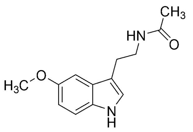
Melatonin (N-acetyl-5-methoxytryptamine, Scheme 1) is an amazing molecule with multiple proven benefits. It not only regulates circadian and seasonal rhythms [2,3,4], but also possesses many other functions in living organisms [5], as summarized in Section 6. Therefore, it is logical to think that the molecular framework of melatonin is an excellent candidate for slight modification to obtain new molecules with even wider benefits. Such research is usually conducted with the premise that, because the structural changes are small, the new derivatives will retain most of the activity of the parent molecule. Although this assumption may seem naïve, nature itself has proven that there are molecules that can be considered as relatives of melatonin that exhibit similar benefits and even improved properties for specific purposes. These are reviewed in Section 7.1.
Scheme 1.
Melatonin (N-acetyl-5-methoxytryptamine).
The main goal of this review is to discuss, at least partially, the beneficial activities of melatonin derivatives reported to date; to consider some of the characteristics of neurodegenerative disorders and why these derivatives may be useful in treating them; and to put into perspective the current stage of development of medical drugs based on melatonin. It is hoped that this review will promote more studies in this field from diverse areas of expertise, which are essential in the development and application of chemicals with pharmacological potential.
2. Multifactorial Diseases
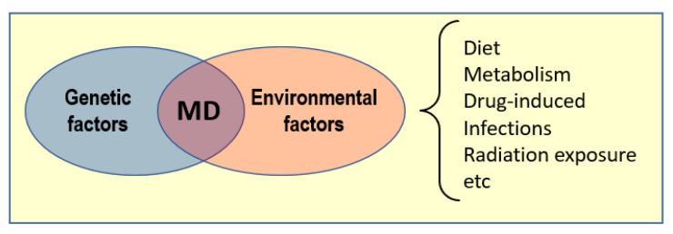
Most of the chronic and progressive diseases that lead to high morbidity and mortality are multifactorial in nature and involve the accumulation of biological disfunctions that result in diverse tissue and organ damages [6]. Some of these are cardiovascular, cerebrovascular and neurodegenerative disorders, and immune-, metabolic-, and infection-induced diseases [7,8,9,10,11,12,13,14,15]. Many factors, both genetic and environmental, can contribute to the onset and development of multifactorial diseases (MD, Figure 1).
Figure 1.
Some factors that can trigger multifactorial diseases (MD).
Neurodegenerative Disorders
This review is focused on the potential benefits of melatonin derivatives in a particular type of MD: neurodegenerative disorders (NDD). There are more than 600 of these [16], including Alzheimer’s (AD), Parkinson’s (PD), and Huntington’s (HD) diseases and amyotrophic lateral sclerosis (ALS). It has been noted that “As the global population ages and the number of individuals expected to develop neurodegenerative conditions increases, the search for an effective cure is becoming progressively more urgent” [17] (p. 1222).
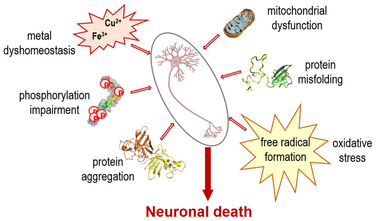
NDD are among the most enigmatic and problematic health disorders, mainly due to their multifactorial nature. Although each has its own molecular mechanisms and clinical manifestations, some general pathways have been recognized [18,19] (Figure 2).
Figure 2.
Some events contributing to the multifactorial nature of neurodegenerative disorders (NDD).
The chronological hierarchy of the events associated with the underlying causes leading to NDD are not fully understood yet [18]. However, the gathered evidence strongly indicates that one can potentiate the other, leading to a self-sustaining cycle that ultimately provokes neuronal death processes [20].
Based on the multifaceted nature of NDD, efficient therapeutic drugs for treating them should be capable of targeting and regulating several pathological aspects, including deactivation of oxidants and metal chelation. Contrary to this expectation, the most common pharmacological research in the past dealt with compounds that have selective activity against specific molecular targets. Although these are effective against diseases with one prevalent alteration, their effectiveness against the majority of multifactorial diseases has been rather disappointing [6]. Thus, there is a new paradigm in drugs designed for the treatment of multifactorial diseases in general and NDD in particular: multifunctional drugs (MFD).
3. Multifunctional Drugs
It has been noted that, because NDD involve a large variety of cellular and biochemical changes, the one-molecule one-target strategy is not adequate for treating them [19]. It was also hypothesized that, because many NDD present similar neuronal disorders, it is possible that a single entity may be potentially used for more than one of these illnesses.
The perspective of such a tactic is promising. The evidence gathered to date indicates that MFD not only have better success at modulating complex diseases, but also do not increase side-effects. Some of the reported advantages of MFD over drug combinations are [1,6,17,18]:
Both palliative and disease modifying actions;
Additive or synergistic therapeutic responses;
Reduced risk of drug-drug interactions;
Improved drug characteristics (for example ADME properties);
More predictable pharmacokinetics and pharmacodynamic relationships;
Prolonged duration of effectiveness;
Simplified therapeutic regimen;
Lower costs.
It has been proposed that “drug design for chronic diseases might be established based on the rational assembly of multiple chemical groups that are putative ligands for the selected cellular macromolecule targets respectively responsible for root cause and symptoms/signs, and therefore generates the desired multi-target effect” [6] (p. 8).
In the particular case of NDD, the multifunctionality of the desired therapeutic one-molecule multiple-target chemicals should include some of the following effects: (i) inhibition of acetylcholinesterase (AChE); (ii) inhibition of monoamine oxidase (MAO); (iii) inhibition of catechol O-methyltransferase (COMT); (iv) antioxidant behavior; (v) free radical scavenging activity; and (vi) metal chelating power [1,21,22,23,24,25,26,27,28,29,30,31,32,33,34].
4. Oxidative Stress
Oxidative stress (OS) has been called the chemical silent killer [35] because it does not produce apparent symptoms and there are no routine tests to detect it yet. Thus, the OS-related damage frequently occurs before the affected person becomes aware of it. In addition, OS can be potentiated by physiological and environmental factors, including physical or mental stress, infections, aging, pollution, radiation, cigarette smoke, and many others [36,37,38,39,40,41,42,43,44,45,46,47,48,49]. There is compelling evidence on the role of OS in the onset and development of a large number of diseases. A few examples are cancer [50,51,52,53], cardiovascular diseases [54,55,56,57,58,59,60,61], diabetes [62,63,64], and neurodegenerative disorders including PD and AD diseases, memory loss, multiple sclerosis, and depression [65,66,67,68,69,70,71,72,73].
More than twenty years ago, the role of reactive oxygen species (ROS) in NDD was proposed to be as important as that of microorganisms in infectious diseases [74]. However, there has been a debate about whether oxidative stress (OS) is a cause or a consequence of the neurodegenerative cascade [75]. Today, there is almost a consensus that OS is an early event of neurodegeneration and one of the major factors of NDD [18].
Neuronal tissue is particularly sensitive to OS. The imbalance in pro-oxidant vs. antioxidant homeostasis in the nervous central system results in the production of ROS, which are involved in the initiation or propagation of radical chain reactions [18]. In AD, PD, HD, and ALS, most biomolecules, including lipids, proteins, and DNA, are oxidatively damaged [76]. Nonetheless, the administration of antioxidants as a treatment has been characterized as being too simplistic, and several clinical studies have demonstrated that they have only modest success in the treatment of NDD [77].
It is possible that antioxidants with other protective effects against this kind of disease may have a better impact. Although transition metals are crucial to biological processes, alterations in their homeostasis increase free radical production, which is frequently catalyzed by traces of redox active metals such as iron, zinc, and copper [78,79]. In addition, metal ions promote fibril generation and deposition [80] and metal-induced OS is associated with mitochondrial dysfunction [81,82,83,84]. Thus, a compound capable of exerting both radical scavenging activity and metal-chelating power may be a better protector against OS than a molecule with only one of these properties [18]. Such a compound may also be a good antioxidant candidate to fight NDD.
5. Chemical Antioxidants
The word “antioxidant” involves a large variety of possible actions. Thus, antioxidants can be classified according to their means of action against OS:
Primary antioxidants (Type I, chain breaking or free radical scavengers): directly react with free radicals yielding less reactive species that are unable to damage biological targets, or end the radical chain reaction.
Secondary antioxidants (Type II, or preventive): exert their protection by chemical routes that do not involve direct reactions with free radicals, such as metal chelation, absorption of UV radiation, deactivation of singlet oxygen, repair of primary antioxidants, and decomposition of hydroperoxide into nonradical species.
Tertiary antioxidants (Type III, or fixers): capable of repairing, mainly through H or electron transfer, biomolecules that are oxidatively damaged and restore their pristine structures.
Multifunctional antioxidants (Type IV, or versatile): can combine more than one of the above-mentioned means of action, or one of them with enhancing enzymatic protection or restoring pathways in the endogenous antioxidative defense system.
Based on this classification, the latter have higher potential to have beneficial effects on the treatment of NDD.
6. Melatonin
Melatonin is a ubiquitous and versatile molecule. In addition to being produced by the pineal gland, it is also found in many other organs, including the skin, retina, cerebellum, kidneys, liver, pancreas, and ovaries [85,86,87,88,89,90]. It has immune-enhancing and anti-inflammatory properties [91,92,93], can inhibit cancer progression [94,95,96], and plays homeostatic roles in the mitochondria [97,98,99].
Melatonin is particularly efficient as an antioxidant [100,101,102]. There are several reasons for its exceptional performance in such a role:
Melatonin has very low toxicity [103].
Melatonin can cross physiologic barriers easily. This is because of its size and solubility (partially soluble in water and highly soluble in non-polar aprotic solvents, including lipids [104,105]).
Metabolisms do not cause a decline in melatonin’s protection against OS, because its metabolites are capable of offering the same kind of protection [106,107,108].
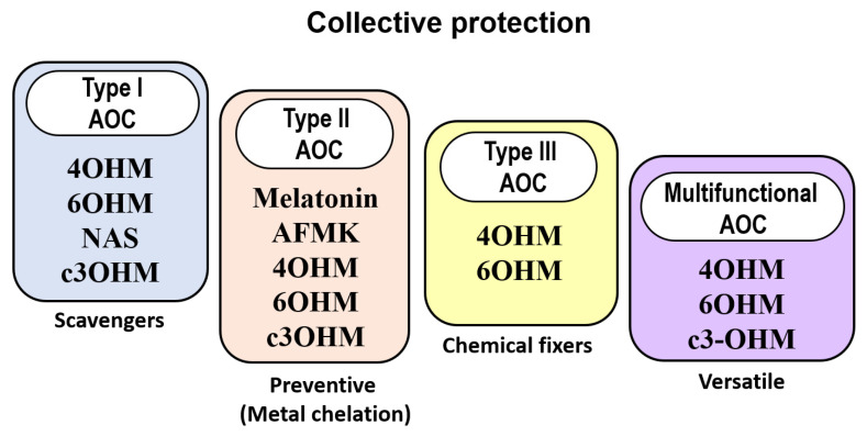
The combined protection of melatonin and its metabolites is widely known [109] and usually referred to as a cascade-like protection [110,111,112]. This collective antioxidant capacity (AOC) implies that melatonin can deactivate more than one equivalent of oxidants. This is a multifaceted process that involves free radical scavenging processes (Type I AOC) and metal chelation (Type II AOC) [113]. It has been proposed that the collective action of melatonin and its metabolites takes place in a “task-division” manner (Figure 3). Some members of this family are expected to perform particularly well as free radical scavengers, whereas others (including melatonin itself) would be more efficient as metal chelators [114].
Figure 3.
Antioxidant protection exerted by melatonin and its metabolites. AOC: antioxidant activity; c3OHM: cyclic 3-hydroxymelatonin; 6OHM: 6-hydroxymelatonin; 4OHM: 4-hydroxymelatonin; AFMK: N1-acetyl-N2-formyl-5-methoxykynuramine; NAS: N-acetyl serotonin.
Studies have also shown that melatonin and its metabolites can protect from OS as Type III antioxidants, particularly by preventing DNA damage. The shielding effect of these compounds against the structural modification of DNA, and the consequent health issues, is believed to be mediated by their capability to repair OS-induced damage to DNA sites. The repairing process was hypothesized to involve various chemical routes. The guanine-centered radical cations are expected to be repaired via electron transfer, at very high rates; C-centered free radicals (in the sugar moiety of 2′-deoxyguanosine), via formal H atom transfer; and OH adducts (in the imidazole ring), by a two-step route: an H atom transfer followed by dehydration [115].
There is abundant evidence supporting the role of melatonin as a protector against OS and derived damages [101,102,116,117,118,119,120,121,122,123,124]. In fact, such a role has been proposed as responsible for many of its numerous benefits for human health [125,126,127,128,129,130,131,132]. It has been even hypothesized that melatonin’s main function in living systems is to prevent them from oxidative damage [133]. The AOC of melatonin acquires particular relevance regarding NDD. The brain is highly susceptible to oxidation and the BBB prevents many chemicals from entering the region. Melatonin is an efficient antioxidant generated in situ and released into the cerebrospinal fluid [134]. Therefore, melatonin is available when needed and capable of protecting against OS. In fact, there is evidence that melatonin has beneficial effects on OS-related disorders that affect the brain [135]. Some examples are Parkinson’s and Alzheimer’s diseases [136,137,138,139,140,141,142,143]. This does not mean that melatonin’s protection against OS is exclusive to the brain. It also protects other organs from this chemical stress, including the stomach, the heart, the liver, and the skin [144,145,146,147,148,149,150,151,152,153,154].
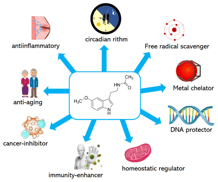
Melatonin is capable of reducing the molecular damage arising from high amounts of free radicals in vivo [155]. It has been reported that melatonin is also capable of scavenging diverse oxidants. Some of these are: •OOCCl3 [156], •OH [157,158,159,160], •OR [161,162], •NO [163,164], and 1O2 [165,166]. Melatonin protects low-density lipoprotein from oxidation [167] and binds copper, which prevents lipid peroxidation induced by this metal [168]. Based on the chelating ability of melatonin, it has been suggested that the neuroprotection (and also the AOC) of this molecule is a consequence of its ability to remove redox metals from the CNN [168]. This hypothesis is supported by other findings. Melatonin can act as a ligand, forming complexes with diverse metals, including not only copper, but also iron, cadmium, aluminum, zinc, and lead [169]. Melatonin significantly lessens the production of free radicals induced by the interaction of Cu(II), Fe(II) Al(III), Mn(II), and Zn(II) with the β-amyloid peptide [170]. In addition, melatonin is also capable of counteracting the oxidative damage to proteins induced by Cu(II)/H2O2 mixtures [171]. The protective effects of melatonin against molecular damage catalyzed by redox metals were recently thoroughly reviewed [172]. Thus, melatonin itself can be classified a multifunctional antioxidant, and a multifunctional molecule in general (Figure 4).
Figure 4.
Multifunctionality of melatonin.
7. Derivatives
7.1. From Nature
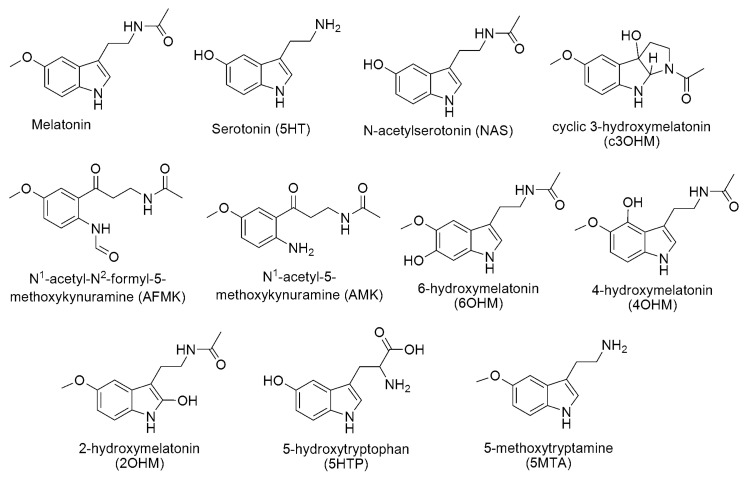
Among the naturally-occurring melatonin derivatives, its metabolites and precursors stand out (Scheme 2). These are all involved in the tryptophan enzymatic metabolism. The direct precursor of melatonin is serotonin (also referred to as normelatonin). It is produced from 5-hydroxytryptophan in mammals and from tryptamine in plants, which are the immediate products of tryptophan in the respective routes.
Scheme 2.
Melatonin and some related natural compounds.
N1-acetyl-5-methoxykynuramine (AMK), N1-acetyl-N2-formyl-5-methoxykynuramine (AFMK), cyclic 3-hydroxymelatonin (c3OHM), 5-methoxytryptamine (5MTA), and 6-hydroxymelatonin (6OHM) are melatonin’s metabolites. These are produced not only by metabolic routes, but also from the melatonin-mediated free radicals’ detoxification. It is assumed that c3OHM is formed as a result of the reaction between melatonin and the hydroxyl radical [173]. AFMK is yielded when c3OHM further reacts with other free radicals. The virtuous AOC process continues and AMK is a product derived from the radical scavenging reactions of AFMK [174]. By comparison, in mammals, 2-hydroxymelatonin (2OHM) and 4-hydroxymelatonin (4OHM) are the result of the melatonin’s metabolism induced by UV radiation [175]. In plants, 2OHM is enzymatically produced and the most abundant metabolite of melatonin [176].
The AOC of melatonin-derived compounds that are naturally produced has been widely demonstrated. The different manifestations of this activity are summarized in Table 1. They clearly show that all of these compounds are versatile antioxidants.
Table 1.
Evidence of the AOC of some natural melatonin derivatives.
| Actions | Ref. | |
|---|---|---|
| AFMK | Hydroxyl radical scavenger. Radiation protector. Inhibitor of lipid peroxidation and OS-induced neuronal damage. Inhibitor of OS induced by Cu(II)-ascorbate mixtures. ROS scavenger. RNS scavenger. Protector against diverse oxidants. |
[177,178,179] [180] [177,178,179,181] [113] [182,183,184] [185,186,187,188,189] [183,190,191] |
| Serotonin | Inhibitor of lipid peroxidation. ROS scavenger. Fe(II) chelator. Inhibitor of •OH production. |
[123] [121] [192] [125] |
| NAS | Neuroprotector. Antioxidant and anti-aging protector. Inhibitor of DNA oxidation induced by H2O2 and Cr(III). Inhibitor of UV-induced cataracts. Inhibitor of Cu-induced lipoproteins oxidation. Inhibitor of lipid peroxidation. Keeper of optimal fluidity of the biological membranes. |
[193,194] [195] [196,197,198] [44] [199,200] [201,202,203,204] [205] |
| 6OHM | Inhibitor of quinolinic-acid induced neurotoxicity. Inhibitor of UV-induced OS. Inhibitor of KCN-induced O2•− production. Inhibitor of iron-induced lipid peroxidation. Inhibitor of cyanide-induced OS. Inhibitor of thiobarbituric acid-induced lipid peroxidation. |
[206] [183] [207] [208,209] [207] [210] |
| c3OHM | Scavenger of ABTS•+ (2,2′-azino-bis(3-ethylbenzothiazoline-6-sulfonic acid) radical cation). | [211] |
| Peroxyl radicals scavenger. | [212] | |
| Hydroxyl radical scavenger. | [173,212] | |
| Inhibitor of OS induced by Cu(II)-ascorbate mixtures. | [113] | |
| 5HTP | Anti-inflammatory. Free radical scavenger. Inhibitor of iron-induced lipid peroxidation. Inhibitor of Fe/ascorbate-induced oxidation. Metal chelator. Inhibitor of UV-induced apoptosis in human monocytes. |
[114] [115] [116] [117] [120] [119] |
| 5MTA | Hydroxyl radical scavenger. Inhibitor of lipid peroxidation. Hypochlorous acid deactivator. Inhibitor of malondialdehyde formation. Inhibitor of exercise-induced cellular oxidative changes |
[212] [213,214] [201] [215] [216] |
7.2. From the Lab
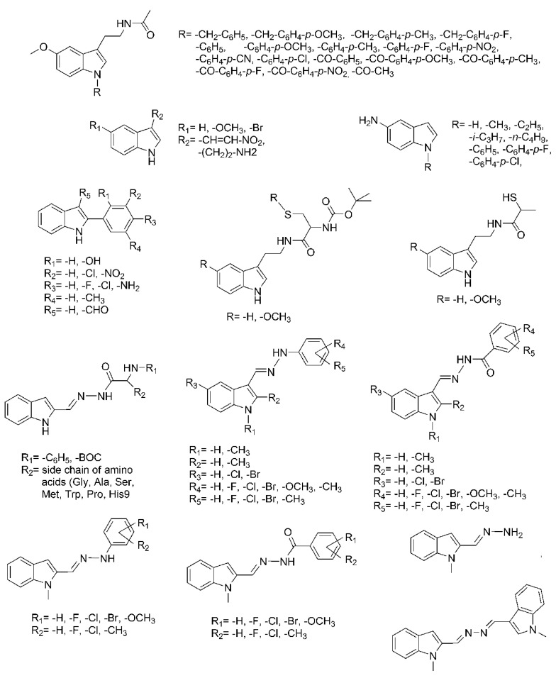
The appealing properties of melatonin have inspired the search for synthetic derivatives of this molecule intended for a variety of purposes, including AOC protection (Scheme 3) [217,218]. The main strategy behind the search has been to modify the indole ring by functionalization at different sites. Some of the groups that have been used in the structural modifications of the melatonin framework are benzoyl and phenyl groups [219,220]. The produced derivatives were demonstrated to surpass melatonin in their anticonvulsant, anxiolytic, and sedative properties. The anti-inflammatory effects of acetyl- and benzoyl-melatonin were reported to be higher than those of melatonin [221]. In addition, they all were found to exhibit significant AOC [222].
Scheme 3.
Structures of some synthetic melatonin derivatives. BOC stands for the t-butyloxycarbonyl group.
The AOC of several melatonin derivatives obtained from functionalization with the sulfhydryl group has been tested, and some of these were proven to have improved antioxidant protection compared to melatonin [223]. Melatonin derivatives containing retinamide [224], 2-phenylindole [225], indole-3-propionamides [226], and N-methylindole plus hydrazide/hydrazone [227] moieties were reported to inhibit lipid peroxidation. Chloroindole hydrazide/hydrazone melatonin derivatives were found to be highly efficient as free radical scavengers [228]. Modifications in the methoxy and acylaminoethyl groups of melatonin have been also pursued as promising synthetic routes to produce compounds with enhanced AOC compared to melatonin [229]. Indole amino acid derivatives of melatonin have been produced and tested for AOC. Their performance as DPPH (2,2-difenil-1-picrylhydrazyl) scavengers was found to be similar to that of melatonin, whereas their efficiency as inhibitors of lipid peroxidation was higher [230].
Thus, there is a large amount of evidence supporting the hypothesis that modest chemical modifications (functionalization) to the melatonin’s molecular framework may lead to the discovery of novel compounds with a wide variety of desired properties. Moreover, they may retain and even surpass the health benefits of the parent molecule.
7.3. Computational Designed
When designing multifunctional drugs, three key aspects must be addressed:
-
(i)
Building the candidates;
-
(ii)
Sampling the search space;
-
(iii)
Evaluating their potential for the desired purpose.
The sampling and evaluation stages should simultaneously include: (a) positive design controls that help identifying the chemical space where there is a higher probability of finding drug-like molecules, and (b) negative design controls (‘taboo zones’) that define unwanted properties and/or structures [231]. A unified quantity, allowing direct comparisons among a large number of candidates, is also required. For this purpose, a weighted scoring function is frequently used. This consists of a summation of terms (as many as needed) weighted by factors that are proportional to their relative importance: f(p) = w1p1 + w2p2 + … Part of the challenge here is setting such weighting factors. It appears worthwhile to mention that properties considered in the terms of the summation are diverse and go further than the binding affinity. Some important properties to consider are ADME (absorption, distribution, metabolism, excretion), toxicity, and synthetic accessibility.
A computational protocol known as CADMA-Chem has been recently proposed for the design of antioxidants with multifunctional behavior [232]. This is based on a diversity of chemical properties and involves building a modest number of candidates (a few hundred). The approach used to build them is to add functional groups (1 to 3) to a molecular framework that is already known for a desired purpose. The computational strategies used to design MFD can be roughly grouped into library screening or rational design [17]. The CADMA-Chem protocol is similar to the latter. The difference is that classical rational design combines two (or more) molecular scaffolds (with the target properties) into a single molecular entity, whereas CADMA-Chem slightly modifies a single molecular scaffold (with the desirable pharmaceutical behavior) by adding a few functional groups. This addition is intended to potentiate AOC and improve the physicochemical properties that would allow the candidates to cross lipid barriers (via passive diffusion). In this manner, CADMA-Chem is intended to take advantage of the current knowledge regarding pharmaceuticals that are known to be efficient for the intended purpose, in particular as therapeutic agents in the treatment of OS-related diseases. CADMA-Chem is in line with the three aspects of rational design, as mentioned above. To define negative and positive spaces, this protocol considers AOC, toxicity, ADME properties, and manufacturability.
The hypothesis behind this protocol is to design multifunctional antioxidants with potential as neuroprotectors, although it can be used for other purposes. The desirable properties of the designed molecules are:
Free radical scavenging capability;
Metal chelation properties (OH inactivating ligand behavior);
Low toxicity;
Adequate permeation and bioavailability;
Non-difficult manufacturability;
No pro-oxidant behavior;
Efficient for repairing oxidatively damaged biological targets;
Inhibition of COMT, AChE and/or MAO.
It is then assumed that a chemical with most of these properties should have neuroprotective effects.
The CADMA-Chem protocol was used to construct melatonin derivatives that are expected to be multifunctional antioxidants [233]. The target multifunctionality consisted of different types of AOC plus possible neuroprotection. By adding different functional groups (-OH, -SH, -NH2 and -COOH) to the melatonin scaffold, 116 derivatives were built; 16 with only one functional group, 96 with two functional groups, and four with three functional groups.
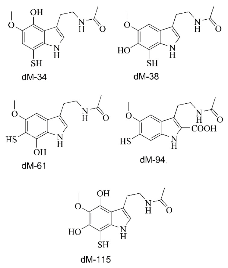
The selection score used to sample the search space included eight terms for ADME properties, two for toxicity, and one for synthetic accessibility; each of these had equivalent weight factors. Using this selection score, a first subset (20 candidates) was selected. These were the derivatives with the best drug-like behavior. In a second stage of the search, the pKas of these 20 molecules, in addition to various reactivity indices, were calculated. The reactivity indices were chosen to account for H and electron donor capabilities, which are expected to be good indicators of AOC (type I, free radical scavengers) when involving single electron transfer (SET) and/or formal hydrogen transfer (HAT) chemical routes. Five melatonin derivatives were identified as the most likely candidates to be chemical antioxidants, namely: dM-34, dM-115, dM-38, dM-61, and dM-94 (Scheme 4), in that order. The efficiency of each of these, as type I antioxidants, was predicted to be higher than those of Trolox and melatonin.
Scheme 4.
Some computationally designed melatonin derivatives.
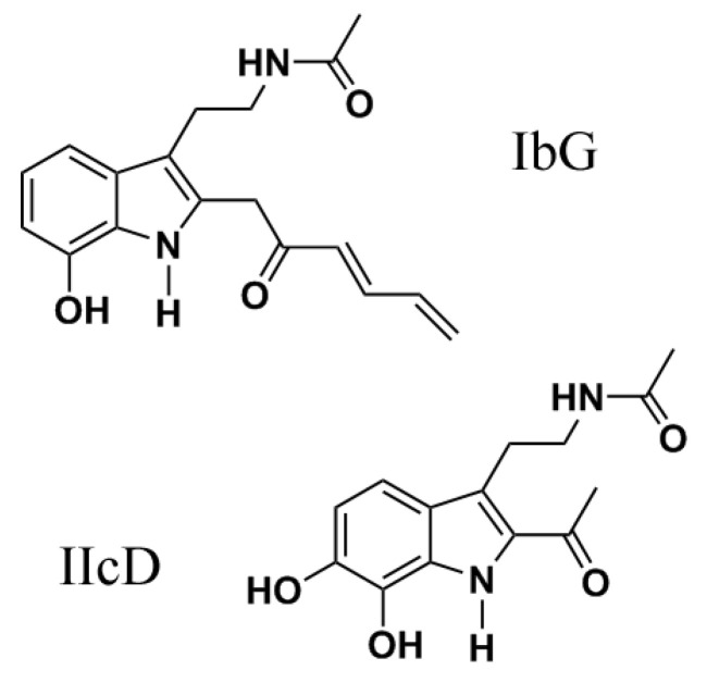
A different approach was followed in a previous work [234]. To build the candidates, it was considered that the structural features enhancing AOC type I and type II are not necessarily the same. Accordingly, in a first stage of the investigation, two sets of melatonin derivatives were built and tested, one for each type of AOC. The main structural modification in the first set (eight candidates) was to add phenolic OH groups, based on the good free radical scavenging activity of phenolic compounds (including natural melatonin derivatives such as NAS and 6OHM) [113]. The second set of melatonin derivatives (seven candidates) was designed to mimic a particular molecular topology that is assumed to promote metal chelation and the associated type II AOC [234]. In addition, it was proposed that the presence of such topology may be considered as a key structural feature of therapeutic agents in the treatment of AD [235].
The separated analyses of the above-mentioned two sets of molecules allowed identification of the most promising structural features in the designed melatonin derivatives that were assumed to independently promote type I and type II AOC. These features were then combined in a third set (four candidates). These were intended to simultaneously act as free radical scavengers (type I AOC) and redox metal chelators capable of inhibiting OH radical production, via Fenton-like reactions (type II AOC). Two of these were proposed as the most promising candidates as multifunctional antioxidants (Scheme 5). The selection was made based on the finding that they are highly efficient as OH inhibitors, via metal chelation, and also excellent for scavenging free radicals.
Scheme 5.
Other computationally designed melatonin derivatives.
Further investigations of other properties of the newly designed melatonin derivatives discussed in this section are still needed from both experimental and theoretical approaches. Some of these are to explore their possible pro-oxidant behavior in the absence and the presence of redox metal ions, their efficiency for repairing oxidatively damaged biological targets, and their inhibition potential of COMT, AChE, and/or MAO.
8. Summary
Most chronic and progressive health disorders leading to high morbidity and mortality are multifactorial diseases. In the particular case of neurodegenerative disorders, efficient therapeutic drugs for treatment should be capable of targeting and regulating several pathological aspects including AChE-inhibition, MAO-inhibition, COMT-inhibition, antioxidant behavior, free radical scavenging activity, and metal chelating power. Thus, antioxidant molecules are good initial candidates.
Melatonin is an astonishing molecule, but it is possible that chemistry will help to identify new compounds derived from it that may exceed our expectations regarding antioxidant protection and perhaps even neuroprotection. This review briefly summarizes the significant amount of data gathered to date regarding the multiple health benefits of melatonin and related compounds. It also highlights some of the recent directions in the discovery of multifunctional pharmaceuticals intended to act as one-molecule multiple-target drugs with potential use in multifactorial diseases, including neurodegenerative disorders. It is hoped that this review will promote more investigations on the subject from both experimental and theoretical perspectives.
Acknowledgments
We gratefully thank the Laboratorio de Visualización y Cómputo Paralelo at Universidad Autónoma Metropolitana-Iztapalapa for computing time. E.G.G.L. acknowledges CONACyT for a Doctoral fellowship. This investigation is inserted in the research projects IFC-2016/1828 and MX18MO10.
Abbreviations
| AChE | acetylcholinesterase |
| AD | Alzheimer’s disease |
| ADME | absorption, distribution, metabolism, and excretion |
| ALS | amyotrophic lateral sclerosis |
| AOC | antioxidant capacity |
| BBB | brain blood barrier |
| CNN | central nervous system |
| CADMA-Chem | Computer-assisted Design of Multifunctional Antioxidants, based on chemical properties |
| COMT | catechol-O-methyltransferase |
| HD | Huntington’s disease |
| MAO | monoamine oxidase |
| MD | multifactorial diseases |
| MFD | multifunctional drugs |
| NDD | neurodegenerative disorders |
| OS | oxidative stress |
| PD | Parkinson’s disease |
| RNS | reactive oxnitrogen species |
| ROS | reactive oxygen species |
Funding
This research received no external funding.
Institutional Review Board Statement
Not applicable.
Informed Consent Statement
Not applicable.
Data Availability Statement
Not applicable.
Conflicts of Interest
The authors declare no conflict of interest.
Footnotes
Publisher’s Note: MDPI stays neutral with regard to jurisdictional claims in published maps and institutional affiliations.
References
- 1.Bansal Y., Silakari O. Multifunctional compounds: Smart molecules for multifactorial diseases. Eur. J. Med. Chem. 2014;76:31–42. doi: 10.1016/j.ejmech.2014.01.060. [DOI] [PubMed] [Google Scholar]
- 2.Vriend J., Reiter R.J. Melatonin feedback on clock genes: A theory involving the proteasome. J. Pineal Res. 2015;58:1–11. doi: 10.1111/jpi.12189. [DOI] [PubMed] [Google Scholar]
- 3.Reiter R.J. The melatonin rhythm: Both a clock and a calendar. Experientia. 1993;49:654–664. doi: 10.1007/BF01923947. [DOI] [PubMed] [Google Scholar]
- 4.Reiter R.J. Circannual reproductive rhythms in mammals related to photoperiod and pineal function: A review. Chronobiologia. 1974;1:365–395. [PubMed] [Google Scholar]
- 5.Reiter R.J., Tan D.X., Fuentes-Broto L. Melatonin: A multi-tasking molecule. Prog. Brain Res. 2010;181:127–151. doi: 10.1016/S0079-6123(08)81008-4. [DOI] [PubMed] [Google Scholar]
- 6.Kong W.J., Vernieri C., Foiani M., Jiang J.D. Berberine in the treatment of metabolism-related chronic diseases: A drug cloud (dCloud) effect to target multifactorial disorders. Pharmacol. Ther. 2020;209:1596. doi: 10.1016/j.pharmthera.2020.107496. [DOI] [PubMed] [Google Scholar]
- 7.Wan M.L., Wang Y., Zeng Z., Deng B., Zhu B.S., Cao T., Li Y.K., Xiao J., Han Q., Wu Q. Colorectal cancer (CRC) as a multifactorial disease and its causal correlations with multiple signaling pathways. Biosci. Rep. 2020;40:BSR20200265. doi: 10.1042/BSR20200265. [DOI] [PMC free article] [PubMed] [Google Scholar]
- 8.Badimon J.J., Fuster V., Chesebro J.H., Badimon L. Coronary atherosclerosis—a multifactorial disease. Circulation. 1993;87:3–16. [PubMed] [Google Scholar]
- 9.Jones G.H., Vecera C.M., Pinjari O.F., Machado-Vieira R. Inflammatory signaling mechanisms in bipolar disorder. J. Biomed. Sci. 2021;28:45. doi: 10.1186/s12929-021-00742-6. [DOI] [PMC free article] [PubMed] [Google Scholar]
- 10.Fleckenstein M., Keenan T.D.L., Guymer R.H., Chakravarthy U., Schmitz-Valckenberg S., Klaver C.C., Wong W.T., Chew E.Y. Age-related macular degeneration. Nat. Rev. Dis. Primers. 2021;7:31. doi: 10.1038/s41572-021-00265-2. [DOI] [PubMed] [Google Scholar]
- 11.Park H.R., Yang E.J. Oxidative stress as a therapeutic target in amyotrophic lateral sclerosis: Opportunities and limitations. Diagnostics. 2021;11:1546. doi: 10.3390/diagnostics11091546. [DOI] [PMC free article] [PubMed] [Google Scholar]
- 12.Munoz-Pinto M.F., Empadinhas N., Cardoso S.M. The neuromicrobiology of Parkinson’s disease: A unifying theory. Aging Res. Rev. 2021;70:101396. doi: 10.1016/j.arr.2021.101396. [DOI] [PubMed] [Google Scholar]
- 13.Koklesova L., Samec M., Liskova A., Zhai K., Büsselberg D., Giordano F.A., Kubatka P., Golunitschaja O. Mitochondrial impairments in aetiopathology of multifactorial diseases: Common origin but individual outcomes in context of 3P medicine. EPMA J. 2021;12:27–40. doi: 10.1007/s13167-021-00237-2. [DOI] [PMC free article] [PubMed] [Google Scholar]
- 14.Uddin M.S., Al Mamun A., Kabir M.T., Ashraf G.M., Bin-Jumah M.N., Abdel-Daim M.M. Multi-Target Drug Candidates for Multifactorial Alzheimer’s Disease: AChE and NMDAR as Molecular Targets. Mol. Neurobiol. 2021;58:281–303. doi: 10.1007/s12035-020-02116-9. [DOI] [PubMed] [Google Scholar]
- 15.Katselou M.G., Matralis A.N., Kourounakis A.P. Multi-target drug design approaches for multifactorial diseases: From neurodegenerative to cardiovascular applications. Curr. Med. Chem. 2014;21:2743–2787. doi: 10.2174/0929867321666140303144625. [DOI] [PubMed] [Google Scholar]
- 16.Trippier P.C., Jansen Labby K., Hawker D.D., Mataka J.J., Silverman R.B. Target- and mechanism-based therapeutics for neurodegenerative diseases: Strength in numbers. J. Med. Chem. 2013;56:3121–3147. doi: 10.1021/jm3015926. [DOI] [PMC free article] [PubMed] [Google Scholar]
- 17.Savelieff M.G., Nam G., Kang J., Lee H.J., Lee M., Lim M.H. Development of multifunctional molecules as potential therapeutic candidates for Alzheimer’s disease, Parkinson’s disease, and amyotrophic lateral sclerosis in the last decade. Chem. Rev. 2018;119:1221–1322. doi: 10.1021/acs.chemrev.8b00138. [DOI] [PubMed] [Google Scholar]
- 18.Cavalli A., Bolognesi M.L., Mìnarini A., Rosini M., Tumiatti V., Recanatini M., Melchiorre C. Multi-target-directed ligands to combat neurodegenerative diseases. J. Med. Chem. 2008;51:347–372. doi: 10.1021/jm7009364. [DOI] [PubMed] [Google Scholar]
- 19.Jellinger K.A. General aspects of neurodegeneration. J. Neural Transm. Suppl. 2003;15:101–144. doi: 10.1007/978-3-7091-0643-3_7. [DOI] [PubMed] [Google Scholar]
- 20.Zeevalk G.D., Bernard L.P., Song C., Gluck M., Ehrhart J. Mitochondrial inhibition and oxidative stress: Reciprocating players in neurodegeneration. Antioxid. Redox Signal. 2005;7:1117–1139. doi: 10.1089/ars.2005.7.1117. [DOI] [PubMed] [Google Scholar]
- 21.Finberg J.P.M. Inhibitors of MAO-B and COMT: Their effects on brain dopamine levels and uses in Parkinson’s disease. J. Neural Transm (Vienna) 2019;126:433–448. doi: 10.1007/s00702-018-1952-7. [DOI] [PubMed] [Google Scholar]
- 22.Patel C.N., Georrge J.J., Modi K.M., Narechania M.B., Patel D.P., Gonzalez F.J., Pandya H.A. Pharmacophore-based virtual screening of catechol-o-methyltransferase (COMT) inhibitors to combat Alzheimer’s disease. J. Biomol. Struct. Dyn. 2018;36:3938–3957. doi: 10.1080/07391102.2017.1404931. [DOI] [PMC free article] [PubMed] [Google Scholar]
- 23.Lichtenstein M.P., Carriba P., Masgrau R., Pujol A., Galea E. Staging anti-inflammatory therapy in Alzheimer’s disease. Front. Aging Neurosci. 2010;2:142. doi: 10.3389/fnagi.2010.00142. [DOI] [PMC free article] [PubMed] [Google Scholar]
- 24.Geldenhuys W.J., Youdim M.B.H., Carroll R.T., Van der Schyf C.J. The emergence of designed multiple ligands for neurodegenerative disorders. Prog. Neurobiol. 2011;94:347–359. doi: 10.1016/j.pneurobio.2011.04.010. [DOI] [PubMed] [Google Scholar]
- 25.Zhang H.Y. One-compound-multiple-targets strategy to combat Alzheimer’s disease. FEBS Lett. 2005;579:5260–5264. doi: 10.1016/j.febslet.2005.09.006. [DOI] [PubMed] [Google Scholar]
- 26.Van der Schyf C.J., Geldenhuys W.J. Multimodal drugs and their future for Alzheimer’s and Parkinson’s disease. Int. Rev. Neurobiol. 2011;100:107–125. doi: 10.1016/B978-0-12-386467-3.00006-6. [DOI] [PubMed] [Google Scholar]
- 27.Youdim M.B.H., Geldenhuys W.J., Van der Schyf C.J. Why should we use multifunctional neuroprotective and neurorestorative drugs for Parkinson’s disease? Parkinsonism Relat. Disord. 2007;13((Suppl. 3)):S281–S291. doi: 10.1016/S1353-8020(08)70017-8. [DOI] [PubMed] [Google Scholar]
- 28.Birla H., Minocha T., Kumar G., Misra A., Singh S.K. Role of Oxidative Stress and Metal Toxicity in the Progression of Alzheimer’s Disease. Curr. Neuropharmacol. 2020;18:552–562. doi: 10.2174/1570159X18666200122122512. [DOI] [PMC free article] [PubMed] [Google Scholar]
- 29.Cassidy L., Fernandez F., Johnson J.B., Naiker M., Owoola A.G., Broszczak D.A. Oxidative stress in alzheimer’s disease: A review on emergent natural polyphenolic therapeutics. Complement. Ther. Med. 2020;49:102294. doi: 10.1016/j.ctim.2019.102294. [DOI] [PubMed] [Google Scholar]
- 30.Jurcau A., Simion A. Oxidative stress in the pathogenesis of Alzheimer’s disease and cerebrovascular disease with therapeutic implications. CNS Neurol. Disord. Drug Targets. 2020;19:94–108. doi: 10.2174/1871527319666200303121016. [DOI] [PubMed] [Google Scholar]
- 31.Kowalska M., Wize K., Prendecki M., Lianeri M., Kozubski W., Dorszewska J. Genetic variants and oxidative stress in alzheimer’s disease. Curr. Alzheimer Res. 2020;17:208–223. doi: 10.2174/1567205017666200224121447. [DOI] [PubMed] [Google Scholar]
- 32.Plascencia-Villa G., Perry G. Preventive and Therapeutic Strategies in Alzheimer’s Disease: Focus on Oxidative Stress, Redox Metals, and Ferroptosis. Antioxid. Redox Signal. 2021;34:591–610. doi: 10.1089/ars.2020.8134. [DOI] [PMC free article] [PubMed] [Google Scholar]
- 33.Sharma C., Kim S.R. Linking oxidative stress and proteinopathy in alzheimer’s disease. Antioxidants. 2021;10:1231. doi: 10.3390/antiox10081231. [DOI] [PMC free article] [PubMed] [Google Scholar]
- 34.Zarrouk A., Hammouda S., Ghzaiel I., Hammami S., Khamlaoui W., Ahmed S.H., Lizard G., Hammami M. Association between oxidative stress and altered cholesterol metabolism in alzheimer’s disease patients. Curr. Alzheimer Res. 2020;17:823–834. doi: 10.2174/1567205017666201203123046. [DOI] [PubMed] [Google Scholar]
- 35.Galano A., Raúl Alvarez-Idaboy J. Computational strategies for predicting free radical scavengers’ protection against oxidative stress: Where are we and what might follow? Int. J. Quantum Chem. 2018;119:e25665. doi: 10.1002/qua.25665. [DOI] [Google Scholar]
- 36.Pham-Huy L.A., He H., Pham-Huy C. Free radicals, antioxidants in disease and health. Int. J. Biomed. Sci. 2008;4:89–96. [PMC free article] [PubMed] [Google Scholar]
- 37.Pacher P., Beckman J.S., Liaudet L. Nitric oxide and peroxynitrite in health and disease. Physiol. Rev. 2007;87:315–424. doi: 10.1152/physrev.00029.2006. [DOI] [PMC free article] [PubMed] [Google Scholar]
- 38.Genestra M. Oxyl radicals, redox-sensitive signalling cascades and antioxidants. Cell. Signal. 2007;19:1807–1819. doi: 10.1016/j.cellsig.2007.04.009. [DOI] [PubMed] [Google Scholar]
- 39.Valko M., Leibfritz D., Moncol J., Cronin M.T.D., Mazur M., Telser J. Free radicals and antioxidants in normal physiological functions and human disease. Int. J. Biochem. Cell Biol. 2007;39:44–84. doi: 10.1016/j.biocel.2006.07.001. [DOI] [PubMed] [Google Scholar]
- 40.Valko M., Rhodes C.J., Moncol J., Izakovic M., Mazur M. Free radicals, metals and antioxidants in oxidative stress-induced cancer. Chem. Biol. Interact. 2006;160:1–40. doi: 10.1016/j.cbi.2005.12.009. [DOI] [PubMed] [Google Scholar]
- 41.Dröge W. Free radicals in the physiological control of cell function. Physiol. Rev. 2002;82:47–95. doi: 10.1152/physrev.00018.2001. [DOI] [PubMed] [Google Scholar]
- 42.Young I.S., Woodside J.V. Antioxidants in health and disease. J. Clin. Pathol. 2001;54:176–186. doi: 10.1136/jcp.54.3.176. [DOI] [PMC free article] [PubMed] [Google Scholar]
- 43.Halliwell B. Biochemistry of oxidative stress. Biochem. Soc. Trans. 2007;35:1147–1150. doi: 10.1042/BST0351147. [DOI] [PubMed] [Google Scholar]
- 44.Willcox J.K., Ash S.L., Catignani G.L. Antioxidants and prevention of chronic disease. Crit. Rev. Food Sci. Nutr. 2004;44:275–295. doi: 10.1080/10408690490468489. [DOI] [PubMed] [Google Scholar]
- 45.Parthasarathy S., Santanam N., Ramachandran S., Meilhac O. Oxidants and antioxidants in atherogenesis: An appraisal. J. Lipid Res. 1999;40:2143–2157. doi: 10.1016/S0022-2275(20)32089-7. [DOI] [PubMed] [Google Scholar]
- 46.Lowe F.J., Cemeli E. Biomarkers of oxidative stress and the relationship to cigarette smoking. Mini Rev. Org. Chem. 2011;8:377–386. doi: 10.2174/157019311797440308. [DOI] [Google Scholar]
- 47.Valko M., Morris H., Cronin M.T.D. Metals, toxicity and oxidative stress. Curr. Med. Chem. 2005;12:1161–1208. doi: 10.2174/0929867053764635. [DOI] [PubMed] [Google Scholar]
- 48.Reiter R.J., Tan D.X., Sainz R.M., Mayo J.C., Lopez-Burillo S. Melatonin: Reducing the toxicity and increasing the efficacy of drugs. J. Pharm. Pharmacol. 2002;54:1299–1321. doi: 10.1211/002235702760345374. [DOI] [PubMed] [Google Scholar]
- 49.Reiter R.J., Manchester L.C., Tan D.X. Neurotoxins: Free radical mechanisms and melatonin protection. Curr. Neuropharmacol. 2010;8:194–210. doi: 10.2174/157015910792246236. [DOI] [PMC free article] [PubMed] [Google Scholar]
- 50.Tekiner-Gulbas B., Westwell A.D., Suzen S. Oxidative stress in carcinogenesis: New synthetic compounds with dual effects upon free radicals and cancer. Curr. Med. Chem. 2013;20:4451–4459. doi: 10.2174/09298673113203690142. [DOI] [PubMed] [Google Scholar]
- 51.Thanan R., Oikawa S., Hiraku Y., Ohnishi S., Ma N., Pinlaor S., Yongvanit P., Kawanishi S., Murata M. Oxidative stress and its significant roles in neurodegenerative diseases and cancer. Int. J. Mol. Sci. 2014;16:193–217. doi: 10.3390/ijms16010193. [DOI] [PMC free article] [PubMed] [Google Scholar]
- 52.Filaire E., Dupuis C., Galvaing G., Aubreton S., Laurent H., Richard R., Filaire M. Lung cancer: What are the links with oxidative stress, physical activity and nutrition. Lung Cancer. 2013;82:383–389. doi: 10.1016/j.lungcan.2013.09.009. [DOI] [PubMed] [Google Scholar]
- 53.Paschos A., Pandya R., Duivenvoorden W.C.M., Pinthus J.H. Oxidative stress in prostate cancer: Changing research concepts towards a novel paradigm for prevention and therapeutics. Prostate Cancer Prostatic Dis. 2013;16:217–225. doi: 10.1038/pcan.2013.13. [DOI] [PubMed] [Google Scholar]
- 54.Matsuda M., Shimomura I. Roles of adiponectin and oxidative stress in obesity-associated metabolic and cardiovascular diseases. Rev. Endocr. Metab. Disord. 2014;15:1–10. doi: 10.1007/s11154-013-9271-7. [DOI] [PubMed] [Google Scholar]
- 55.Eren E., Ellidag H.Y., Cekin Y., Ayoglu R.U., Sekercioglu A.O., Yilmaz N. Heart valve disease: The role of calcidiol deficiency, elevated parathyroid hormone levels and oxidative stress in mitral and aortic valve insufficiency. Redox Rep. 2014;19:34–39. doi: 10.1179/1351000213Y.0000000069. [DOI] [PMC free article] [PubMed] [Google Scholar]
- 56.Siti H.N., Kamisah Y., Kamsiah J. The role of oxidative stress, antioxidants and vascular inflammation in cardiovascular disease (a review) Vascul. Pharmacol. 2015;71:40–56. doi: 10.1016/j.vph.2015.03.005. [DOI] [PubMed] [Google Scholar]
- 57.Popolo A., Autore G., Pinto A., Marzocco S. Oxidative stress in patients with cardiovascular disease and chronic renal failure. Free Radic. Res. 2013;47:346–356. doi: 10.3109/10715762.2013.779373. [DOI] [PubMed] [Google Scholar]
- 58.Miller M.R., Shaw C.A., Langrish J.P. From particles to patients: Oxidative stress and the cardiovascular effects of air pollution. Future Cardiol. 2012;8:577–602. doi: 10.2217/fca.12.43. [DOI] [PubMed] [Google Scholar]
- 59.Tousoulis D., Psaltopoulou T., Androulakis E., Papageorgiou N., Papaioannou S., Oikonomou E., Synetos A., Stefanadis C. Oxidative stress and early atherosclerosis: Novel antioxidant treatment. Cardiovasc. Drugs Ther. 2015;29:75–88. doi: 10.1007/s10557-014-6562-5. [DOI] [PubMed] [Google Scholar]
- 60.Li H., Horke S., Förstermann U. Vascular oxidative stress, nitric oxide and atherosclerosis. Atherosclerosis. 2014;237:208–219. doi: 10.1016/j.atherosclerosis.2014.09.001. [DOI] [PubMed] [Google Scholar]
- 61.Peluso I., Morabito G., Urban L., Ioannone F., Serafini M. Oxidative stress in atherosclerosis development: The central role of LDL and oxidative burst. Endocr. Metab. Immune Disord. Drug Targets. 2012;12:351–360. doi: 10.2174/187153012803832602. [DOI] [PubMed] [Google Scholar]
- 62.Rosales-Corral S., Tan D.X., Manchester L., Reiter R.J. Diabetes and Alzheimer disease, two overlapping pathologies with the same background: Oxidative stress. Oxid. Med. Cell. Longev. 2015;2015:985845. doi: 10.1155/2015/985845. [DOI] [PMC free article] [PubMed] [Google Scholar]
- 63.Zephy D., Ahmad J. Type 2 diabetes mellitus: Role of melatonin and oxidative stress. Diabetes Metab. Syndr. 2015;9:127–131. doi: 10.1016/j.dsx.2014.09.018. [DOI] [PubMed] [Google Scholar]
- 64.Maiese K. New insights for oxidative stress and diabetes mellitus. Oxid. Med. Cell. Longev. 2015;2015:875961. doi: 10.1155/2015/875961. [DOI] [PMC free article] [PubMed] [Google Scholar]
- 65.Halliwell B. Role of free radicals in the neurodegenerative diseases: Therapeutic implications for antioxidant treatment. Drugs Aging. 2001;18:685–716. doi: 10.2165/00002512-200118090-00004. [DOI] [PubMed] [Google Scholar]
- 66.Pohanka M. Alzheimer’s disease and oxidative stress: A review. Curr. Med. Chem. 2014;21:356–364. doi: 10.2174/09298673113206660258. [DOI] [PubMed] [Google Scholar]
- 67.Pimentel C., Batista-Nascimento L., Rodrigues-Pousada C., Menezes R.A. Oxidative stress in Alzheimer’s and Parkinson’s diseases: Insights from the yeast Saccharomyces cerevisiae. Oxid. Med. Cell. Longev. 2012;2012:132146. doi: 10.1155/2012/132146. [DOI] [PMC free article] [PubMed] [Google Scholar]
- 68.Blesa J., Trigo-Damas I., Quiroga-Varela A., Jackson-Lewis V.R. Oxidative stress and Parkinson’s disease. Front. Neuroanat. 2015;9:91. doi: 10.3389/fnana.2015.00091. [DOI] [PMC free article] [PubMed] [Google Scholar]
- 69.Sharma N., Nehru B. Characterization of the lipopolysaccharide induced model of Parkinson’s disease: Role of oxidative stress and neuroinflammation. Neurochem. Int. 2015;87:92–105. doi: 10.1016/j.neuint.2015.06.004. [DOI] [PubMed] [Google Scholar]
- 70.Gaki G.S., Papavassiliou A.G. Oxidative stress-induced signaling pathways implicated in the pathogenesis of Parkinson’s disease. Neuromol. Med. 2014;16:217–230. doi: 10.1007/s12017-014-8294-x. [DOI] [PubMed] [Google Scholar]
- 71.Swomley A.M., Butterfield D.A. Oxidative stress in Alzheimer disease and mild cognitive impairment: Evidence from human data provided by redox proteomics. Arch. Toxicol. 2015;89:1669–1680. doi: 10.1007/s00204-015-1556-z. [DOI] [PubMed] [Google Scholar]
- 72.Mota S.I., Costa R.O., Ferreira I.L., Santana I., Caldeira G.L., Padovano C., Fonseca A.C., Baldeiras I., Cunha C., Letra L., et al. Oxidative stress involving changes in Nrf2 and ER stress in early stages of Alzheimer’s disease. Biochim. Biophys. Acta. 2015;1852:1428–1441. doi: 10.1016/j.bbadis.2015.03.015. [DOI] [PubMed] [Google Scholar]
- 73.Meraz-Ríos M.A., Franco-Bocanegra D., Toral Rios D., Campos-Peña V. Early onset Alzheimer’s disease and oxidative stress. Oxid. Med. Cell. Longev. 2014;2014:375968. doi: 10.1155/2014/375968. [DOI] [PMC free article] [PubMed] [Google Scholar]
- 74.Bray T.M. Antioxidants and oxidative stress in health and disease: Introduction. Proc. Soc. Exp. Biol. Med. 1999;222:195. doi: 10.1046/j.1525-1373.1999.d01-135.x. [DOI] [PubMed] [Google Scholar]
- 75.Andersen J.K. Oxidative stress in neurodegeneration: Cause or consequence? Nat. Med. 2004;10:S18. doi: 10.1038/nrn1434. [DOI] [PubMed] [Google Scholar]
- 76.Contestabile A. Oxidative stress in neurodegeneration: Mechanisms and therapeutic perspectives. Curr. Top. Med. Chem. 2001;1:553–568. doi: 10.2174/1568026013394723. [DOI] [PubMed] [Google Scholar]
- 77.Wang J.Y., Wen L.L., Huang Y.N., Chen Y.T., Ku M.C. Dual effects of antioxidants in neurodegeneration: Direct neuroprotection against oxidative stress and indirect protection via suppression of glia-mediated inflammation. Curr. Pharm. Des. 2006;12:3521–3533. doi: 10.2174/138161206778343109. [DOI] [PubMed] [Google Scholar]
- 78.Gaggelli E., Kozlowski H., Valensin D., Valensin G. Copper homeostasis and neurodegenerative disorders (Alzheimer’s, prion, and Parkinson’s diseases and amyotrophic lateral sclerosis) Chem. Rev. 2006;106:1995–2044. doi: 10.1021/cr040410w. [DOI] [PubMed] [Google Scholar]
- 79.Kozlowski H., Janicka-Klos A., Brasun J., Gaggelli E., Valensin D., Valensin G. Copper, iron, and zinc ions homeostasis and their role in neurodegenerative disorders (metal uptake, transport, distribution and regulation) Coord. Chem. Rev. 2009;253:2665–2685. doi: 10.1016/j.ccr.2009.05.011. [DOI] [Google Scholar]
- 80.Gaeta A., Hider R.C. The crucial role of metal ions in neurodegeneration: The basis for a promising therapeutic strategy. Br. J. Pharmacol. 2005;146:1041–1059. doi: 10.1038/sj.bjp.0706416. [DOI] [PMC free article] [PubMed] [Google Scholar]
- 81.Di Carlo M., Giacomazza D., Picone P., Nuzzo D., San Biagio P.L. Are oxidative stress and mitochondrial dysfunction the key players in the neurodegenerative diseases? Free Radic. Res. 2012;46:1327–1338. doi: 10.3109/10715762.2012.714466. [DOI] [PubMed] [Google Scholar]
- 82.Lin M.T., Beal M.F. Mitochondrial dysfunction and oxidative stress in neurodegenerative diseases. Nature. 2006;443:787–795. doi: 10.1038/nature05292. [DOI] [PubMed] [Google Scholar]
- 83.Okazawa H., Ikawa M., Tsujikawa T., Kiyono Y., Yoneda M. Brain imaging for oxidative stress and mitochondrial dysfunction in neurodegenerative diseases. Q. J. Nucl. Med. Mol. Imaging. 2014;58:387–397. [PubMed] [Google Scholar]
- 84.Trushina E., McMurray C.T. Oxidative stress and mitochondrial dysfunction in neurodegenerative diseases. Neuroscience. 2007;145:1233–1248. doi: 10.1016/j.neuroscience.2006.10.056. [DOI] [PubMed] [Google Scholar]
- 85.Wiechmann A.F., Sherry D.M. Role of melatonin and its receptors in the vertebrate retina. Int. Rev. Cell Mol. Biol. 2013;300:211–242. doi: 10.1016/B978-0-12-405210-9.00006-0. [DOI] [PubMed] [Google Scholar]
- 86.Kim T.K., Kleszczynśki K., Janjetovic Z., Sweatman T., Lin Z., Li W., Reiter R.J., Fischer T.W., Slominski A.T. Metabolism of melatonin and biological activity of intermediates of melatoninergic pathway in human skin cells. FASEB J. 2013;27:2742–2755. doi: 10.1096/fj.12-224691. [DOI] [PMC free article] [PubMed] [Google Scholar]
- 87.Pinato L., da Silveira Cruz-Machado S., Franco D.G., Campos L.M.G., Cecon E., Fernandes P.A.C.M., Bittencourt J.C., Markus R.P. Selective protection of the cerebellum against intracerebroventricular LPS is mediated by local melatonin synthesis. Brain Struct. Funct. 2013;220:1–14. doi: 10.1007/s00429-013-0686-4. [DOI] [PMC free article] [PubMed] [Google Scholar]
- 88.Lahiri D.K., Ge Y.W., Sharman E.H., Bondy S.C. Age-related changes in serum melatonin in mice: Higher levels of combined melatonin and 6-hydroxymelatonin sulfate in the cerebral cortex than serum, heart, liver and kidney tissues. J. Pineal Res. 2004;36:217–223. doi: 10.1111/j.1600-079X.2004.00120.x. [DOI] [PubMed] [Google Scholar]
- 89.Cruz M.H.C., Leal C.L.V., Cruz J.F., Tan D.X., Reiter R.J. Essential actions of melatonin in protecting the ovary from oxidative damage. Theriogenology. 2014;82:925–932. doi: 10.1016/j.theriogenology.2014.07.011. [DOI] [PubMed] [Google Scholar]
- 90.Acuña-Castroviejo D., Escames G., Venegas C., Díaz-Casado M.E., Lima-Cabello E., López L.C., Rosales-Corral S., Tan D.X., Reiter R.J. Extrapineal melatonin: Sources, regulation, and potential functions. Cell. Mol. Life Sci. 2014;71:2997–3025. doi: 10.1007/s00018-014-1579-2. [DOI] [PMC free article] [PubMed] [Google Scholar]
- 91.Jung K.H., Hong S.W., Zheng H.M., Lee H.S., Lee H., Lee D.H., Lee S.Y., Hong S.S. Melatonin ameliorates cerulein-induced pancreatitis by the modulation of nuclear erythroid 2-related factor 2 and nuclear factor-kappaB in rats. J. Pineal Res. 2010;48:239–250. doi: 10.1111/j.1600-079X.2010.00748.x. [DOI] [PubMed] [Google Scholar]
- 92.Chahbouni M., Escames G., Venegas C., Sevilla B., García J.A., López L.C., Muñoz-Hoyos A., Molina-Carballo A., Acuna-Castroviejo D. Melatonin treatment normalizes plasma pro-inflammatory cytokines and nitrosative/oxidative stress in patients suffering from Duchenne muscular dystrophy. J. Pineal Res. 2010;48:282–289. doi: 10.1111/j.1600-079X.2010.00752.x. [DOI] [PubMed] [Google Scholar]
- 93.Carrillo-Vico A., Guerrero J.M., Lardone P.J., Reiter R.J. A review of the multiple actions of melatonin on the immune system. Endocrine. 2005;27:189–200. doi: 10.1385/ENDO:27:2:189. [DOI] [PubMed] [Google Scholar]
- 94.Wang J., Xiao X., Zhang Y., Shi D., Chen W., Fu L., Liu L., Xie F., Kang T., Huang W., et al. Simultaneous modulation of COX-2, p300, Akt, and Apaf-1 signaling by melatonin to inhibit proliferation and induce apoptosis in breast cancer cells. J. Pineal Res. 2012;53:77–90. doi: 10.1111/j.1600-079X.2012.00973.x. [DOI] [PubMed] [Google Scholar]
- 95.Jung-Hynes B., Reiter R.J., Ahmad N. Sirtuins, melatonin and circadian rhythms: Building a bridge between aging and cancer. J. Pineal Res. 2010;48:9–19. doi: 10.1111/j.1600-079X.2009.00729.x. [DOI] [PMC free article] [PubMed] [Google Scholar]
- 96.Jung-Hynes B., Huang W., Reiter R.J., Ahmad N. Melatonin resynchronizes dysregulated circadian rhythm circuitry in human prostate cancer cells. J. Pineal Res. 2010;49:60–68. doi: 10.1111/j.1600-079X.2010.00767.x. [DOI] [PMC free article] [PubMed] [Google Scholar]
- 97.Jou M.J., Peng T.I., Hsu L.F., Jou S.B., Reiter R.J., Yang C.M., Chiao C.C., Lin Y.F., Chen C.C. Visualization of melatonin’s multiple mitochondrial levels of protection against mitochondrial Ca2+-mediated permeability transition and beyond in rat brain astrocytes. J. Pineal Res. 2010;48:20–38. doi: 10.1111/j.1600-079X.2009.00721.x. [DOI] [PubMed] [Google Scholar]
- 98.Paradies G., Petrosillo G., Paradies V., Reiter R.J., Ruggiero F.M. Melatonin, cardiolipin and mitochondrial bioenergetics in health and disease. J. Pineal Res. 2010;48:297–310. doi: 10.1111/j.1600-079X.2010.00759.x. [DOI] [PubMed] [Google Scholar]
- 99.Milczarek R., Hallmann A., Sokołowska E., Kaletha K., Klimek J. Melatonin enhances antioxidant action of α-tocopherol and ascorbate against NADPH- and iron-dependent lipid peroxidation in human placental mitochondria. J. Pineal Res. 2010;49:149–155. doi: 10.1111/j.1600-079X.2010.00779.x. [DOI] [PubMed] [Google Scholar]
- 100.Ramis M.R., Esteban S., Miralles A., Tan D.X., Reiter R.J. Protective effects of melatonin and mitochondria-targeted antioxidants against oxidative stress: A review. Curr. Med. Chem. 2015;22:2690–2711. doi: 10.2174/0929867322666150619104143. [DOI] [PubMed] [Google Scholar]
- 101.Galano A., Tan D.X., Reiter R.J. Melatonin as a natural ally against oxidative stress: A physicochemical examination. J. Pineal Res. 2011;51:1–16. doi: 10.1111/j.1600-079X.2011.00916.x. [DOI] [PubMed] [Google Scholar]
- 102.Hardeland R. Antioxidative protection by melatonin: Multiplicity of mechanisms from radical detoxification to radical avoidance. Endocrine. 2005;27:119–130. doi: 10.1385/ENDO:27:2:119. [DOI] [PubMed] [Google Scholar]
- 103.Jahnke G., Marr M., Myers C., Wilson R., Travlos G., Price C. Maternal and developmental toxicity evaluation of melatonin administered orally to pregnant Sprague-Dawley rats. Toxicol. Sci. 1999;50:271–279. doi: 10.1093/toxsci/50.2.271. [DOI] [PubMed] [Google Scholar]
- 104.Ceraulo L., Ferrugia M., Tesoriere L., Segreto S., Livrea M.A., Turco Liveri V. Interactions of melatonin with membrane models: Portioning of melatonin in AOT and lecithin reversed micelles. J. Pineal Res. 1999;26:108–112. doi: 10.1111/j.1600-079X.1999.tb00570.x. [DOI] [PubMed] [Google Scholar]
- 105.Bonnefont-Rousselot D., Collin F. Melatonin: Action as antioxidant and potential applications in human disease and aging. Toxicology. 2010;278:55–67. doi: 10.1016/j.tox.2010.04.008. [DOI] [PubMed] [Google Scholar]
- 106.Gurer-Orhan H., Suzen S. Melatonin, its metabolites and its synthetic analogs as multi-faceted compounds: Antioxidant, prooxidant and inhibitor of bioactivation reactions. Curr. Med. Chem. 2015;22:490–499. doi: 10.2174/0929867321666141215095259. [DOI] [PubMed] [Google Scholar]
- 107.Galano A., Tan D.X., Reiter R.J. On the free radical scavenging activities of melatonin’s metabolites, AFMK and AMK. J. Pineal Res. 2013;54:245–257. doi: 10.1111/jpi.12010. [DOI] [PubMed] [Google Scholar]
- 108.Reiter R.J., Tan D.X., Jou M.J., Korkmaz A., Manchester L.C., Paredes S.D. Biogenic amines in the reduction of oxidative stress: Melatonin and its metabolites. Neuro Endocrinol. Lett. 2008;29:391–398. [PubMed] [Google Scholar]
- 109.Galano A., Reiter R.J. Melatonin and its metabolites vs oxidative stress: From individual actions to collective protection. J. Pineal Res. 2018;65:e12514. doi: 10.1111/jpi.12514. [DOI] [PubMed] [Google Scholar]
- 110.Tan D.X., Manchester L.C., Reiter R.J., Qi W.B., Karbownik M., Calvo J.R. Significance of melatonin in antioxidative defense system: Reactions and products. Biol. Signals Recept. 2000;9:137–159. doi: 10.1159/000014635. [DOI] [PubMed] [Google Scholar]
- 111.Reiter R.J., Tan D.X., Galano A. Melatonin: Exceeding expectations. Physiology. 2014;29:325–333. doi: 10.1152/physiol.00011.2014. [DOI] [PubMed] [Google Scholar]
- 112.Zhang H.M., Zhang Y. Melatonin: A well-documented antioxidant with conditional pro-oxidant actions. J. Pineal Res. 2014;57:131–146. doi: 10.1111/jpi.12162. [DOI] [PubMed] [Google Scholar]
- 113.Galano A., Medina M.E., Tan D.X., Reiter R.J. Melatonin and its metabolites as copper chelating agents and their role in inhibiting oxidative stress: A physicochemical analysis. J. Pineal Res. 2015;58:107–116. doi: 10.1111/jpi.12196. [DOI] [PubMed] [Google Scholar]
- 114.Álvarez-Diduk R., Galano A., Tan D.X., Reiter R.J. N-Acetylserotonin and 6-hydroxymelatonin against oxidative stress: Implications for the overall protection exerted by melatonin. J. Phys. Chem. B. 2015;119:8535–8543. doi: 10.1021/acs.jpcb.5b04920. [DOI] [PubMed] [Google Scholar]
- 115.Pérez-González A., Castañeda-Arriaga R., Álvarez-Idaboy J.R., Reiter R.J., Galano A. Melatonin and its metabolites as chemical agents capable of directly repairing oxidized DNA. J. Pineal Res. 2019;66:e12539. doi: 10.1111/jpi.12539. [DOI] [PubMed] [Google Scholar]
- 116.Tan D.X., Reiter R.J., Manchester L.C., Yan M.T., El-Sawi M., Sainz R.M., Mayo J.C., Kohen R., Allegra M., Hardeland R. Chemical and physical properties and potential mechanisms: Melatonin as a broad spectrum antioxidant and free radical scavenger. Curr. Top. Med. Chem. 2002;2:181–197. doi: 10.2174/1568026023394443. [DOI] [PubMed] [Google Scholar]
- 117.Miller E., Walczak A., Majsterek I., Kedziora J. Melatonin reduces oxidative stress in the erythrocytes of multiple sclerosis patients with secondary progressive clinical course. J. Neuroimmunol. 2013;257:97–101. doi: 10.1016/j.jneuroim.2013.02.012. [DOI] [PubMed] [Google Scholar]
- 118.Souza L.C., Wilhelm E.A., Bortolatto C.F., Nogueira C.W., Boeira S.P., Jesse C.R. The protective effect of melatonin against brain oxidative stress and hyperlocomotion in a rat model of mania induced by ouabain. Behav. Brain Res. 2014;271:316–324. doi: 10.1016/j.bbr.2014.06.030. [DOI] [PubMed] [Google Scholar]
- 119.Manchester L.C., Coto-Montes A., Boga J.A., Andersen L.P.H., Zhou Z., Galano A., Vriend J., Tan D.X., Reiter R.J. Melatonin: An ancient molecule that makes oxygen metabolically tolerable. J. Pineal Res. 2015;59:403–419. doi: 10.1111/jpi.12267. [DOI] [PubMed] [Google Scholar]
- 120.Bejarano I., Monllor F., Marchena A.M., Ortiz A., Lozano G., Jiménez M.I., Gaspar P., García J.F., Pariente J.A., Rodríguez A.B., et al. Exogenous melatonin supplementation prevents oxidative stress-evoked DNA damage in human spermatozoa. J. Pineal Res. 2014;57:333–339. doi: 10.1111/jpi.12172. [DOI] [PubMed] [Google Scholar]
- 121.Sharma R.D., Katkar G.D., Sundaram M.S., Paul M., NaveenKumar S.K., Swethakumar B., Hemshekhar M., Girish K.S., Kemparaju K. Oxidative stress-induced methemoglobinemia is the silent killer during snakebite: A novel and strategic neutralization by melatonin. J. Pineal Res. 2015;59:240–254. doi: 10.1111/jpi.12256. [DOI] [PubMed] [Google Scholar]
- 122.Kaya Y., Savas K., Sarikcioglu L., Yaras N., Angelov D.N. Melatonin leads to axonal regeneration, reduction in oxidative stress, and improved functional recovery following sciatic nerve injury. Curr. Neurovasc. Res. 2015;12:53–62. doi: 10.2174/1567202612666150102151900. [DOI] [PubMed] [Google Scholar]
- 123.Colín-González A.L., Aguilera G., Serratos I.N., Escribano B.M., Santamaría A., Túnez I. On the relationship between the light/dark cycle, melatonin and oxidative stress. Curr. Pharm. Des. 2015;21:3477–3488. doi: 10.2174/1381612821666150706110940. [DOI] [PubMed] [Google Scholar]
- 124.Borges L.D.S., Dermargos A., Junior E.P.d.S., Weimann E., Lambertucci R.H., Hatanaka E. Melatonin decreases muscular oxidative stress and inflammation induced by strenuous exercise and stimulates growth factor synthesis. J. Pineal Res. 2015;58:166–172. doi: 10.1111/jpi.12202. [DOI] [PubMed] [Google Scholar]
- 125.Tengattini S., Reiter R.J., Tan D.X., Terron M.P., Rodella L.F., Rezzani R. Cardiovascular diseases: Protective effects of melatonin. J. Pineal Res. 2008;44:16–25. doi: 10.1111/j.1600-079X.2007.00518.x. [DOI] [PubMed] [Google Scholar]
- 126.Sánchez-Barceló E.J., Mediavilla M.D., Tan D.X., Reiter R.J. Clinical uses of melatonin: Evaluation of human trials. Curr. Med. Chem. 2010;17:2070–2095. doi: 10.2174/092986710791233689. [DOI] [PubMed] [Google Scholar]
- 127.Agorastos A., Huber C.G. The role of melatonin in glaucoma: Implications concerning pathophysiological relevance and therapeutic potential. J. Pineal Res. 2011;50:1–7. doi: 10.1111/j.1600-079X.2010.00816.x. [DOI] [PubMed] [Google Scholar]
- 128.Gitto E., Aversa S., Reiter R.J., Barberi I., Pellegrino S. Update on the use of melatonin in pediatrics. J. Pineal Res. 2011;50:21–28. doi: 10.1111/j.1600-079X.2010.00814.x. [DOI] [PubMed] [Google Scholar]
- 129.Russcher M., Koch B., Nagtegaal E., Van Der Putten K., Wee P., Gaillard C. The role of melatonin treatment in chronic kidney disease. Front. Biosci. 2011;17:2644–2656. doi: 10.2741/4075. [DOI] [PubMed] [Google Scholar]
- 130.Motilva V., García-Mauriño S., Talero E., Illanes M. New paradigms in chronic intestinal inflammation and colon cancer: Role of melatonin. J. Pineal Res. 2011;51:44–60. doi: 10.1111/j.1600-079X.2011.00915.x. [DOI] [PubMed] [Google Scholar]
- 131.Gitto E., Marseglia L., Manti S., D’Angelo G., Barberi I., Salpietro C., Reiter R.J. Protective role of melatonin in neonatal diseases. Oxid. Med. Cell. Longev. 2013;2013:980374. doi: 10.1155/2013/980374. [DOI] [PMC free article] [PubMed] [Google Scholar]
- 132.Escribano B.M., Colín-González A.L., Santamaría A., Túnez I. The role of melatonin in multiple sclerosis, huntington’s disease and cerebral ischemia. CNS Neurol. Disord. Drug Targets. 2014;13:1096–1119. doi: 10.2174/1871527313666140806160400. [DOI] [PubMed] [Google Scholar]
- 133.Tan D.X., Hardeland R., Manchester L.C., Paredes S.D., Korkmaz A., Sainz R.M., Mayo J.C., Fuentes-Broto L., Reiter R.J. The changing biological roles of melatonin during evolution: From an antioxidant to signals of darkness, sexual selection and fitness. Biol. Rev. Camb. Philos. Soc. 2010;85:607–623. doi: 10.1111/j.1469-185X.2009.00118.x. [DOI] [PubMed] [Google Scholar]
- 134.Reiter R.J., Tan D.X., Kim S.J., Cruz M.H. Delivery of pineal melatonin to the brain and SCN: Role of canaliculi, cerebrospinal fluid, tanycytes and Virchow-Robin perivascular spaces. Brain. Struct. Funct. 2014;219:1873–1887. doi: 10.1007/s00429-014-0719-7. [DOI] [PubMed] [Google Scholar]
- 135.Samantaray S., Das A., Thakore N.P., Matzelle D.D., Reiter R.J., Ray S.K., Banik N.L. Therapeutic potential of melatonin in traumatic central nervous system injury. J. Pineal Res. 2009;47:134–142. doi: 10.1111/j.1600-079X.2009.00703.x. [DOI] [PMC free article] [PubMed] [Google Scholar]
- 136.Rudnitskaya E.A., Muraleva N.A., Maksimova K.Y., Kiseleva E., Kolosova N.G., Stefanova N.A. Melatonin attenuates memory impairment, amyloid- β accumulation, and neurodegeneration in a rat model of sporadic Alzheimer’s disease. J. Alzheimer’s Dis. 2015;47:103–116. doi: 10.3233/JAD-150161. [DOI] [PubMed] [Google Scholar]
- 137.Rosales-Corral S.A., Reiter R.J., Tan D.X., Manchester L.C., Liu X. Aging: Oxidative Stress and Dietary Antioxidants. Academic Press; Cambridge, MA, USA: 2014. Antioxidant and anti-inflammatory role of melatonin in Alzheimer’s neurodegeneration; pp. 177–193. [DOI] [Google Scholar]
- 138.Lin L., Huang Q.X., Yang S.S., Chu J., Wang J.Z., Tian Q. Melatonin in Alzheimer’s disease. Int. J. Mol. Sci. 2013;14:14575–14593. doi: 10.3390/ijms140714575. [DOI] [PMC free article] [PubMed] [Google Scholar]
- 139.Rosales-Corral S.A., Acuña-Castroviejo D., Coto-Montes A., Boga J.A., Manchester L.C., Fuentes-Broto L., Korkmaz A., Ma S., Tan D.X., Reiter R.J. Alzheimer’s disease: Pathological mechanisms and the beneficial role of melatonin. J. Pineal Res. 2012;52:167–202. doi: 10.1111/j.1600-079X.2011.00937.x. [DOI] [PubMed] [Google Scholar]
- 140.Cardinali D.P., Furio A.M., Brusco L.I. Clinical aspects of melatonin intervention in Alzheimer’s disease progression. Curr. Neuropharmacol. 2010;8:218–227. doi: 10.2174/157015910792246209. [DOI] [PMC free article] [PubMed] [Google Scholar]
- 141.He H., Dong W., Huang F. Anti-amyloidogenic and anti-apoptotic role of melatonin in alzheimer disease. Curr. Neuropharmacol. 2010;8:211–217. doi: 10.2174/157015910792246137. [DOI] [PMC free article] [PubMed] [Google Scholar]
- 142.Srinivasan V., Srinivasan U.S., Kaur C., Zakaria R., Othman Z., Lauterbach E.C., Acuña-Castroviejo D. Melatonin and Melatonergic Drugs in Clinical Practice. Springer; New Delhi, India: 2014. Melatonin in Parkinson’s disease and its therapeutic potential; pp. 249–261. [DOI] [Google Scholar]
- 143.Yildirim F.B., Ozsoy O., Tanriover G., Kaya Y., Ogut E., Gemici B., Dilmac S., Ozkan A., Agar A., Aslan M. Mechanism of the beneficial effect of melatonin in experimental Parkinson’s disease. Neurochem. Int. 2014;79:1–11. doi: 10.1016/j.neuint.2014.09.005. [DOI] [PubMed] [Google Scholar]
- 144.Amin A.H., El-Missiry M.A., Othman A.I. Melatonin ameliorates metabolic risk factors, modulates apoptotic proteins, and protects the rat heart against diabetes-induced apoptosis. Eur. J. Pharmacol. 2015;747:166–173. doi: 10.1016/j.ejphar.2014.12.002. [DOI] [PubMed] [Google Scholar]
- 145.Gürses I., Özeren M., Serin M., Yücel N., Erkal H.Ş. Histopathological evaluation of melatonin as a protective agent in heart injury induced by radiation in a rat model. Pathol. Res. Pract. 2014;210:863–871. doi: 10.1016/j.prp.2014.08.006. [DOI] [PubMed] [Google Scholar]
- 146.Simko F., Bednarova K.R., Krajcirovicova K., Hrenak J., Celec P., Kamodyova N., Gajdosechova L., Zorad S., Adamcova M. Melatonin reduces cardiac remodeling and improves survival in rats with isoproterenol-induced heart failure. J. Pineal Res. 2014;57:177–184. doi: 10.1111/jpi.12154. [DOI] [PubMed] [Google Scholar]
- 147.Yang Y., Sun Y., Yi W., Li Y., Fan C., Xin Z., Jiang S., Di S., Qu Y., Reiter R.J., et al. A review of melatonin as a suitable antioxidant against myocardial ischemia-reperfusion injury and clinical heart diseases. J. Pineal Res. 2014;57:357–366. doi: 10.1111/jpi.12175. [DOI] [PubMed] [Google Scholar]
- 148.Konturek P.C., Konturek S.J., Celinski K., Slomka M., Cichoz-Lach H., Bielanski W., Reiter R.J. Role of melatonin in mucosal gastroprotection against aspirin-induced gastric lesions in humans. J. Pineal Res. 2010;48:318–323. doi: 10.1111/j.1600-079X.2010.00755.x. [DOI] [PubMed] [Google Scholar]
- 149.Jaworek J., Brzozowski T., Konturek S.J. Melatonin as an organoprotector in the stomach and the pancreas. J. Pineal Res. 2005;38:73–83. doi: 10.1111/j.1600-079X.2004.00179.x. [DOI] [PubMed] [Google Scholar]
- 150.Abdel-Wahab M.H., Arafa H.M.M., El-Mahdy M.A., Abdel-Naim A.B. Potential protective effect of melatonin against dibromoacetonitrile-induced oxidative stress in mouse stomach. Pharmacol. Res. 2002;46:287–293. doi: 10.1016/S1043-6618(02)00093-2. [DOI] [PubMed] [Google Scholar]
- 151.Fischer T.W., Kleszczyński K., Hardkop L.H., Kruse N., Zillikens D. Melatonin enhances antioxidative enzyme gene expression (CAT, GPx, SOD), prevents their UVR-induced depletion, and protects against the formation of DNA damage (8-hydroxy-2’-deoxyguanosine) in ex vivo human skin. J. Pineal Res. 2013;54:303–312. doi: 10.1111/jpi.12018. [DOI] [PubMed] [Google Scholar]
- 152.Sierra A.F., Ramírez M.L.G., Campmany A.C.C., Martínez A.R., Naveros B.C. In vivo and in vitro evaluation of the use of a newly developed melatonin loaded emulsion combined with UV filters as a protective agent against skin irradiation. J. Dermatol. Sci. 2013;69:202–214. doi: 10.1016/j.jdermsci.2012.10.013. [DOI] [PubMed] [Google Scholar]
- 153.Cho Y.A., Noh K., Jue S.S., Lee S.Y., Kim E.C. Melatonin promotes hepatic differentiation of human dental pulp stem cells: Clinical implications for the prevention of liver fibrosis. J. Pineal Res. 2015;58:127–135. doi: 10.1111/jpi.12198. [DOI] [PubMed] [Google Scholar]
- 154.Reiter R.J., Rosales-Corral S.A., Manchester L.C., Liu X., Tan D.X. Melatonin in the biliary tract and liver: Health implications. Curr. Pharm. Des. 2014;20:4788–4801. doi: 10.2174/1381612819666131119105826. [DOI] [PubMed] [Google Scholar]
- 155.Melchiorri D., Reiter R.J., Attia A.M., Hara M., Burgos A., Nistico G. Potent protective effect of melatonin on in vivo paraquat-induced oxidative damage in rats. Life Sci. 1994;56:83–89. doi: 10.1016/0024-3205(94)00928-7. [DOI] [PubMed] [Google Scholar]
- 156.Galano A. On the direct scavenging activity of melatonin towards hydroxyl and a series of peroxyl radicals. Phys. Chem. Chem. Phys. 2011;13:7178–7188. doi: 10.1039/c0cp02801k. [DOI] [PubMed] [Google Scholar]
- 157.Matuszak Z., Reszka K.J., Chignell C.F. Reaction of melatonin and related indoles with hydroxyl radicals: EPR and spin trapping investigations. Free Radic. Biol. Med. 1997;23:367–372. doi: 10.1016/S0891-5849(96)00614-4. [DOI] [PubMed] [Google Scholar]
- 158.Stasica P., Ulanski P., Rosiak J.M. Melatonin as a hydroxyl radical scavenger. J. Pineal Res. 1998;25:65–66. doi: 10.1111/j.1600-079X.1998.tb00387.x. [DOI] [PubMed] [Google Scholar]
- 159.Sofic E., Rimpapa Z., Kundurovic Z., Sapcanin A., Tahirovic I., Rustembegovic A., Cao G. Antioxidant capacity of the neurohormone melatonin. J. Neural Transm. 2005;112:349–358. doi: 10.1007/s00702-004-0270-4. [DOI] [PubMed] [Google Scholar]
- 160.Velkov Z.A., Velkov Y.Z., Galunska B.T., Paskalev D.N., Tadjer A.V. Melatonin: Quantum-chemical and biochemical investigation of antioxidant activity. Eur. J. Med. Chem. 2009;44:2834–2839. doi: 10.1016/j.ejmech.2008.12.017. [DOI] [PubMed] [Google Scholar]
- 161.Scaiano J.C. Exploratory laser flash photolysis study of free radical reactions and magnetic field effects in melatonin chemistry. J. Pineal Res. 1995;19:189–195. doi: 10.1111/j.1600-079X.1995.tb00188.x. [DOI] [PubMed] [Google Scholar]
- 162.Reiter R.J., Acuña-Castroviejo D., Tan D.X., Burkhardt S. Free radical-mediated molecular damage: Mechanisms for the protective actions of melatonin in the central nervous system. Ann. N. Y. Acad. Sci. 2001;939:200–215. doi: 10.1111/j.1749-6632.2001.tb03627.x. [DOI] [PubMed] [Google Scholar]
- 163.Escames G., Guerrero J.M., Reiter R.J., Garcia J.J., Munoz-Hoyos A., Ortiz G.G., Oh C.S. Melatonin and vitamin E limit nitric oxide-induced lipid peroxidation in rat brain homogenates. Neurosci. Lett. 1997;230:147–150. doi: 10.1016/S0304-3940(97)00498-9. [DOI] [PubMed] [Google Scholar]
- 164.Siu A.W., Ortiz G.G., Benitez-King G., To C.H., Reiter R.J. Effect of melatonin on the nitric oxide treated retina. Br. J. Ophthalmol. 2004;88:1078–1081. doi: 10.1136/bjo.2003.037879. [DOI] [PMC free article] [PubMed] [Google Scholar]
- 165.Cagnoli C.M., Atabay C., Kharlamova E., Manev H. Melatonin protects neurons from singlet oxygen-induced apoptosis. J. Pineal Res. 1995;18:222–226. doi: 10.1111/j.1600-079X.1995.tb00163.x. [DOI] [PubMed] [Google Scholar]
- 166.Matuszak Z., Bilska M.A., Reszkat K.J., Chignell C.F., Bilski P. Interaction of Singlet Molecular Oxygen with Melatonin and Related Indoles. Photochem. Photobiol. 2003;78:449–455. doi: 10.1562/0031-8655(2003)078<0449:IOSMOW>2.0.CO;2. [DOI] [PubMed] [Google Scholar]
- 167.Wakatsuki A., Okatani Y., Ikenoue N., Izumiya C., Kaneda C. Melatonin inhibits oxidative modification of low-density lipoprotein particles in normolipidemic post-menopausal women. J. Pineal Res. 2000;28:136–142. doi: 10.1034/j.1600-079X.2001.280302.x. [DOI] [PubMed] [Google Scholar]
- 168.Parmar P., Limson J., Nyokong T., Daya S. Melatonin protects against copper-mediated free radical damage. J. Pineal Res. 2002;32:237–242. doi: 10.1034/j.1600-079X.2002.01859.x. [DOI] [PubMed] [Google Scholar]
- 169.Limson J., Nyokong T., Daya S. The interaction of melatonin and its precursors with aluminium, cadmium, copper, iron, lead, and zinc: An adsorptive voltammetric study. J. Pineal Res. 1998;24:15–21. doi: 10.1111/j.1600-079X.1998.tb00361.x. [DOI] [PubMed] [Google Scholar]
- 170.Zatta P., Tognon G., Carampin P. Melatonin prevents free radical formation due to the interaction between β-amyloid peptides and metal ions [Al(III), Zn(II), Cu(II), Mn(II), Fe(II)] J. Pineal Res. 2003;35:98–103. doi: 10.1034/j.1600-079X.2003.00058.x. [DOI] [PubMed] [Google Scholar]
- 171.Mayo J.C., Tan D.X., Sainz R.M., Natarajan M., Lopez-Burillo S., Reiter R.J. Protection against oxidative protein damage induced by metal-catalyzed reaction or alkylperoxyl radicals: Comparative effects of melatonin and other antioxidants. Biochim Biophys Acta. 2003;1620:139–150. doi: 10.1016/S0304-4165(02)00527-5. [DOI] [PubMed] [Google Scholar]
- 172.Romero A., Ramos E., De Los Ríos C., Egea J., Del Pino J., Reiter R.J. A review of metal-catalyzed molecular damage: Protection by melatonin. J. Pineal Res. 2014;56:343–370. doi: 10.1111/jpi.12132. [DOI] [PubMed] [Google Scholar]
- 173.Tan D.X., Hardeland R., Manchester L.C., Galano A., Reiter R.J. Cyclic-3-hydroxymelatonin (C3HOM), a potent antioxidant, scavenges free radicals and suppresses oxidative reactions. Curr. Med. Chem. 2014;21:1557–1565. doi: 10.2174/0929867321666131129113146. [DOI] [PubMed] [Google Scholar]
- 174.Reiter R.J., Tan D.X., Galano A. Melatonin reduces lipid peroxidation and membrane viscosity. Front. Physiol. 2014;5:195. doi: 10.3389/fphys.2014.00377. [DOI] [PMC free article] [PubMed] [Google Scholar]
- 175.Fischer T.W., Sweatman T.W., Semak I., Sayre R.M., Wortsman J., Slominski A. Constitutive and UV-induced metabolism of melatonin in keratinocytes and cell-free systems. FASEB J. 2006;20:E897–E907. doi: 10.1096/fj.05-5227fje. [DOI] [PubMed] [Google Scholar]
- 176.Byeon Y., Tan D.X., Reiter R.J., Back K. Predominance of 2-hydroxymelatonin over melatonin in plants. J. Pineal Res. 2015;59:448–454. doi: 10.1111/jpi.12274. [DOI] [PubMed] [Google Scholar]
- 177.Tan D.X., Manchester L.C., Burkhardt S., Sainz R.M., Mayo J.C., Kohen R., Shohami E., Huo Y.S., Hardeland R., Reiter R.J. N1-acetyl-N2-formyl-5-methoxykynuramine, a biogenic amine and melatonin metabolite, functions as a potent antioxidant. FASEB J. 2001;15:2294–2296. doi: 10.1096/fj.01-0309fje. [DOI] [PubMed] [Google Scholar]
- 178.Burkhardt S., Reiter R.J., Tan D.X., Hardeland R., Cabrera J., Karbownik M. DNA oxidatively damaged by chromium(III) and H2O2 is protected by the antioxidants melatonin, N1-acetyl-N2-formyl-5-methoxykynuramine, resveratrol and uric acid. Int. J. Biochem. Cell Biol. 2001;33:775–783. doi: 10.1016/S1357-2725(01)00052-8. [DOI] [PubMed] [Google Scholar]
- 179.Manda K., Ueno M., Anzai K. AFMK, a melatonin metabolite, attenuates X-ray-induced oxidative damage to DNA, proteins and lipids in mice. J. Pineal Res. 2007;42:386–393. doi: 10.1111/j.1600-079X.2007.00432.x. [DOI] [PubMed] [Google Scholar]
- 180.Manda K., Ueno M., Anzai K. Space radiation-induced inhibition of neurogenesis in the hippocampal dentate gyrus and memory impairment in mice: Ameliorative potential of the melatonin metabolite, AFMK. J. Pineal Res. 2008;45:430–438. doi: 10.1111/j.1600-079X.2008.00611.x. [DOI] [PubMed] [Google Scholar]
- 181.Liu X., Chen Z., Chua C.C., Ma Y.S., Youngberg G.A., Hamdy R., Chua B.H.L. Melatonin as an effective protector against doxorubicin-induced cardiotoxicity. Am. J. Physiol. Heart Circ. Physiol. 2002;283:H254–H263. doi: 10.1152/ajpheart.01023.2001. [DOI] [PubMed] [Google Scholar]
- 182.Maharaj D.S., Anoopkumar-Dukie S., Glass B.D., Antunes E.M., Lack B., Walker R.B., Daya S. The identification of the UV degradants of melatonin and their ability to scavenge free radicals. J. Pineal Res. 2002;32:257–261. doi: 10.1034/j.1600-079X.2002.01866.x. [DOI] [PubMed] [Google Scholar]
- 183.Ressmeyer A.R., Mayo J.C., Zelosko V., Sáinz R.M., Tan D.X., Poeggeler B., Antolín I., Zsizsik B.K., Reiter R.J., Hardeland R. Antioxidant properties of the melatonin metabolite N1-acetyl-5-methoxykynuramine (AMK): Scavenging of free radicals and prevention of protein destruction. Redox Rep. 2003;8:205–213. doi: 10.1179/135100003225002709. [DOI] [PubMed] [Google Scholar]
- 184.Schaefer M., Hardeland R. The melatonin metabolite N1-acetyl-5-methoxykynuramine is a potent singlet oxygen scavenger. J. Pineal Res. 2009;46:49–52. doi: 10.1111/j.1600-079X.2008.00614.x. [DOI] [PubMed] [Google Scholar]
- 185.Guenther A.L., Schmidt S.I., Laatsch H., Fotso S., Ness H., Ressmeyer A.R., Poeggeler B., Hardeland R. Reactions of the melatonin metabolite AMK (N1-acetyl-5- methoxykynuramine) with reactive nitrogen species: Formation of novel compounds, 3-acetamidomethyl-6-methoxycinnolinone and 3-nitro-AMK. J. Pineal Res. 2005;39:251–260. doi: 10.1111/j.1600-079X.2005.00242.x. [DOI] [PubMed] [Google Scholar]
- 186.León J., Escames G., Rodríguez M.I., López L.C., Tapias V., Entrena A., Camacho E., Carrión M.D., Gallo M.A., Espinosa A., et al. Inhibition of neuronal nitric oxide synthase activity by N 1-acetyl-5-methoxykynuramine, a brain metabolite of melatonin. J. Neurochem. 2006;98:2023–2033. doi: 10.1111/j.1471-4159.2006.04029.x. [DOI] [PubMed] [Google Scholar]
- 187.Hardeland R., Backhaus C., Fadavi A. Reactions of the NO redox forms NO+, NO and HNO (protonated NO-) with the melatonin metabolite N1-acetyl- 5-methoxykynuramine. J. Pineal Res. 2007;43:382–388. doi: 10.1111/j.1600-079X.2007.00489.x. [DOI] [PubMed] [Google Scholar]
- 188.Hardeland R., Backhaus C., Fadavi A., Hess M. N1-acetyl-5-methoxykynuramine contrasts with other tryptophan metabolites by a peculiar type of NO scavenging: Cyclization to a cinnolinone prevents formation of unstable nitrosamines. J. Pineal Res. 2007;43:104–105. doi: 10.1111/j.1600-079X.2007.00431.x. [DOI] [PubMed] [Google Scholar]
- 189.Tapias V., Escames G., López L.C., López A., Camacho E., Carrión M.D., Entrena A., Gallo M.A., Espinosa A., Acuña-Castroviejo D. Melatonin and its brain metabolite N1-acetyl-5-methoxykynuramine prevent mitochondrial nitric oxide synthase induction in Parkinsonian mice. J. Neurosci. Res. 2009;87:3002–3010. doi: 10.1002/jnr.22123. [DOI] [PubMed] [Google Scholar]
- 190.Behrends A., Hardeland R., Ness H., Grube S., Poeggeler B., Haldar C. Photocatalytic actions of the pesticide metabolite 2-hydroxyquinoxaline: Destruction of antioxidant vitamins and biogenic amines—Implications of organic redox cycling. Redox Rep. 2004;9:279–288. doi: 10.1179/135100004225006759. [DOI] [PubMed] [Google Scholar]
- 191.Kuesel J.T., Hardeland R., Pfoertner H., Aeckerle N. Reactions of the melatonin metabolite N1-acetyl-5- methoxykynuramine with carbamoyl phosphate and related compounds. J. Pineal Res. 2010;48:47–54. doi: 10.1111/j.1600-079X.2009.00723.x. [DOI] [PubMed] [Google Scholar]
- 192.Sarikaya S.B.Ö., Gülçin I. Radical scavenging and antioxidant capacity of serotonin. Curr. Bioact. Comp. 2013;9:143–152. doi: 10.2174/22115528112019990006. [DOI] [Google Scholar]
- 193.Zhou H., Wang J., Jiang J., Stavrovskaya I.G., Li M., Li W., Wu Q., Zhang X., Luo C., Zhou S., et al. N-acetyl-serotonin offers neuroprotection through inhibiting mitochondrial death pathways and autophagic activation in experimental models of ischemic injury. J. Neurosci. 2014;34:2967–2978. doi: 10.1523/JNEUROSCI.1948-13.2014. [DOI] [PMC free article] [PubMed] [Google Scholar]
- 194.Bachurin S., Oxenkrug G.F., Lermontova N., Afanasiev A., Beznosko B., Vankin G., Shevtzova E., Mukhina T., Serkova T. N-acetylserotonin, melatonin and their derivatives improve cognition and protect against β-amyloid-induced neurotoxicity. Ann. N. Y. Acad. Sci. 1999;890:155–166. doi: 10.1111/j.1749-6632.1999.tb07990.x. [DOI] [PubMed] [Google Scholar]
- 195.Oxenkrug G., Requintina P., Bachurin S. Antioxidant and antiaging activity of N-acetylserotonin and melatonin in the in vivo models. Ann. N. Y. Acad. Sci. 2001;939:190–199. doi: 10.1111/j.1749-6632.2001.tb03626.x. [DOI] [PubMed] [Google Scholar]
- 196.Qi W., Reiter R.J., Tan D.X., Manchester L.C., Siu A.W., Garcia J.J. Increased levels of oxidatively damaged DNA induced by chromium(III) and H2O2: Protection by melatonin and related molecules. J. Pineal Res. 2000;29:54–61. doi: 10.1034/j.1600-079X.2000.290108.x. [DOI] [PubMed] [Google Scholar]
- 197.Jiang J., Yu S., Jiang Z., Liang C., Yu W., Li J., Du X., Wang H., Gao X., Wang X. N-acetyl-serotonin protects HepG2 cells from oxidative stress injury induced by hydrogen peroxide. Oxid. Med. Cell. Longev. 2014;2014:310504. doi: 10.1155/2014/310504. [DOI] [PMC free article] [PubMed] [Google Scholar]
- 198.Chang C.L., Sung P.H., Sun C.K., Chen C.H., Chiang H.J., Huang T.H., Chen Y.L., Zhen Y.Y., Chai H.T., Chung S.Y., et al. Protective effect of melatonin-supported adipose-derived mesenchymal stem cells against small bowel ischemia-reperfusion injury in rat. J. Pineal Res. 2015;59:206–220. doi: 10.1111/jpi.12251. [DOI] [PubMed] [Google Scholar]
- 199.Seeger H., Mueck A.O., Lippert T.H. Effect of melatonin and metabolites on copper-mediated oxidation of low density lipoprotein. Br. J. Clin. Pharmacol. 1997;44:283–284. doi: 10.1046/j.1365-2125.1997.00648.x. [DOI] [PMC free article] [PubMed] [Google Scholar]
- 200.Gozzo A., Lesieur D., Duriez P., Fruchart J.C., Teissier E. Structure-activity relationships in a series of melatonin analogues with the low-density lipoprotein oxidation model. Free Radic. Biol. Med. 1999;26:1538–1543. doi: 10.1016/S0891-5849(99)00020-9. [DOI] [PubMed] [Google Scholar]
- 201.Chan T.Y., Tang P.L. Characterization of the antioxidant effects of melatonin and related indoleamines in vitro. J. Pineal Res. 1996;20:187–191. doi: 10.1111/j.1600-079X.1996.tb00257.x. [DOI] [PubMed] [Google Scholar]
- 202.Longoni B., Pryor W.A., Marchiafava P. Inhibition of lipid peroxidation by N-acetylserotonin and its role in retinal physiology. Biochem. Biophys. Res. Commun. 1997;233:778–780. doi: 10.1006/bbrc.1997.6563. [DOI] [PubMed] [Google Scholar]
- 203.Lezoualc’h F., Sparapani M., Behl C. N-acetyl-serotonin (normelatonin) and melatonin protect neurons against oxidative challenges and suppress the activity of the transcription factor NF-κB. J. Pineal Res. 1998;24:168–178. doi: 10.1111/j.1600-079X.1998.tb00530.x. [DOI] [PubMed] [Google Scholar]
- 204.García J.J., Reiter R.J., Karbownik M., Calvo J.R., Ortiz G.G., Tan D.X., Martínez-Ballarín E., Acua-Castroviejo D. N-acetylserotonin suppresses hepatic microsomal membrane rigidity associated with lipid peroxidation. Eur. J. Pharmacol. 2001;428:169–175. doi: 10.1016/S0014-2999(01)01342-5. [DOI] [PubMed] [Google Scholar]
- 205.García J.J., Lõpez-Pingarrõn L., Almeida-Souza P., Tres A., Escudero P., García-Gil F.A., Tan D.X., Reiter R.J., Ramírez J.M., Bernal-Pérez M. Protective effects of melatonin in reducing oxidative stress and in preserving the fluidity of biological membranes: A review. J. Pineal Res. 2014;56:225–237. doi: 10.1111/jpi.12128. [DOI] [PubMed] [Google Scholar]
- 206.Maharaj D.S., Maharaj H., Antunes E.M., Maree D.M., Nyokong T., Glass B.D., Daya S. 6-Hydroxymelatonin protects against quinolinic-acid-induced oxidative neurotoxicity in the rat hippocampus. J. Pharm. Pharmacol. 2005;57:877–881. doi: 10.1211/0022357056424. [DOI] [PubMed] [Google Scholar]
- 207.Maharaj D.S., Walker R.B., Glass B.D., Daya S. 6-Hydroxymelatonin protects against cyanide induced oxidative stress in rat brain homogenates. J. Chem. Neuroanat. 2003;26:103–107. doi: 10.1016/S0891-0618(03)00034-6. [DOI] [PubMed] [Google Scholar]
- 208.Maharaj D.S., Limson J.L., Daya S. 6-Hydroxymelatonin converts Fe (III) to Fe (II) and reduces iron-induced lipid peroxidation. Life Sci. 2003;72:1367–1375. doi: 10.1016/S0024-3205(02)02405-0. [DOI] [PubMed] [Google Scholar]
- 209.Maharaj D.S., Maharaj H., Daya S., Glass B.D. Melatonin and 6-hydroxymelatonin protect against iron-induced neurotoxicity. J. Neurochem. 2006;96:78–81. doi: 10.1111/j.1471-4159.2005.03532.x. [DOI] [PubMed] [Google Scholar]
- 210.Pierrefiche G., Topall G., Courboin G., Henriet I., Laborit H. Antioxidant activity of melatonin in mice. Res. Commun. Chem. Pathol. Pharmacol. 1993;80:211–224. [PubMed] [Google Scholar]
- 211.Tan D.X., Hardeland R., Manchester L.C., Poeggeler B., Lopez-Burillo S., Mayo J.C., Sainz R.M., Reiter R.J. Mechanistic and comparative studies of melatonin and classic antioxidants in terms of their interactions with the ABTS cation radical. J. Pineal Res. 2003;34:249–259. doi: 10.1034/j.1600-079X.2003.00037.x. [DOI] [PubMed] [Google Scholar]
- 212.Galano A., Tan D.X., Reiter R.J. Cyclic 3-hydroxymelatonin, a key metabolite enhancing the peroxyl radical scavenging activity of melatonin. RSC Adv. 2014;4:5220–5227. doi: 10.1039/c3ra44604b. [DOI] [Google Scholar]
- 213.Tan D.X., Chen L.D., Poeggeler B., Manchester L.C., Reiter R.J. Melatonin: A potent, endogenous hydroxyl radical scavenger. Endocr. J. 1993;1:57–60. [Google Scholar]
- 214.Ng T.B., Liu F., Zhao L. Antioxidative and free radical scavenging activities of pineal indoles. J. Neural Transm. 2000;107:1243–1251. doi: 10.1007/s007020070014. [DOI] [PubMed] [Google Scholar]
- 215.Wang H.X., Liu F., Ng T.B. Examination of pineal indoles and 6-methoxy-2-benzoxazolinone for antioxidant and antimicrobial effects. Comp. Biochem. Physiol. C Toxicol. Pharmacol. 2001;130:379–388. doi: 10.1016/S1532-0456(01)00264-2. [DOI] [PubMed] [Google Scholar]
- 216.Hara M., Iigo M., Ohtani-Kaneko R., Nakamura N., Suzuki T., Reiter R.J., Hirata K. Administration of melatonin and related indoles prevents exercise-induced cellular oxidative changes in rats. Biol. Signals. 1997;6:90–100. doi: 10.1159/000109113. [DOI] [PubMed] [Google Scholar]
- 217.Suzen S. Melatonin and synthetic analogs as antioxidants. Curr. Drug Del. 2013;10:71–75. doi: 10.2174/1567201811310010013. [DOI] [PubMed] [Google Scholar]
- 218.Lira-Rocha A., Espejo-González O., Naranjo-Rodríguez E.B. Receptor-binding studies of 1-N-substituted melatonin analogues. Eur. J. Med. Chem. 2002;37:945–951. doi: 10.1016/S0223-5234(02)01410-1. [DOI] [PubMed] [Google Scholar]
- 219.Bustamante-García R., Naranjo-Rodríguez E.B., Lira-Rocha A.S., Ortiz-Osornio A., Picazo O. Behavioural actions of two new 1-N substituted analogues of melatonin. Behav. Brain Res. 2013;236:148–156. doi: 10.1016/j.bbr.2012.08.045. [DOI] [PubMed] [Google Scholar]
- 220.Bustamante-García R., Lira-Rocha A.S., Espejo-González O., Gómez-Martínez A.E., Picazo O. Anxiolytic-like effects of a new 1-N substituted analog of melatonin in pinealectomized rats. Prog. Neuro-Psychopharmacol. Biol. Psychiatry. 2014;51:133–139. doi: 10.1016/j.pnpbp.2014.01.015. [DOI] [PubMed] [Google Scholar]
- 221.Phiphatwatcharaded C., Topark-Ngarm A., Puthongking P., Mahakunakorn P. Anti-inflammatory activities of melatonin derivatives in lipopolysaccharide-stimulated RAW 264.7 cells and antinociceptive effects in mice. Drug Dev. Res. 2014;75:235–245. doi: 10.1002/ddr.21177. [DOI] [PubMed] [Google Scholar]
- 222.Johns J.R., Platts J.A. Theoretical insight into the antioxidant properties of melatonin and derivatives. Org. Biomol. Chem. 2014;12:7820–7827. doi: 10.1039/C4OB01396D. [DOI] [PubMed] [Google Scholar]
- 223.Tsia P.L., Hu M.K. Free radical scavenging and antioxidative activity of melatonin derivatives. J. Pharm Pharm. 2003;55:1655–1660. doi: 10.1211/0022357022250. [DOI] [PubMed] [Google Scholar]
- 224.Ates-Alagoz Z., Coban T., Buyukbingol E. Synthesis and antioxidant activity of new tetrahydro-naphthalene-indole derivatives as retinoid and melatonin analogs. Arch. Pharm. 2006;339:193–200. doi: 10.1002/ardp.200500177. [DOI] [PubMed] [Google Scholar]
- 225.Suzen S., Bozkaya P., Coban T., Nebioǧlu D. Investigation of the in vitro antioxidant behaviour of some 2-phenylindole derivatives: Discussion on possible antioxidant mechanisms and comparison with melatonin. J. Enzym. Inhib. Med. Chem. 2006;21:405–411. doi: 10.1080/14756360500381210. [DOI] [PubMed] [Google Scholar]
- 226.Ateş-Alagöz Z., Coban T., Suzen S. A comparative study: Evaluation of antioxidant activity of melatonin and some indole derivatives. Med. Chem. Res. 2005;14:169–179. doi: 10.1007/s00044-005-0132-0. [DOI] [Google Scholar]
- 227.Shirinzadeh H., Eren B., Gurer-Orhan H., Suzen S., Özden S. Novel indole-based analogs of melatonin: Synthesis and in vitro antioxidant activity studies. Molecules. 2010;15:2187–2202. doi: 10.3390/molecules15042187. [DOI] [PMC free article] [PubMed] [Google Scholar]
- 228.Yilmaz A.D., Coban T., Suzen S. Synthesis and antioxidant activity evaluations of melatonin-based analogue indole-hydrazide/hydrazone derivatives. J. Enzym. Inhib. Med. Chem. 2012;27:428–436. doi: 10.3109/14756366.2011.594048. [DOI] [PubMed] [Google Scholar]
- 229.Gürkök G., Coban T., Suzen S. Melatonin analogue new indole hydrazide/hydrazone derivatives with antioxidant behavior: Synthesis and structureactivity relationships. J. Enzym. Inhib. Med. Chem. 2009;24:506–515. doi: 10.1080/14756360802218516. [DOI] [PubMed] [Google Scholar]
- 230.Suzen S., Cihaner S.S., Coban T. Synthesis and comparison of antioxidant properties of indole-based melatonin analogue indole amino acid derivatives. Chem. Biol. Drug Des. 2012;79:76–83. doi: 10.1111/j.1747-0285.2011.01216.x. [DOI] [PubMed] [Google Scholar]
- 231.Schneider G., Fechner U. Computer-based de novo design of drug-like molecules. Nat. Rev. Drug Discov. 2005;4:649–663. doi: 10.1038/nrd1799. [DOI] [PubMed] [Google Scholar]
- 232.Reina M., Guzmán-López E.G., Romeo I., Marino T., Russo N., Galano A. Computationally designed p-coumaric acid analogs: Searching for neuroprotective antioxidants. New J. Chem. 2021;45:14369–14380. doi: 10.1039/D1NJ01235E. [DOI] [Google Scholar]
- 233.Reina M., Castañeda-Arriaga R., Pérez-González A., Guzman-Lopez E., Tan D.-X., Reiter R., Galano A. A Computer-Assisted Systematic Search for Melatonin Derivatives with High Potential as Antioxidants. Melatonin Res. 2018;1:27–58. doi: 10.32794/mr11250003. [DOI] [Google Scholar]
- 234.Galano A. Computational-aided design of melatonin analogues with outstanding multifunctional antioxidant capacity. RSC Adv. 2016;6:22951–22963. doi: 10.1039/C6RA00549G. [DOI] [Google Scholar]
- 235.Rodriguez-Rodriguez C., Sanchez de Groot N., Rimola A., Alvarez-Larena A., Lloveras V., Vidal-Gancedo J., Ventura S., Vendrell J., Sodupe M., Gonzalez-Duarte P. Design, selection, and characterization of thioflavin-based intercalation compounds with metal chelating properties for application in alzheimer’s disease. J. Am. Chem. Soc. 2009;131:1436–1451. doi: 10.1021/ja806062g. [DOI] [PubMed] [Google Scholar]
Associated Data
This section collects any data citations, data availability statements, or supplementary materials included in this article.
Data Availability Statement
Not applicable.


