Abstract
Aim
This retrospective study aimed to analyze dental traumatic injuries and their management in children up to 16 years of age.
Materials and methods
Records of the patients who sustained dental trauma from 2013 to 2018 were evaluated for age, gender, etiology, type of injuries, and their management. Children were divided into three groups—primary (0–5 years), mixed (6–11 years), and permanent dentition group (12–16 years). Dental trauma was assessed by Ellis and Davey's classification of tooth fracture along with other associated injuries.
Results
Total records of 466 children with 750 injured teeth (665 permanent and 85 primary) were evaluated. Males were reported twice as females. Fall was noted as the major etiological factor (93.1%). The highest frequency of dental trauma was observed in the permanent dentition group (54.7%). Ellis class IV fracture was the most common dental injury and maxillary central incisor was the most frequently injured tooth. Soft tissue injury was noted as the most commonly associated injury. Most of the dental traumatic injuries in permanent teeth were treated by root canal treatment while the majority of primary dentitions were managed by observation and wound care.
Conclusion
Ellis class IV fracture was noted as the most frequent type of dental injury and fall was a major etiological factor. The permanent dentition group of children was more affected and a male predominance was observed.
Clinical significance
The information gained from the present study would help in providing various preventive modalities to parents, caregivers, and teachers regarding these injuries in the future and also facilitate several new researches in this field.
How to cite this article
Patidar D, Sogi S, Patidar DC, et al. Traumatic Dental Injuries in Pediatric Patients: A Retrospective Analysis. Int J Clin Pediatr Dent 2021;14(4):506–511.
Keywords: Dental trauma, Fracture, Management
Introduction
Traumatic dental injuries have now become a leading health issue due to their high prevalence and their significant impact on children's activities. This may lead to physical discomfort, pain, and other psychological implications like the tendency to keep away from laughing or smiling, which can influence their social interactions. Among all maxillofacial trauma in pediatric patients, the majority accounts for soft tissue and dentoalveolar injuries, while the occurrence of facial bone fractures is markedly low.1
On a global scale, a prevalence of 22% and 15% has been observed for traumatic dental injuries in the primary and permanent dentition, respectively, along with an incidence rate of 28.2 cases per 1,000 per year. Generally, children are likely to injure their primary teeth at around 3 to 4 years and permanent dentition at around 13 to 14 years of age.2 Accidental falls, road traffic accidents, and various sports activities have been reported as the most frequent causes of traumatic dental injuries in children worldwide. Both primary and permanent maxillary incisors are observed as the most compromised teeth in various researches because of their position in the dental arch. These oral injuries may result in esthetic, psychosocial, functional, therapeutic problems and may even lead to irreversible damage to dentition and supporting structure. For these reasons, appropriate treatment is of primary importance. An avulsed permanent tooth is one of the few real emergency situations in dentistry. The incidence of tooth avulsion ranges from 1 to 16% of all traumatic injuries to the permanent teeth and 7 to 13% of injuries to the primary dentition.3–8
Dental trauma may compromise oral function, esthetics, self-confidence and also vary the long-term care for the pediatric patient. The consequences of dental trauma may comprise pulpal necrosis, resorption of hard tissue, pulp obliteration, and seldom loss of the affected tooth/teeth. Management of traumatic dental injury is complex and necessitates a complete and correct diagnosis and treatment plan as well. Dental surgeons and physicians must work together as a team to educate the people regarding the prevention and management of traumatic dental injuries.5,7,9 Hence, the present study was carried out to evaluate traumatic dental injuries and their management in children up to 16 years of age.
Materials and Methods
The study was retrospectively conducted to evaluate the data collected related to children up to 16 years of age, who sustained traumatic dental injuries and reported to the Outpatient Department of Pediatric and Preventive Dentistry and Oral and Maxillofacial Surgery, MM College of Dental Sciences and Research, Mullana, from January 2013 to December 2018. Ethical approval was taken from Institutional Research Committee and Ethics Committee, MM Deemed to be University, Mullana, Haryana.
Information regarding age, gender distribution, etiology, type/pattern of injury, and their management were recorded. Children were divided into three groups—primary dentition group (0–5 years), mixed dentition group (6–11 years), and permanent dentition group (12–16 years). The type of tooth fracture was classified based on Ellis and Davey's classification (1960).10 Along with dental trauma, other associated injuries were also recorded as midface fracture, mandibular fracture, panfacial trauma, and soft tissue injury. Data collected were subjected to statistical analysis.
Statistical Analysis
The data were tabulated and analyzed in Microsoft Office Excel worksheet (version 2007) and Statistical Package for Social Sciences (SPSS) version 19.0 was used for the data analysis. Descriptive statistics and bivariate analysis were performed through Pearson's Chi-squared test (χ2).
Results
As reported in Table 1, a total record of 466 children aged up to 16 years (mean age 11.27 ± 3.31 years) comprising 321 (68.8%) males and 145 (31.1%) females with a total of 750 injured teeth (665 permanent and 85 primary) were analyzed. Most of the dental trauma cases were recorded in permanent dentition group 252 cases, followed by mixed and primary dentition group 179 and 35, respectively.
Table 1.
Frequency distribution table
| Group | n | % |
|---|---|---|
| Primary dentition | 35 | 7.51 |
| Mixed dentition | 179 | 38.4 |
| Permanent dentition | 252 | 54.0 |
| Total | 466 | 100 |
Etiology
Fall was found (93.1%) as the major etiological factor for the trauma in 434 cases out of a total of 466, showing a statistically significant p value of 0.002 with Chi-square test followed by sports injury (5.5%). In the primary dentition group, fall was a main etiologic factor for all 35 cases; however, in the mixed and permanent dentition group, fall accounts for 175 (97.7%) and 224 (88.8%) cases, respectively (Fig. 1). 92.3% of all sports injury cases (24 out of 26) were recorded alone in the permanent dentition group and no sports injury was noted in children with primary dentition.
Fig. 1.
Etiology distribution for dental trauma
Tooth Fracture
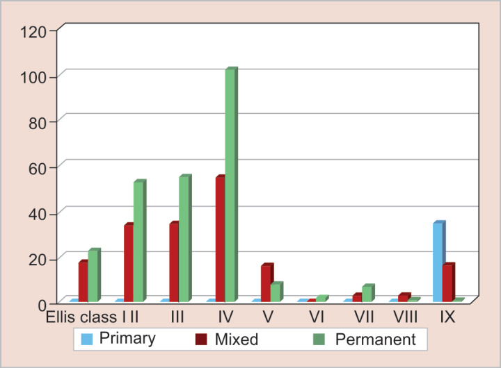
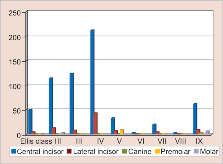
Table 2 and Figure 2 show most of the cases 158 (33.9%) were recorded under class IV group of Ellis and Davey's classification followed by 90 class III (19.3%) and 87 (18.6%) class II and least with class VI fracture (0.42%). A significant p value of 0.0001 was found with the Chi-square test. In the permanent dentition group, 103 cases (40.8%) were seen as Ellis class IV fracture followed by 55 cases of class III fractures (21.8%). Similarly among the mixed dentition group, class IV and class III fractures were found in 55 (30.7%) and 35 (19.5%) cases, respectively. However, all 35 cases in the primary dentition group were categorized under the Ellis class IX group of fracture. In terms of gender distribution, male predominance is clearly seen among all classes of Ellis fracture with a significant p value of 0.01 (Table 3). Overall 620 (64 primary and 556 permanent teeth) central incisors in either jaw emerged as the most frequently traumatized tooth in all classes of Ellis tooth fracture in the study (Fig. 3 and Table 4). The permanent teeth most affected by the trauma were the maxillary central incisors and accounts for (512 teeth) 76.8% of all injured teeth. And the primary teeth mostly affected by trauma were also maxillary central primary teeth (64 teeth) comprising 8.53% of all injured teeth but 75.2% of all traumatized primary teeth. The detailed distribution of dental trauma according to Ellis class of fracture is given in Table 4.
Table 2.
Intergroup distribution of Ellis class of tooth fracture
| Groups | Ellis class I | II | III | IV | V | VI | VII | VIII | IX | Total (n) |
|---|---|---|---|---|---|---|---|---|---|---|
| Primary | – | – | – | – | – | – | – | – | 35 | 35 |
| Mixed | 17 | 34 | 35 | 55 | 16 | – | 3 | 3 | 16 | 179 |
| Permanent | 23 | 53 | 55 | 103 | 8 | 2 | 7 | 1 | – | 252 |
| Total | 40 | 87 | 90 | 158 | 24 | 2 | 10 | 4 | 51 | 466 |
p value 0.0001 statistically significant
n = no. of cases
Fig. 2.
Intergroup comparison of Ellis class of tooth fracture
Table 3.
Gender distribution according to Ellis class of tooth fracture
| Gender | Tooth fracture (Ellis) | Total | ||||||||
|---|---|---|---|---|---|---|---|---|---|---|
| I | II | III | IV | V | VI | VII | VIII | IX | ||
| Male | 21 | 64 | 56 | 116 | 19 | 2 | 10 | 4 | 29 | 321 |
| Female | 19 | 23 | 34 | 43 | 5 | 0 | 0 | 0 | 21 | 145 |
| Total | 40 | 87 | 90 | 159 | 24 | 2 | 10 | 4 | 50 | 466 |
Statistically significant p value 0.01
Fig. 3.
Distribution of teeth according to Ellis class of fracture
Table 4.
Distribution of dental trauma according to Ellis class of fracture
| Ellis class | Central incisor | Lateral incisor | Canine | Premolar | Molar | Total (n) | |||||
|---|---|---|---|---|---|---|---|---|---|---|---|
| Max | Mand | Max | Mand | Max | Mand | Max | Mand | Max | Mand | ||
| I | 47 | 3 | 3 | 2 | – | – | – | – | – | – | 55 |
| II | 110 | 6 | 11 | 2 | – | – | 1 | – | – | 4 | 134 |
| III | 107 | 9 | 7 | 2 | – | – | – | – | 1 | 1 | 127 |
| IV | 194 | 19 | 37 | 7 | – | – | 1 | – | 2 | – | 260 |
| V | 30 | 4 | 2 | 6 | – | – | 10 | – | – | – | 52 |
| VI | 3 | – | – | – | – | – | – | – | – | – | 3 |
| VII | 19 | 2 | 4 | 1 | – | 1 | 1 | 1 | – | – | 29 |
| VIII | 2 | 1 | 1 | 1 | – | – | – | – | – | – | 5 |
| IX Primary | 64 | – | 8 | 2 | 3 | 1 | – | – | 2 | 5 | 85 |
| Total | 576 | 44 | 73 | 23 | 3 | 2 | 13 | 1 | 5 | 10 | 750 |
n = no. of teeth
Associated Injuries
Soft tissue injury accounts for the most commonly associated injury with dental trauma. For 31 (6.6%) soft tissue injuries, a higher proportion was found among the primary dentition group with 11 (31%) out of 35 cases and at least 9 cases (3.5%) among the permanent dentition group out of 252 cases. Two cases of midface fracture, one each in mixed and permanent dentition group were observed while 16 cases of mandible fracture were also noted with 3, 8, and 5 cases in primary, mixed, and permanent dentition groups, respectively.
Management
The majority of the dental injuries (220 cases) were treated with root canal treatment (RCT), showing a statistically significant p value <0.001. The majority being done in 252 teeth from 157 permanent dentition cases. However, the mixed dentition group showed a higher proportion of 18 revascularizations done on 20 teeth and 12 re-implantations on 18 teeth. In the primary dentition, most of the dental traumatic injuries were managed by observation and wound care (48.5%). The detail of group-wise treatment procedures done for traumatic dental injuries is presented in Table 5.
Table 5.
Intergroup distribution of management of dental trauma
| Management | Primary dentition | Mixed dentition | Permanent dentition | Total | ||||
|---|---|---|---|---|---|---|---|---|
| (n) | Teeth | (n) | Teeth | (n) | Teeth | (n) | Teeth | |
| RCT | – | – | 63 | 83 | 157 | 252 | 220 | 335 |
| Pulpectomy | 7 | 8 | – | – | – | – | 7 | 8 |
| Revascularization | – | – | 18 | 20 | 7 | 8 | 25 | 28 |
| Apexification | – | – | 34 | 39 | 60 | 79 | 94 | 118 |
| Reimplantation | – | – | 12 | 18 | 3 | 5 | 15 | 23 |
| Pulpotomy | – | – | 4 | 4 | 1 | 1 | 5 | 5 |
| IPC | – | – | 1 | 1 | 2 | 2 | 3 | 3 |
| DPC | 1 | 1 | 5 | 7 | 4 | 4 | 10 | 12 |
| Restoration | 2 | 2 | 35 | 43 | 40 | 51 | 77 | 96 |
| Extraction | 3 | 3 | 8 | 13 | 1 | 1 | 12 | 17 |
| Wound care/observation | 17 | – | 11 | – | 3 | – | 31 | – |
| Close reduction/splinting | 4 | – | 8 | – | 9 | – | 21 | – |
| No treatment | 6 | – | 31 | – | 49 | – | 86 | – |
n, no. of cases; RCT, root canal treatment; IPC, indirect pulp capping; DPC, direct pulp capping
Chi-square test, p value <0.001* (significant)
The majority of the soft tissue injuries were managed through proper wound care and observation, although suturing was performed in one case of the permanent dentition group. Midface fractures in the mixed and permanent group were managed with open reduction and internal fixation (ORIF) and splinting, respectively, and most of the mandible fractures were managed through close reduction/cap splint and conservative approach, however, ORIF and interpositional arthroplasty were executed in one case each.
Discussion
The present study witnessed a higher frequency of dental trauma in the 12 to 16 years of age group and a considerably lower incidence in children <5 years, which is in agreement with Unal et al.11 and Kargul et al.12 who also noticed the highest frequency of dental trauma in 12 to 14 years of age group. The preponderance of males in all age groups in the present study was in concurrence with the results of prior studies.2,3,9,11,13–16 This may be associated with higher physical activities, involvement in more aggressive sports among male children, and several cultural, customs, and socioeconomic conditions as well. In contrast, younger children with primary dentition have less gender discrepancy owing to inadequate motor coordination, leaving them unaware of various further complications.2
Accidental falls were found to be the most common cause of traumatic dental injuries in children as shown in many studies.2,3,11–18 Additionally, it has been observed that in infants and preschool children, falls in the home are the most common cause of dental trauma while 63.2% of school-aged children having mixed dentition accounted for more dental injuries at school. These findings are credited to poor motor coordination, particularly in children <3 years of age, and as the age increases the motor skills improve, sports-related injuries become more often as well.3 This is in accordance with our study in which fall was the predominant etiological factor for dental trauma in children of all age-groups followed by a sports injury and it was also noted that 92.3% of all sports injury cases were recorded in 12 to 16 years of age group alone (Fig. 1).
Traumatic dental injuries may vary from minimal enamel loss to complex fractures affecting the pulp tissue and even loss of the crown structure. These injuries may damage not only the hard tissues and the pulp of the tooth but also involve the supporting periodontal structures, which lead to an entirely different prognosis.11 Hence, an accurate diagnosis is essential for accurate therapy. It was noticed in the present study that the most common dental traumatic injury overall was Ellis class IV fracture, in which a traumatized tooth becomes non-vital, with or without the loss of the tooth structure10 and maxillary central incisor (both primary and permanent) was the noted as the most frequently injured tooth corresponding to several previous researches done on pediatric dental injuries2–4,11,12,14–17 (Tables 2 and 4, Figs 2 and 3). Previous history of dental trauma, negligence, lack of awareness, and delayed reporting to the hospital could be few reasons for the higher proportion of non-vital/devitalized teeth in the present study. Usually, dental trauma in children and adolescents is associated with injury to perioral soft tissues, results in bleeding and edema. This may lead to more anxiety in the parents and appears to motivate early emergency management.3 Present study reported 6.6% of soft tissue injuries with a dental injury which is on the lower side than the value obtained by the study done by Diaz et al.3 (39%) among children and adolescents aged 1 to 15 years in Chile.
While formulating the treatment planning, considerations must be given to the patient's health and developmental status, and the extent of injuries. It is also important to consider the biological, functional, esthetic, economic aspect and patient's willingness as well for the management of traumatic dental injuries. It has been observed in the present study that RCT was the most common treatment procedure done on permanent teeth while in primary teeth, the majority of the dental trauma were managed by observation and wound care (Table 5). Corresponding with the present study, Unal et al.11 and Kargul et al.12 reported examination and follow-up as the most frequent treatment procedure for primary teeth; additionally, Unal et al.11 noticed 27% direct restoration without any endodontic treatment as the most common treatment given for permanent teeth in their study done on Turkish children.
As all dental traumatic injuries could produce pulp and/or periodontal ligament healing complications, it seems logical to prevent them by timely and appropriate management.3,7,9
Conclusion
Traumatic dental injuries in pediatric patients constitute a critical health issue. In the present study, Ellis class IV fracture was noted as the most common type of dental injury and the permanent dentition group of children was more affected with male predominance. Parents and teachers should be informed about the prevention and immediate management of traumatic dental injuries in children. Additionally, early intervention should be provided to prevent further complications.
Clinical Significance
This study was conducted to evaluate various traumatic dental injuries in the pediatric population. Information and details gained from the present study would help in providing the preventive modalities to parents, caregivers, and teachers regarding these injuries in the future and also facilitate various new researches in this field.
Acknowledgments
We want to acknowledge late Dr Debdutta Das, Former Head of Department of Oral and Maxillofacial Surgery, MMCDSR, Mullana, for his constant support and guidance.
Footnotes
Source of support: Nil
Conflict of interest: None
References
- 1.Gassner R, Tuli T, Hachl O, et al. Craniomaxillofacial trauma in children: a review of 3,385 cases with 6,060 injuries in 10 years. J Oral Maxillofac Surg. 2004;62(4):339–407. doi: 10.1016/j.joms.2003.05.013. [DOI] [PubMed] [Google Scholar]
- 2.Lydia NG, Malandris M, Cheung W. Traumatic dental injuries presenting to a paediatric emergency department in a tertiary children's hospital, Adelaide, Australia. Dent Traumatol. 2020;36(4):360–370. doi: 10.1111/edt.12548. [DOI] [PubMed] [Google Scholar]
- 3.Dıaz JA, Bustos L, Brandt AC, et al. Dental injuries among children and adolescents aged 1–15 years attending to public hospital in Temuco, Chile. Dent Traumatol. 2010;26(3):254–261. doi: 10.1111/j.1600-9657.2010.00878.x. [DOI] [PubMed] [Google Scholar]
- 4.Rocha MJ, Cardoso M. Traumatized permanent teeth in Brazilian children assisted at the Federal University of Santa Catarina, Brazil. Dent Traumatol. 2001;17(6):245–249. doi: 10.1034/j.1600-9657.2001.170601.x. [DOI] [PubMed] [Google Scholar]
- 5.Ashrafullaha,, Pandey RK, Mishra A. The incidence of facial injuries in children in Indian population: a retrospective study. J Oral Biol Craniofac Res. 2018;8(2):82–85. doi: 10.1016/j.jobcr.2017.09.006. [DOI] [PMC free article] [PubMed] [Google Scholar]
- 6.Kumaraswamy S, Madan N, Keerthi R, et al. Pediatric injuries in maxillofacial trauma: a 5 year study. J Maxillofac Oral Surg. 2009;8(2):150–153. doi: 10.1007/s12663-009-0037-4. [DOI] [PMC free article] [PubMed] [Google Scholar]
- 7.Guideline on Management of Acute Dental Trauma. Pediatric sentistry. Reference Manual. 2012/13;34(6):230–238. [Google Scholar]
- 8.Sharma A, Patidar DC, Gandhi G, et al. Mandibular fracture in children: a new approach for management and review of literature. Int J Clin Pediat Dentis. 2019;12(4):357–359. doi: 10.5005/jp-journals-10005-1643. [DOI] [PMC free article] [PubMed] [Google Scholar]
- 9.Casey RP, Bensadigh BM, Lake MT, et al. Dentoalveolar trauma in the pediatric population. J Craniofac Surg. 2010;21(4):1305–1309. doi: 10.1097/SCS.0b013e3181e206c1. [DOI] [PubMed] [Google Scholar]
- 10.Tandon S. 3rd ed., Vol. 2. Hyderabad: Paras Medical Publisher; 2018. Textbook of pedodontics. p. 1005. [Google Scholar]
- 11.Unal M, Oznurhan F, Kapdan A, et al. Traumatic dental injuries in children. Experience of a hospital in the Central Anatolia Region of Turkey. Eur J Paediatr Dent. 2014;15(1):17–22. [PubMed] [Google Scholar]
- 12.Kargul B, Caglar E, Tanboga I. Dental trauma in Turkish children, Istanbul. Dent Traumatol. 2003;19(2):72–75. doi: 10.1034/j.1600-9657.2003.00091.x. [DOI] [PubMed] [Google Scholar]
- 13.Caldas AF, Burgos ME. A retrospective study of traumatic dental injuries in a Brazilian Dental Trauma Clinic. Dent Traumatol. 2001;17(6):250–253. doi: 10.1034/j.1600-9657.2001.170602.x. [DOI] [PubMed] [Google Scholar]
- 14.Marcenes W, Alessi ON, Traebert J. Causes and prevalence of traumatic injuries to the permanent incisors of school children aged 12 years in Jaragua do Sul. Brazil Int Dent J. 2000;50(2):87–92. doi: 10.1002/j.1875-595x.2000.tb00804.x. [DOI] [PubMed] [Google Scholar]
- 15.Marcenes W, Zabot NE, Traebert J. Socio-economic correlates of traumatic injuries to the permanent incisors in school children aged 12 years in Blumenau, Brazil. Endod Dent Traumatol. 2001;17(5):222–226. doi: 10.1034/j.1600-9657.2001.170507.x. [DOI] [PubMed] [Google Scholar]
- 16.Nicolau B, Marcenes W, Sheiham A. Prevalence, causes and correlates of traumatic dental injuries among 13-years-olds in Brazil. Endod Dent Traumatol. 2001;17(5):213–217. doi: 10.1034/j.1600-9657.2001.170505.x. [DOI] [PubMed] [Google Scholar]
- 17.Marcenes W, Beiruti NA, Tayfour D, et al. Epidemiology of traumatic dental injuries to permanent incisors of school children aged 9-12 in Damascus, Syria. Endod Dent Traumatol. 1999;15(3):117–123. doi: 10.1111/j.1600-9657.1999.tb00767.x. [DOI] [PubMed] [Google Scholar]
- 18.Kovacs M, Pacurar M, Petcu B, et al. Prevalence of traumatic dental injuries in children who attended two dental clinics in Targu Mures between 2003 and 2011. Oral Health Dent Manag. 2012;11(3):116–124. [PubMed] [Google Scholar]


