Abstract
目的
探讨青少年特发性脊柱侧弯(adolescent idiopathic scoliosis,AIS)矫形术中机器人辅助植钉的安全性及准确性。
方法
回顾分析 2018 年 6 月—2019 年 12 月采用后路矫形植骨融合内固定术治疗且符合选择标准的 46 例 AIS 患者临床资料。其中,22 例术中采用机器人辅助植钉(机器人组),24 例徒手植钉(徒手组)。两组患者性别、年龄、身体质量指数、Lenke 分型以及术前主弯 Cobb 角、疼痛视觉模拟评分(VAS)、日本骨科协会(JOA)评分等一般资料比较,差异均无统计学意义(P>0.05),具有可比性。记录术中出血量、植钉时间、术中调钉次数以及术后 VAS 评分、JOA 评分,X 线片测量主弯 Cobb 角及计算脊柱矫正率,CT 评估螺钉位置并计算植钉准确率。
结果
两组手术均顺利完成;机器人组术中出血量、植钉时间以及术中调钉次数均少于徒手组(P<0.05)。术后机器人组 1 例切口愈合不良,徒手组 2 例轻度神经根损伤、2 例切口愈合不良,两组并发症发生率比较差异无统计学意义(P=0.667)。两组患者术后均获随访,随访时间 3~9 个月,平均 6.4 个月。两组末次随访时 VAS 评分、JOA 评分均优于术前(P<0.05);VAS 评分、JOA 评分手术前后差值组间比较,差异均无统计学意义(P>0.05)。影像学复查示,术中机器人组植入 343 枚螺钉、徒手组 374 枚螺钉,两组植钉位置分级及植钉准确率(89.5%vs 79.1%)比较,差异均有统计学意义(Z=−3.964,P=0.000;χ2=14.361,P=0.000)。两组末次随访时主弯 Cobb 角均较术前减小,差异有统计学意义(P<0.05);手术前后差值组间比较差异有统计学意义(t=0.999,P=0.323)。机器人组脊柱矫正率为 79.82%±5.33%,徒手组为 79.62%±5.58%,差异无统计学意义(t=0.120,P=0.905)。
结论
与徒手植钉相比,AIS 矫形术中采用机器人辅助植钉安全、微创,而且准确性更高。
Keywords: 青少年特发性脊柱侧弯, 机器人, 椎弓根螺钉, 脊柱矫形
Abstract
Objective
To investigate the safety and accuracy of robot-assisted pedicle screw implantation in the adolescent idiopathic scoliosis (AIS) surgery.
Methods
The clinical data of 46 patients with AIS who were treated with orthopedics, bone graft fusion, and internal fixation via posterior approach between June 2018 and December 2019 were analyzed retrospectively. Among them, 22 cases were treated with robot-assisted pedicle screw implantation (robot group) and 24 cases with manual pedicle screw implantation without robot assistance (control group). There was no significant difference in gender, age, body mass index, Lenke classification, and preoperative Cobb angle of the main curve, pain visual analogue scale (VAS) score, Japanese Orthopaedic Association (JOA) score between the two groups (P>0.05). The intraoperative blood loss, pedicle screw implantation time, intraoperative pedicle screw adjustment times, and VAS and JOA scores after operation were recorded. The Cobb angle of the main curve was measured on X-ray film and the spinal correction rate was calculated. The screw position and the accuracy of screw implantation were evaluated on CT images.
Results
The operation completed successfully in the two groups. The intraoperative blood loss, pedicle screw implantation time, and pedicle screw adjustment times in the robot group were significantly less than those in the control group (P<0.05). There was 1 case of poor wound healing in the robot group and 2 cases of mild nerve root injury and 2 cases of poor incision healing in the control group, and there was no significant difference in the incidence of complications between the two groups (P=0.667). All patients in the two groups were followed up 3-9 months (mean, 6.4 months). The VAS and JOA scores at last follow-up in the two groups were superior to those before operation (P<0.05), but there was no significant difference in the difference of pre- and post-operative scores between the two groups (P>0.05). The imaging review showed that 343 screws were implanted in the robot group and 374 screws in the control group. There were significant differences in pedicle screw implantation classification and accuracy between the two groups (89.5%vs 79.1%)(Z=−3.964,P=0.000; χ2=14.361, P=0.000). At last follow-up, the Cobb angles of the main curve in the two groups were significantly lower than those before operation (P<0.05), and there was significant difference in the difference of pre- and post-operative Cobb angles between the two groups (t=0.999, P=0.323). The spinal correction rateswere 79.82%±5.33% in the robot group and 79.62%±5.58% in the control group, showing no significant difference (t=0.120, P=0.905).
Conclusion
Compared with manual pedicle screw implantation, robot-assisted pedicle screw implantation in AIS surgery is safer, less invasive, and more accurate.
Keywords: Adolescent idiopathic scoliosis, robot, pedicle screw, spinal orthopedics
青少年特发性脊柱侧弯(adolescent idiopathic scoliosis,AIS)是以脊柱某一个或多个节段向侧方弯曲并伴椎体旋转的复杂三维畸形,发病率为 1.05%~4%,随着青少年生长发育,畸形会进行性加重[1-4]。AIS 不仅影响患者体型、心理健康,严重时可引起胸腰背部疼痛、心肺功能障碍以及压迫神经。早期可行支具治疗,对于支具治疗无效且侧弯呈进行性发展或 Cobb 角>40° 者需手术矫正[5-7]。
近年来,临床 AIS 矫形常采用经后路椎弓根钉棒系统内固定术,能获得较好疗效[8-9]。但此类患者通常胸椎椎弓根细小、发育异常,同时伴椎体旋转,术中徒手准确植钉有一定困难[9]。随着计算机导航技术的发展,骨科机器人系统逐渐用于脊柱手术辅助植钉,提高了植钉准确性[10]。现回顾分析后路矫形植骨融合内固定术治疗的 AIS 患者临床资料,比较徒手与机器人辅助植钉的术中出血量、手术时间、植钉准确度及手术相关并发症发生情况,以期进一步明确 AIS 矫形术中机器人辅助植钉是否存在优势。报告如下。
1. 临床资料
1.1. 患者选择标准
纳入标准:① 患者年龄 10~18 岁,其中女性患者需已初潮;② AIS 患者,脊柱全长冠状位 X 线片测量主弯 Cobb 角>40°~50°;③ 经支具治疗无效且侧弯呈进行性发展;④ 行后路矫形植骨融合内固定术,且手术由参与机器人操作 10 次以上的高年资骨科医师完成。
排除标准:① 伴半椎体;② 合并严重心肺功能、凝血功能障碍;③ 合并严重脊髓、神经损伤;④ 合并严重内科疾病,无法耐受手术者。
2018 年 6 月—2019 年 12 月,共 46 例患者符合选择标准纳入研究。其中 22 例术中采用机器人辅助植钉(机器人组),24 例采用徒手植钉(徒手组)。
1.2. 一般资料
机器人组:男 4 例,女 18 例;年龄 11~18 岁,平均 13.9 岁。身体质量指数(18.92±1.01)kg/m2。Lenke 分型:1AN 型 7 例,1A+型 2 例,3CN 型2 例,1BN 型 2 例,4C+型 3 例,5CN 型 6 例。
徒手组:男 5 例,女 19 例;年龄 11~18 岁,平均 14.1 岁。身体质量指数(19.79±1.95)kg/m2。Lenke 分型:1AN 型 9 例,1A+型 2 例,3CN 型 3 例,1BN 型 2 例,5CN 型 6 例,2A−型 2 例。
两组患者性别、年龄、身体质量指数、Lenke 分型以及术前主弯 Cobb 角、疼痛视觉模拟评分(VAS)、日本骨科协会(JOA)评分等一般资料比较,差异均无统计学意义(P>0.05),具有可比性。见表 1、2。
表 1.
Comparison of VAS and JOA scores before and after operation in the two groups (
 )
)
两组手术前后 VAS 及 JOA 评分比较(
 )
)
| 组别
Group |
例数
n |
VAS | JOA | |||||||
| 术前
Preoperative |
末次随访
Last follow-up |
差值
Difference |
统计值
Statistic |
术前
Preoperative |
末次随访
Last follow-up |
差值
Difference |
统计值
Statistic |
|||
| 机器人组
Robot group |
22 | 1.8±0.6 | 1.2±0.4 | 0.6±0.5 |
t=6.062
P=0.000 |
20.4±1.7 | 22.6±1.5 | 45.4±7.5 |
t=−4.136
P=0.000 |
|
| 徒手组
Control group |
24 | 2.1±0.8 | 1.3±0.4 | 0.8±0.8 |
t=5.362
P=0.000 |
21.3±2.0 | 22.6±1.5 | 43.2±7.1 |
t=−4.337
P=0.000 |
|
| 统计值
Statistic |
t=−1.043
P=0.303 |
− |
t=−1.261
P=0.214 |
t=−1.731
P=0.090 |
− |
t=0.999
P=0.323 |
||||
表 2.
Comparison of Cobb angle of main curve before and after operation in the two groups (
 , °)
, °)
两组手术前后主弯 Cobb 角比较(
 ,°)
,°)
| 组别
Group |
例数
n |
术前
Preoperative |
末次随访
Last follow-up |
差值
Difference |
统计值
Statistic |
| 机器人组
Robot group |
22 | 56.64±7.27 | 11.27±2.59 | 45.36±7.51 |
t=28.324
P=0.000 |
| 徒手组
Control group |
24 | 54.00±6.39 | 10.79±2.32 | 43.21±7.11 |
t=29.756
P=0.000 |
| 统计值
Statistic |
t=1.309
P=0.197 |
− |
t=0.999
P=0.323 |
1.3. 手术方法
1.3.1. 机器人组
本组采用“天玑”骨科机器人(北京天智航医疗科技股份有限公司)辅助手术。全麻下,患者取俯卧位。C 臂 X 线机摄脊柱正侧位片,确定远、近端固定椎后,沿固定节段相应水平脊柱后正中线切开皮肤,逐层切开皮下、腰背筋膜,骨膜下剥离椎旁肌,显露固定节段椎体棘突、椎板及关节突关节,在棘突上安装示踪器。
采用 C 臂 X 线机依次扫描需固定节段(每次扫描不超过 4 个椎体);将扫描数据传送至机器人操作工作站,在机器人操作台对每个节段逐一辨识、标记、分割,调整椎弓根扫描层面,设定螺钉进钉点、螺钉尺寸及植入方向,从三维图像的冠状面、矢状面、横断面仔细检查螺钉位置,确定无误后将机械臂末端标定尺替换成导向器,运行机器人。
机械臂被指示移动至选定轨迹相应位置后,使用电钻在导向器辅助下依次植入导针,然后在导针引导下攻丝并拧入螺钉。每植入 4 个椎体的螺钉需行正侧位透视,检查螺钉位置;如螺钉位置明显不当,需在机器人操作台调整规划后重新植入。确定螺钉位置无误后,选取适当长度连接棒,预弯后通过旋棒技术矫正脊柱畸形。透视下明确畸形矫正满意、内固定物位置良好后,依次拧紧螺帽,安装横连。处理植骨床,将切取的棘突、椎板骨质修剪成颗粒状,混合人工骨(山西奥瑞生物材料有限公司)后,行固定节段植骨融合。彻底止血并观察无活动性出血,反复冲洗切口,术区置引流管 1 根,逐层缝合关闭切口,无菌敷料包扎。见图 1。
图 1.
Schematic diagram of robot-assisted pedicle screw implantation
机器人辅助植钉示意图
a. 安装示踪器及合适的导向器;b. 在机器人操作台进行规划;c. 在机器人导向器引导下植入导针;d. 在导针引导下对钉道进行攻丝;e. 在导针引导下植入螺钉
a. Installing tracer and suitable guide; b. Planning on the robot operating platform; c. Implanting the guide needle under the guidance of the robot guide; d. Expanding of the pedicle screw path under the guidance of the guide needle; e. Implanting screws under the guidance of the guide needle
1.3.2. 徒手组
根据影像学检查结果确定截骨节段、螺钉进钉点以及螺钉尺寸。全麻下,患者取俯卧位,C 臂 X 线机摄脊柱正侧位片,确定远、近端固定椎,沿固定节段相应水平脊柱后中线切开皮肤,逐层切开皮下、腰背筋膜,骨膜下剥离椎旁肌,显露固定节段椎体棘突、椎板及关节突关节,通过大致解剖位置判断进钉点,依次植入螺钉。透视下明确螺钉位置无误后拧紧螺帽,安装横连。植骨及切口处理同机器人组。
1.4. 术后处理
两组患者术后处理方法一致。常规给予预防感染、多模式镇痛、消肿以及补充血容量、电解质、营养支持等治疗。待引流量约为 50 mL/d 时拔除引流管,嘱患者开始佩戴支具下地活动。术后定期门诊复查。
1.5. 疗效评价指标
记录术中出血量、植钉时间、术中调钉次数以及末次随访时 VAS 评分、JOA 评分。其中,植钉时间=每例患者全部螺钉植入所需时间/该例患者总螺钉数,全部螺钉植入所需时间为开始作切口至全部螺钉植入完成的时间。
手术前后摄脊柱全长正侧位以及凸侧侧屈位 X 线片,测量主弯 Cobb 角,按以下公式计算脊柱矫正率,(术前主弯 Cobb 角–术后主弯 Cobb 角)/术前主弯 Cobb 角×100%。Cobb 角由 2 名脊柱外科医生测量,取均值。
拔除引流管后对固定节段行 CT 扫描,层厚 1.25 mm,在 PACS 系统上通过 PacsClient 软件按照 Gertzbein 等[11]的分级法评估每枚螺钉位置,共分为 5 级。A 级,椎弓根骨皮质完整;B~E级,螺钉穿透椎弓根骨皮质,穿透厚度分别<2 mm、2~4 mm、4~6 mm、≥6 mm。其中,A、B 级为植钉准确,C、D、E 级为植钉错误,计算植钉准确率。
1.6. 统计学方法
采用 SPSS23.0 统计软件进行分析。计量资料均符合正态分布,以均数±标准差表示,组间比较采用独立样本 t 检验,组内比较采用配对 t 检验;计数资料以率表示,组间比较采用 χ2 检验或 Fisher 确切概率法;等级资料组间比较采用秩和检验;检验水准 α=0.05。
2. 结果
两组手术均顺利完成;机器人组术中出血量、植钉时间以及术中调钉次数均少于徒手组,差异有统计学意义(P<0.05)。见表 3。机器人组 1 例切口愈合不良,徒手组 2 例轻度神经根损伤、2 例切口愈合不良,两组并发症发生率分别为4.5%及16.7%,组间比较差异无统计学意义(P=0.667)。对于切口愈合不良患者,经积极清洁换药、补充营养后,切口愈合;对于神经根损伤患者,给予脱水、营养神经等治疗,2 个月后残留神经症状消失。
表 3.
Comparison of intraoperative blood loss, pedicle screw implantation time, and intraoperative pedicle screw adjustment times between the two groups (
 )
)
两组术中出血量、植钉时间及调钉次数比较(
 )
)
| 组别
Group |
例数
n |
植钉时间(min)
Pedicle screw implantation time (minutes) |
术中出血量(mL)
Intraoperative blood loss (mL) |
术中调钉次数(次)
Intraoperative pedicle screw adjustment times (times) |
| 机器人组
Robot group |
22 | 2.36±0.58 | 625.68±72.51 | 4.13±1.36 |
| 徒手组
Control group |
24 | 3.75±1.03 | 676.25±93.15 | 5.92±1.06 |
| 统计值
Statistic |
t=−2.041
P=0.047 |
t=−5.672
P=0.000 |
t=−4.985
P=0.000 |
两组患者术后均获随访,随访时间 3~9 个月,平均 6.4 个月。两组末次随访时 VAS 评分、JOA 评分均优于术前,差异有统计学意义(P<0.05);VAS 评分、JOA 评分手术前后差值组间比较,差异均无统计学意义(P>0.05)。见表 1。
术中机器人组共植入 343 枚螺钉,螺钉位置分级达 A 级 259 枚、B 级 48 枚、C 级 19 枚、D 级 10 枚、E 级 7 枚,植钉准确率为 89.5%;徒手组植入 374 枚螺钉,其中 A 级 235 枚、B 级 61 枚、C 级 37 枚、D 级 24 枚、E 级 17 枚,植钉准确率为 79.1%。两组植钉位置分级及植钉准确率比较,差异均有统计学意义(Z=−3.964,P=0.000;χ2=14.361,P=0.000)。
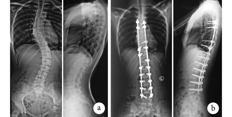
影像学复查显示两组患者脊柱侧弯畸形均获得良好矫形,末次随访时主弯 Cobb 角均较术前减小,差异有统计学意义(P<0.05);手术前后差值组间比较差异无统计学意义(t=0.999,P=0.323)。见表2。机器人组脊柱矫正率为 79.82%±5.33%,徒手组为 79.62%±5.58%,差异无统计学意义(t=0.120,P=0.905)。见图 2、3。
图 2.
Spinal full-length anteroposterior and lateral X-ray films of a 15-year-old female patient with AIS (Lenke classification of type 1AN) in the robot group
机器人组患者,女,15 岁,AIS(Lenke 1AN 型)脊柱全长正侧位 X 线片
a. 术前;b. 术后 3 个月
a. Before operation; b. At 3 months after operation
图 3.
Spinal full-length anteroposterior and lateral X-ray films of a 16-year-old male patient with AIS (Lenke classification of type 5CN) in the control group
徒手组患者,男,16 岁,AIS(Lenke 5CN 型)脊柱全长正侧位 X 线片
a. 术前;b. 术后 3 个月
a. Before operation; b. At 3 months after operation
3. 讨论
3.1. 徒手及机器人辅助植钉疗效比较
AIS 矫形术中成功、安全植入椎弓根螺钉是一项具有挑战性的技术,徒手植钉难度较大,尤其伴侧弯、椎体旋转、椎弓根过细、脊髓偏移时,极易发生螺钉误植[12]。研究报道 AIS 术中徒手植钉引起的骨皮质穿破率为 1.25%~14%,AIS 翻修患者中 20% 存在螺钉穿破骨皮质[11, 13-17]。Kwan 等[18]研究发现脊柱侧弯手术中,腰椎椎弓根螺钉误植率达 5%~41%、胸椎达 3%~51%。植钉错误可引起神经、脊髓及主动脉损伤,是 AIS 翻修的主要原因之一。本研究徒手组术后出现 2 例轻度神经根损伤,患者单侧下肢放射性疼痛,CT 复查见相应节段螺钉突破椎弓根骨皮质,经营养神经、脱水治疗后症状消失。
随着数字骨科技术不断发展,骨科机器人也逐渐应用于 AIS 矫形手术中。翟功伟等[19]的对比研究显示机器人辅助 AIS 手术可提高椎弓根植钉准确性、降低辐射量、减少术中出血,缩短患者术后住院时间,有利于患者恢复。Devito 等[20]在 80 例 AIS 手术中采用机器人辅助植钉,其中 A 级植钉率达 95%,术后无植钉相关并发症发生。
“天玑”骨科机器人系统是我国第一台具有完全自主知识产权的骨科手术机器人,可辅助脊柱及创伤骨科内固定定位,具有微创、精准、操作安全便捷等优点。本研究机器人组即在该机器人辅助下顺利植钉,术后影像学评价显示植钉分级及准确率均明显高于徒手组。一项研究结果显示与传统植钉技术相比,计算机导航辅助植钉可以明显缩短植钉时间[21]。本研究机器人组螺钉植入时间也较徒手组明显缩短,术中调钉次数也减少,差异有统计学意义;分析原因为机器人组首先在操作台上精准地模拟螺钉轨迹及其长度、直径,降低了术中植钉失误可能性以及因螺钉尺寸不合适造成的二次植钉。此外,在调钉过程中存在的钉道出血不可忽视,由于机器人组调整植钉次数减少,故术中出血量也明显少于徒手组。
3.2. 机器人辅助植钉优缺点
优点:① 机器人可将脊柱图像三维立体化输出完成术前规划,并且机械臂稳定性强,操作简单及可重复性好,可以消除因医师技术水平差异造成的手术效果差异;② 提前规划螺钉直径、长度及植钉角度,使植钉更精准、快捷,减少了因植钉失误造成的血管、神经损伤,从而降低手术相关并发症发生率;③ 提高了一次性植钉成功率,缩短植钉时间,避免了改道过程中钉道出血,有效减少术中出血。
缺点:① 术前螺钉位置规划需术者在机器人软件中完成,对术者机器人操作经验要求高;② 机器人成本及设备相关维护费用较高;③ 缺乏实时跟踪系统,患者体位发生微动就会影响植钉准确性,尤其是 AIS 患者椎旁肌肉松解后部分椎体可能发生移动,与提前规划的螺钉位置有一定偏差,此时需重新调整规划;③ AIS 患者脊柱畸形复杂多样,无论是冠状面、矢状面、横断面都存在一定旋转畸形且固定节段长,因此术中需采用机器人三维模式采集固定脊柱节段三维影像数据至少 2 次及以上,增加了辐射暴露。
3.3. 机器人辅助植钉注意事项
① 机械臂位置锁定后,C 臂 X 线机摄片时注意避免碰撞机械臂;② 为了降低机器人辅助植钉错误率,每次 CT 椎体数据采集不应超过 4 个节段;③ 定位套筒紧贴骨面,避免肌肉牵拉导致套筒滑移影响导针植入准确性;④ 对于严重旋转椎体,在机器人辅助植入导针后应摄正侧位 X 线片,以及时验正螺钉位置;⑤ 患者体位始终保持不变,避免因体位改变导致与螺钉规划位置产生偏差,影响植钉准确度。
综上述,AIS 患者后路矫形术中机器人辅助植钉不仅更精准、微创,而且安全、节约手术时间。但本研究病例数有限,随访时间较短,结论需要大样本前瞻性随机对照研究进一步验证。
作者贡献:陈豪杰、董亮负责研究设计及实施、数据收集整理及统计分析、论文撰写;刘团江负责研究实施,论文修改;朱贤友负责文章审核。
利益冲突:所有作者声明,在课题研究和文章撰写过程中不存在利益冲突。
机构伦理问题:研究方案经西安交通大学附属红会医院伦理委员会批准(202103060)。
References
- 1.赵树山. 特发性脊柱侧凸流行病学、遗传易感性及治疗的系统评价. 长沙: 中南大学, 2014.
- 2.邱贵兴, 庄乾宇 青少年特发性脊柱侧弯的流行病学研究进展. 中华医学杂志. 2006;86(11):790–792. doi: 10.3760/j:issn:0376-2491.2006.11.019. [DOI] [Google Scholar]
- 3.Cheng JC, Castelein RM, Chu WC, et al. Adolescent idiopathic scoliosis. Nat Rev Dis Primers, 2015, 24: 15030. doi: 10.1136/bmj.2.6184.274-c.
- 4.丛琳, 朱海涛, 崔璀, 等 机器人辅助皮下经肌间置钉技术微创治疗青少年特发性脊柱侧凸病例报告. 中华骨科杂志. 2018;38(22):1392–1394. doi: 10.3760/cma.j.issn.0253-2352.2018.22.006. [DOI] [Google Scholar]
- 5.买硕, 曾岩, 袁磊, 等 青少年特发性脊柱侧凸支具治疗效果影响因素的研究现状. 中国矫形外科杂志. 2020;28(11):942–945. [Google Scholar]
- 6.郭立民, 舒钧, 劳汉昌, 等 青少年特发性脊柱侧凸支具治疗研究分析. 中国矫形外科杂志. 2015;23(11):967–971. [Google Scholar]
- 7.龚磊, 方亮, 叶晓凌, 等 青少年特发性脊柱侧凸的治疗进展. 中国骨伤. 2020;33(2):184–189. [Google Scholar]
- 8.Ayvaz M, Olgun ZD, Demirkiran HG, et al Posterior all-pedicle screw instrumentation combined with multiple chevron and concave rib osteotomies in the treatment of adolescent congenital kyphoscoliosis. Spine J. 2014;14(1):11–19. doi: 10.1016/j.spinee.2012.10.016. [DOI] [PubMed] [Google Scholar]
- 9.Pan Y, Lü GH, Kuang L, et al Accuracy of thoracic pedicle screw placement in adolescent patients with severe spinal deformities: a retrospective study comparing drill guide template with free-hand technique. Eur Spine J. 2018;27(2):319–326. doi: 10.1007/s00586-017-5410-2. [DOI] [PubMed] [Google Scholar]
- 10.Gao S, Lv Z, Fang H Robot-assisted and conventional freehand pedicle screw placement: a systematic review and meta-analysis of randomized controlled trials. Eur Spine J. 2018;27(4):921–930. doi: 10.1007/s00586-017-5333-y. [DOI] [PubMed] [Google Scholar]
- 11.Gertzbein SD, Robbins SE Accuracy of pedicular screw placement in vivo . Spine (Phila Pa 1976) 1990;15(1):11–14. doi: 10.1097/00007632-199001000-00004. [DOI] [PubMed] [Google Scholar]
- 12.Sato T, Yonezawa I, Akimoto T, et al. Novel hump measurement system with a 3D camera for early diagnosis of patients with adolescent idiopathic scoliosis: A study of accuracy and reliability. Cureus, 2020, 12(5): e8229. doi: 10.7759/cureus.8229.
- 13.Samdani AF, Ranade A, Sciubba DM, et al Accuracy of free-hand placement of thoracic pedicle screws in adolescent idiopathic scoliosis: how much of a difference does surgeon experience make? Eur Spine J. 2010;19(1):91–95. doi: 10.1007/s00586-009-1183-6. [DOI] [PMC free article] [PubMed] [Google Scholar]
- 14.Sarlak AY, Tosun B, Atmaca H, et al Evaluation of thoracic pedicle screw placement in adolescent idiopathic scoliosis. Eur Spine J. 2009;18(12):1892–1897. doi: 10.1007/s00586-009-1065-y. [DOI] [PMC free article] [PubMed] [Google Scholar]
- 15.Dede O, Ward WT, Bosch P, et al Using the freehand pedicle screw placement technique in adolescent idiopathic scoliosis surgery: what is the incidence of neurological symptoms secondary to misplaced screws? Spine (Phila Pa 1976) 2014;39(4):286–290. doi: 10.1097/BRS.0000000000000127. [DOI] [PubMed] [Google Scholar]
- 16.Abul-Kasim K, Ohlin A The rate of screw misplacement in segmental pedicle screw fixation in adolescent idiopathic scoliosis. Acta Orthop. 2011;82(1):50–55. doi: 10.3109/17453674.2010.548032. [DOI] [PMC free article] [PubMed] [Google Scholar]
- 17.Ahmed SI, Bastrom TP, Yaszay B, et al 5-year reoperation risk and causes for revision after idiopathic scoliosis surgery. Spine (Phila Pa 1976) 2017;42(13):999–1005. doi: 10.1097/BRS.0000000000001968. [DOI] [PubMed] [Google Scholar]
- 18.Kwan MK, Chiu CK, Gani SMA, et al Accuracy and safety of pedicle screw placement in adolescent idiopathic scoliosis patients: a review of 2020 screws using computed tomography assessment. Spine (Phila Pa 1976) 2017;42(5):326–335. doi: 10.1097/BRS.0000000000001738. [DOI] [PubMed] [Google Scholar]
- 19.翟功伟, 高延征, 高坤, 等 脊柱机器人辅助与传统后路椎弓根螺钉内固定矫形术治疗脊柱侧凸的效果比较. 中华实用诊断与治疗杂志. 2019;33(7):636–640. [Google Scholar]
- 20.Devito DP, Kaplan L, Dietl R, et al Clinical acceptance and accuracy assessment of spinal implants guided with SpineAssist surgical robot: retrospective study. Spine (Phila Pa 1976) 2010;35(24):2109–2115. doi: 10.1097/BRS.0b013e3181d323ab. [DOI] [PubMed] [Google Scholar]
- 21.Vissarionov S, Schroeder JE, Novikov SN, et al The utility of 3-dimensional-navigation in the surgical treatment of children with idiopathic scoliosis. Spine Deform. 2014;2(4):270–275. doi: 10.1016/j.jspd.2014.03.004. [DOI] [PubMed] [Google Scholar]


