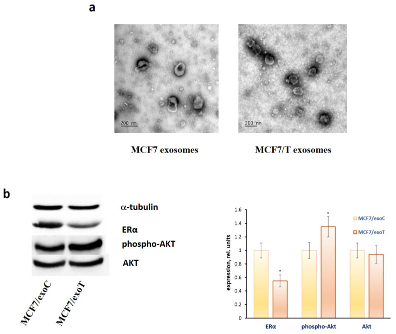
Figure 5.
Exosome influence on the ERα and Akt level in MCF7 cells. (a) The transmission electron microscopy of the exosomes. Exosomes were collected from the MCF7 and MCF7/T conditioned medium by the differential ultracentrifugation, labelled by the gold nanoparticles and imaged as described in Methods. (b) ERα and Akt expression. MCF7 cells were treated with exosomes from MCF7 and MCF7/T for 1 month and Western blot analysis of ERα and Akt was performed in the cell extracts. Protein loading was controlled by membrane hybridization with α-tubulin antibodies. Densitometry for immunoblotting data (right diagram) was carried out using ImageJ software; * p < 0.05 versus scrambled (scr).

