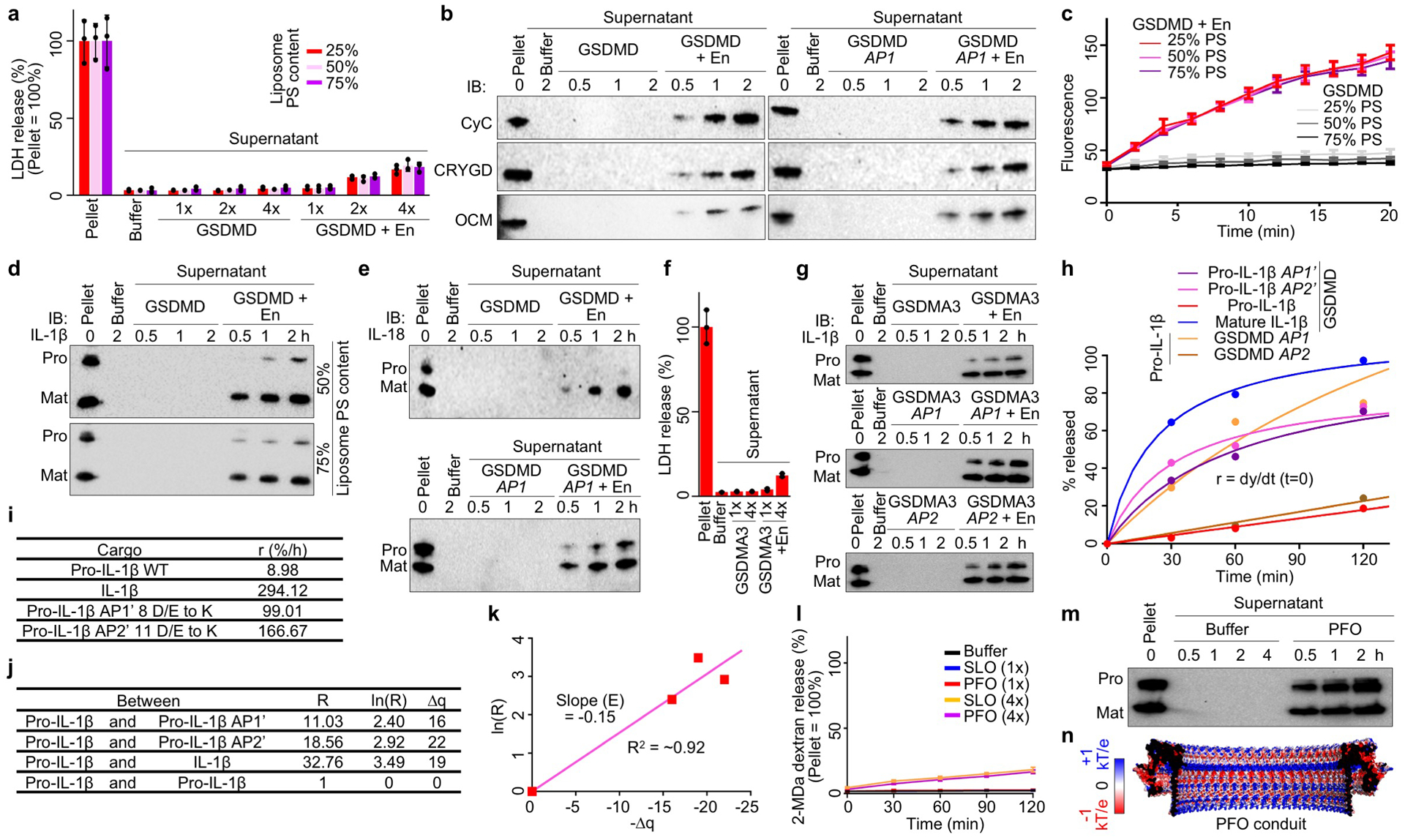
Extended Data Fig. 8 |. Liposome experiments and electrostatics analysis.

a, Unlysed liposomes (25–75% PS) evident in minimal LDH release when GSDMD was added at a sub-lytic concentration (1x = 0.5 μM) (n = 3 biological replicates). En: Activating enzyme. b, Release of CyC, CRYGD, and OCM from liposomes permeabilized by GSDMD shown by immunoblotting. c, Similar rates of GSDMD pore formation on liposomes of various acidic lipid contents (25–75% PS), according to Tb3+ release (n = 3 biological replicates). d, Preferential IL-1β release from liposomes (50% and 75% PS) perforated by GSDMD shown by immunoblotting. e, Release of pro-IL-18 and IL-18 from liposomes permeabilized by GSDMD shown by immunoblotting. f, Unlysed liposomes evident in minimal LDH release when GSDMA3 was activated at a sub-lytic concentration (1x = 0.5 μM) (n = 3 biological replicates). g, Release of pro- and mature IL-1β from liposomes perforated by GSDMA3 shown by immunoblotting. h, Release rates of IL-1β cargoes through GSDMD pores shown by fitted hyperbolic functions. i, Initial release rates (lowercase r) extrapolated from h. j, Charge differences among the cargoes (Δq) and rate ratios (uppercase R). k, Plot of ln(R) versus Δq for estimating the electrostatic potential (E) of the GSDMD pore conduit. l, Unlysed liposomes shown by lack of release of encapsulated bulky FITC-labelled dextrans (2 MDa) when SLO or PFO was added at a sub-lytic concentration (1x = 0.1 μM) (n = 3 biological replicates). m, Similar release of pro- and mature IL-1β from liposomes permeabilized by PFO. n, Electrostatics surfaces of the modelled PFO pore conduit. Data shown in a, c, f, and l are mean ± s.d.. Data shown in b are representative of three, and data shown in d, e, g, and m two, independent experiments.
