Abstract
Background and study aims Feasibility of EUS-guided choledochoduodenostomy (EUS-CDS) using available lumen-apposing stents (LAMS) is limited by the size of the common bile duct (CBD) (≤ 12 mm, cut-off for experts; 15 mm, cut-off for non-experts). We aimed to assess the prevalence and predictive factors associated with CBD size ≥ 12 and 15 mm in naïve patients with malignant distal biliary obstruction (MDBO).
Patients and methods This was a prospective cohort study involving 22 centers with assessment of CBD diameter and subjective feasibility of the EUS-CDS performance in naïve jaundiced patients undergoing EUS evaluation for MDBO.
Results A total of 491 patients (mean age 69 ± 12 years) with mean serum bilirubin of 12.7 ± 6.6 mg/dL entered the final analysis. Dilation of the CBD ≥ 12 and 15 mm was detected in 78.8 % and 51.9 % of cases, respectively. Subjective feasibility of EUS-CDS was expressed by endosonographers in 91.2 % for a CBD ≥ 12 mm and in 96.5 % for a CBD ≥ 15 mm. On multivariate analysis, age ( P < 0.01) and bilirubin level ( P ≤ 0.001) were the only factors associated with both CBD dilation ≥ 12 and ≥ 15 mm. These variables were poorly associated with the extent of duct dilation; however, based on them a prediction model could be constructed that satisfactorily predicted CBD size ≥ 12 mm in patients at least 70 years and a bilirubin level ≥ 7 mg/dL.
Conclusions Our study showed that at presentation in a large cohort of patients with MDBO, EUS-CDS can be potentially performed in three quarters to half of cases by expert and less experienced endosonographers, respectively. Dedicated stents or devices with different designs able to overcome the limitations of existing electrocautery-enhanced LAMS for EUS-CDS are needed.
Introduction
Endoscopic ultrasound-guided biliary drainage (EUS-BD) is increasingly employed after endoscopic retrograde cholangiopancreatography (ERCP) failure in patients with malignant bile duct obstruction 1 2 . Biliary drainage can be accomplished with a transgastric approach by performing a hepatico-gastrostomy (HGS) to the left intrahepatic biliary system, an antegrade stenting or rendezvous procedure if the guidewire can be negotiated through the papilla, or a transduodenal approach to the extrahepatic biliary system carrying out a choledochoduodenostomy (CDS).
As recently reported in a systematic review with meta-analysis, including nine studies with 483 patients, EUS-BD is associated with better clinical success, fewer adverse events (AEs), lower need for reintervention, and lower costs as compared to percutaneous biliary drainage 3 . More recently, three randomized controlled trials (RCTs) directly compared EUS-BD with ERCP as primary drainage for malignant distal biliary obstruction 4 5 6 . The first two studies were performed using a multistep procedure with needle puncture, guidewire placement, tract dilation and, finally, placement of a partially 4 or a fully covered 5 biliary self-expandable metal stent (SEMS). Both studies showed no differences between the two groups, but their conclusions were limited by the small sample size 7 .
In the third study 6 , 61 and 64 patients underwent ERCP and EUS-BD, respectively. In the latter group after puncture and guidewire placement in the biliary system, a dedicated device for both CDS (32 patients) and HGS (32 patients) was used. No difference in technical and clinical success rates was found. However, AEs (19.7 % vs. 6.3 %, P = 0.03), 6-month stent patency (48.9 % vs. 85.1 %, P = 0.001) and re-intervention rates (42.6 % vs. 15.6 %, P = 0.001) were all significantly in favor for the EUS-BD approach, after a mean follow-up of 165 days 6 .
One available dedicated system for a single-step EUS-CDS procedure, avoiding the need for needle puncture and guidewire placement, is the AXIOS stent and electrocautery-enhanced delivery system in which a lumen apposing SEMS is mounted on an electrocautery-enhanced tip catheter (LA-SEMS). Because the delivery catheter acts as a cystotome, the system allows direct stent insertion into the common bile duct (CBD) lumen, where it is then released under complete EUS guidance or with partial endoscopic control 8 9 .
Different studies using the Axios stent reported EUS-CDS to be highly technically and clinically effective 9 10 11 12 13 14 15 16 17 18 19 20 . The smallest sizes of the Axios stent (saddle diameter and length of 6 mm × 8 mm or 8 mm × 8 mm) are preferentially used for this procedure. Because of the risk of bile duct wall injury and misdeployment of the stent during release of the first flange, it is recommended that they be used in CBD diameters larger than 12 mm in expert hands and 15 mm in non-expert ones. This would theoretically limit the number of patients in whom the procedure can be safely performed when they first present with jaundice. However, up to now, no data on the prevalence rate of patients with a CBD diameter sufficient to accomplish an EUS-CDS at the presumed site using these stents are available.
To fill this gap, we conducted a prospective, multicenter cohort study in naïve patients with jaundice undergoing EUS to evaluate malignant distal biliary obstruction to assess the prevalence and predictive factors associated with a CBD ≥ 12 and ≥ 15 mm at the presumed site where EUS-CDS would be performed.
Patients and methods
This was an international, prospective, multicenter, observational study for which consecutive jaundiced patients who underwent EUS to evaluate suspected malignant distal biliary obstruction were enrolled between September 2018 and December 2019. Table 1 shows the study selection criteria. Only patients with a confirmed diagnosis of malignancy were included in the final analysis.
Table 1. Inclusion and exclusion criteria for the patients in the study.
| Inclusion criteria |
|
|
|
|
|
| Exclusion criteria |
|
|
|
|
|
|
|
EUS, endoscopic ultrasound.
We estimated that about 20 centers would be needed, each including about 20 patients, to reach the sample size of 400 patients. Some of these centers, however, dropped out during the study period; therefore, a few additional ones were recruited, reaching a total of 22 enrolling centers.
Ethics committee approval was obtained in each study center and informed consent was obtained from all patients before the procedure. This trial was registered online at Clinicaltrials.gov (NCT03494023) prior to enrollment of the first patient.
Study procedures
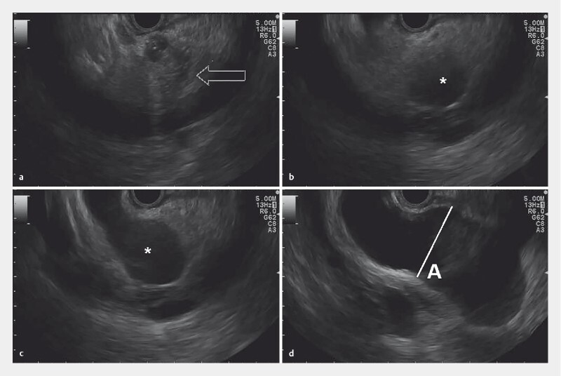
An educational video explaining where and how to perform CBD measurement was sent to all centers before starting the enrollment. During EUS examination, performed with a therapeutic linear echoendoscope (Olympus GF-UCT180, Olympus Medical Corporation Europe, Hamburg, Germany; Pentax EG-3870UTK or EG38-J10UT, Pentax Medical Europe, Hamburg, Germany; or Fujifilm EG-580UT, Fujifilm Medical Systems Europe, Dusseldorf, Germany), careful evaluation of the bile duct obstruction site was performed. After identification and measurement of the mass lesion, the CBD was identified and followed from the point of obstruction up to the liver hilum. The study was centered on the measurement of the CBD diameter and assessment of the subjective feasibility of theoretical performance of EUS-CDS with the appropriate Axios stent ( Fig. 1 ) at the level of the duodenal bulb corresponding to the mid CBD, where EUS-CDS would have been performed. Maximal attention was paid to avoid CBD compression by the EUS transducer during CBD measurement to minimize the risk of underestimating its diameter. The procedure was then continued with tissue acquisition and evaluation of tumor resectability. Tissue sampling and cytologic/histopathological evaluation, when performed, were handled according to local protocols. EUS-guided choledochoduodenostomy was not performed as part of the present study protocol.
Fig. 1.

Measurement of the common bile duct (CBD) diameter at the theoretical site of choledochoduodeno-anastomosis with the AXIOS Stent and Electrocautery Enhanced Delivery System. a Identification of the obstructive tumor (arrow). b, c Tracing of the common bile duct from the obstructive tumor upwards (*). d Identification of the suitable site for EUS-guided choledocoduodenostomy where measurement of the CBD diameter is performed ( a ).
Data collection
All data were collected in a patient case report form including: patient age and sex; interval of time (days) from the onset of jaundice to the day of the EUS procedure; serum levels of total bilirubin, alkaline phosphatase, gamma-GT and CA 19–9 performed within 24 hours before the procedure; and tumor size as measured by EUS.
Endosonographer experience in EUS-CDS was recorded and classified as extensive (> 40 EUS-CDS with Axios), moderate (between 20 and 40 cases) and limited (< 20 cases).
Patients with a positive EUS-FNA/FNB specimen were considered to have a malignant disease. In case of inconclusive or negative cyto-or histopathological results, additional data, such as the histopathological evaluation of the surgical specimen, repeated sampling procedures, or follow-up based on clinical disease evolution and repeat imaging study results were all utilized to establish the malignant nature of the existing biliary stricture.
Statistical analysis
Results were expressed as frequencies for categorical variables (further analyzed by chi-square or Fisher’s exact test when appropriate), mean and standard deviation (SD) for normally distributed continuous variables (analyzed by Student’s t -test), and median and interquartile range (IQR) for non-normally distributed continuous variables (analyzed by Mann-Whitney U test, or Kendall’s tau-b correlation). Univariate and multivariate (logistic regression) analyses were performed to determine whether any of the analyzed variables was associated with the CBD diameter. For logistic regression, independent variables associated with P ≤ 0.10 were included in the model by Enter method. Receiver operating characteristic (ROC) curve analyses were performed to identify cut-off values to be used for prediction. For the prediction model construction, we dichotomized the independent variables favoring the specificity. A two-sided significance of P < 0.05 was used throughout. The statistical analysis was performed using the SPSS for Windows software, version 20.0 (IBM Corp., Armonk, New York, United States).
Results
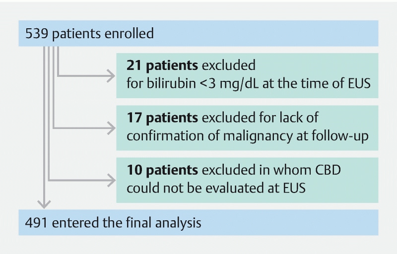
During the study period, 539 patients were assessed for eligibility. Forty-eight patients (8.9 %) were excluded from the overall analysis leading to the inclusion of 491 patients ( Fig. 2 ). The mean number of analyzed patients enrolled per center (22 centers) was 22.3 ± 16.1.
Fig. 2.

Flowchart of the study.
The median interval of time between occurrence of jaundice and EUS examination was 10 days (range, 1–70). Table 2 shows further details on blood test results, tumor characteristics, malignancy type, definitive diagnosis, and diagnostic methods used.
Table 2. Demographics, blood test results, and lesion characteristics of the 491 patients analyzed in the study.
| Variable | Value |
| Gender, no (%) | |
|
277 (56.4 %) |
|
214 (43.6 %%) |
| Mean age (range), years | 69 ± 12, (29–94) |
| Blood test results | |
|
12.7 ± 6.7 (3–38) |
|
4.3 ± 2.6 (0.5–21.8) |
|
11.8 (0.7–89.0) |
|
(0–3172) |
| Median time from occurrence of jaundice (range), days | 10 (1–70) |
| Mean tumor diameter, (mm) | 29.0 ± 10.3 |
| Final diagnoses, no (%) | |
|
423 (86.2 %) |
|
30 (6.1 %) |
|
26 (5.3 %) |
|
4 (0.8 %) |
|
8 (1.6 %) |
| Diagnostic method used to determine malignancy, no (%) | |
|
446 (90.8 %) |
|
21 (4.3 %) |
|
10 (2.0 %) |
|
14 (2.9 %) |
| Resectability assessment, no (%) | |
|
175 (35.6 %) |
|
308 (62.8 %) |
|
8 (1.6 %) |
ULN, upper limit of normal; SD, standard deviation; GGT, gamma-glutamyl transpeptidase; EUS-FNA, endoscopic ultrasound-guided fine-needle aspiration; FNB, fine-needle biopsy.
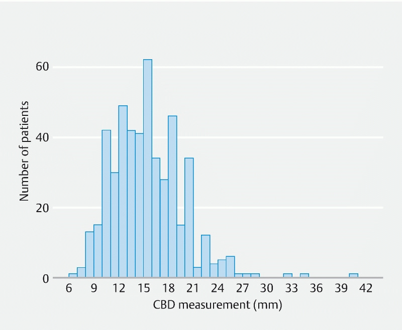
Dilation of the CBD ≥ 12 mm was detected in 387 of 491 patients (78.8 %), whereas a CBD ≥ 15 mm was found in 255 cases (51.9 %) ( Fig. 3 ). Overall, the endosonographers considered EUS-CDS technically feasible in 386 patients (78.6 %). In particular, a positive feasibility was expressed in 353 of 387 cases (91.2 %) for a CBD ≥ 12 mm and in 246 of 255 cases (96.5 %) for a CBD ≥ 15 mm. Interestingly, eight endosonographers indicated a possible attempt at EUS-CDS in 33 patients with CBD smaller than 12 mm. When stratified by endosonographer experience, EUS-CDS was considered possible in 138 of 164 patients (84.1 %) by five endosonographers with extensive expertise, which was significantly higher compared to 104 of 131 patients (79.4 %) evaluated by nine endosonographers with moderate expertise and 144 of 196 patients (73.5 %) evaluated by eight endosonographers with limited experience, respectively ( P = 0.047).
Fig. 3.

Chart detailing the distribution of patients per extent of common bile duct (CBD) dilation.
Univariate analysis revealed that CBD dilation ≥ 12 mm was associated with age (mean 70.1 ± 11.3 vs. 65.6 ± 11.7 years; P = 0.001), bilirubin level (mean of 13.3 ± 6.71 vs. 10.5 ± 5.6 mg/dL; P < 0.001), degree of CA 19–9 elevation (median of 10.8 vs. 5.2 times the ULN; P = 0.012), and increased interval of time between onset of jaundice and EUS procedure (mean of 12.9 ± 9.8 vs. 10.0 ± 8.4 days; P = 0.001). On multivariate analysis, age ( P = 0.002), bilirubin level ( P = 0.001) and interval of time from jaundice to EUS procedure ( P = 0.035) remained independent factors associated with CBD dilation of ≥ 12 mm.
The results of the univariate analysis for CBD dilation ≥ 15 mm were similar to those obtained for CBD dilation ≥ 12 mm, with age (mean of 70.9 ± 11.3 vs. 67.4 ± 11.6 years; P < 0.001), bilirubin level (mean of 13.8 ± 6.99 vs. 11.4 ± 5.90 mg/dL; P < 0.001), degree of CA 19–9 elevation (median of 10.97 vs. 8.19 times the ULN; P = 0.006), and interval of time between jaundice occurrence and EUS procedure (mean of 13.0 ± 9.9 vs. 11.6 ± 9.2 days; P = 0.048) as associated factors. On multivariate analysis, only age ( P = 0.004) and bilirubin level ( P < 0.001) remained associated with CBD dilation ≥ 15 mm.
Age and total bilirubin levels, factors independently associated on multivariate analysis to both CBD ≥ 12 mm and ≥ 15 mm, however, were poorly correlated with the extent of CBD dilation (age; P < 0.001, r = 0.19) (total bilirubin levels; P < 0.001, r = 0.25). AUROCs of 0.624 (95 % CI, 0.564–0.684) and 0.598 (95 % CI, 0.548–0.648) were found for bilirubin level and CBD ≥ 12 and ≥ 15 mm, while considering age and CBD dilation ≥ 12 and ≥ 15 mm, AUROCs of 0.607 (95 % CI, 0.547–0.666) and 0.590 (95 % CI, 0.540–0.641), respectively, were detected. We constructed a model based on cut-offs for age of 70 years and three levels of serum bilirubin (< 7 mg/dL, ≥ 7 mg/dL and < 14 mg/dL, and ≥ 14 mg/dL) in an attempt to predict the extent of CBD dilation for patients with malignant obstructive jaundice ( Table 3 ). A clinically significant (> 80 % of cases) prediction for a CBD diameter ≥ 12 mm was found only for bilirubin ≥ 7 mg/dL and age of more than 70 years.
Table 3. Model for prediction of CBD dilation considering patient’s age and degree of jaundice.
| Total bilirubin (mg/dL) | < 7 | ≥ 7 and < 14 | ≥ 14 | ||||
| Age (years) | < 70 | ≥ 70 | < 70 | ≥ 70 | < 70 | ≥ 70 | |
| No. of patients (%) | 53 (11 %) | 50 (11 %) | 119 (24 %) | 95 (19 %) | 84 (17 %) | 90 (18 %) | |
| Probability of | CBD ≥ 12 mm [95 % CI] |
61 % [57–65 %] |
73 % [61–85 %] |
73 % [64–81 %] |
82 % [73–89 %] |
85 % [77–93 %] |
90 % [84–96 %] |
| CBD ≥ 15 mm [95 % CI] |
31 % [18–43 %] |
42 % [28–57 %] |
48 % [39–57 %] |
60 % [49–70 %] |
53 % [43–65 %] |
65 % [55–75 %] |
|
Finally, concerning the theoretical feasibility of performing EUS-CDS, a correlation with the extent of CBD dilation was found both on univariate and multivariate analysis ( P < 0.001) (mean of 16.07 ± 3.97 mm vs. 11.02 ± 2.69 mm), with an AUROC for CBD dilation extent predicting feasibility of CBD drainage of 0.880 (95 % CI, 0.841–0.918).
Discussion
We performed a prospecitve, multicenter, cohort study to assess the degree of CBD dilation on the possibility to perform EUS-CDS with the Axios stent and the associated predictive factors in a large cohort of jaundiced patients undergoing EUS for evaluation of distal malignant biliary obstruction. Overall, we found that CBD dilation of ≥ 12 mm and ≥ 15 mm, the two cut-offs suggested to safely perform EUS-CDS with the Axios stent, could be observed respectively in 78.8 % and 51.9 % of cases, with no strong predictive factors associated with them.
EUS-CDS, first reported by Giovannini et al. in 2001 21 , along with HGS represent valid alternatives to percutaneous drainage in patients with distal malignant biliary obstruction after failed ERCP 22 . Initial studies are also evaluating the role of EUS-BD versus ERCP as a primary treatment in this clinical setting 4 5 6 . The main theoretical advantage of EUS-BD over ERCP is placement of the stent above the tumor lesion (EUS-CDS) or distant to it (EUS-HGS), with the potential to reduce rates of stent occlusion due to tumor ingrowth or overgrowth. Whether this translates into a significantly better long-term stent patency of the EUS-BD approach is still to be determined.
For distal malignant biliary obstruction, the choice between performing EUS-CDS or EUS-HGS remains at the discretion of the endosonographer. EUS-CDS is certainly technically less demanding than EUS-HGS and appears to be safer 23 . In addition, most of the EUS-CDS studies did not include procedures performed utilizing the Axios stent specifically designed for EUS-BD and associated with a mean rate of AEs of 15.2 % ( Table 4 ).
Table 4. Summary of the studies published as full papers reporting > 2 cases of EUS-guided biliary drainage after failed or not possible ERCP using the Axios stent in patients with distal malignant biliary obstruction.
| Author (year) | Study type | No. of patients | A × ios stents used | Stent size (diameter × length) (mm) | Mean CBD diameter (mm) | Technical success (%) | Clinical success (%) | Mean Follow-up (days) | AEs (number of cases, %) | |
| “Hot” | “Cold” | |||||||||
| Brückner (2015) 10 | RS, SC | 5 | 3 | 2 | 6 × 8 in all | 12.6 ± 3 | 5/5 1 (100) | 5/5 (100) | 9.8 ± 6.8 | 1/5 (20 %): stent dysfunction (1) |
| Anderloni (2016) 11 | CS, SC | 5 2 | 5 | 0 | NR | NR | 5/5 (100) | 5/5 (100) | 7 | NR |
| Kunda (2016) 9 | RS, MC | 57 | 30 | 27 | 6 × 8 in 36 | 17.9 (range 8–35) (median) | 56/57 3 (98.2) | 54/56 (96.4) | 151 ± 145 | 8/56 (14.3 %): duodenal perforation (1); stent migration (1); intraprocedural bleeding (1); transient cholangitis (1); stent occlusion or dysfunction (4) |
| 8 × 8 in 2 | ||||||||||
| 10 × 10 in 16 | ||||||||||
| 15 × 10 in 2 | ||||||||||
| Anderloni (2018) 13 | RS, SC | 16 4 | 16 | 0 | 6 × 8 in 9 | NR | 15/16 (93.8) | 14/15 (93.3) | 138.7 ± 124.6 | 1/15 (6.7 %): fatal severe acute arterial bleeding (1) |
| 8 × 8 in 4 | ||||||||||
| 10 × 10 in 3 | ||||||||||
| Tsuchiya (2018) 14 | PS, MC | 19 | 19 | 0 | 6 × 8 in 10 | 17.3 ± 5.5 | 19/19 (100) | 18/19 (94.7) | 184 (range 12–819) (median) |
7/19 (36.8 %): fever (1); cholangitis (2, in 1 after stent obstruction); stent dysfunction/obstruction (3); stent dislodgement (1) |
| 8 × 8 in 9 | ||||||||||
| Puga (2019) 15 | PS, SC | 7 | 7 | 0 | 6 × 8 in 6 | NR | 7/7 (100) | 5/8 5 (62.5) | 66 6 (95 % CI, 7–127) | 2/7 (28.6 %): fatal bleeding (2) |
| 8 × 8 in 1 | ||||||||||
| Jacques (2019) 16 | RS, MC | 52 7 | 52 | 0 | 6 × 8 in 43 | 17.2 (range 9–25) (median) | 46/52$ (88.5) | 46/46 (100 %) | 155 | 7/46 (15.2 %): cholangitis caused by obstruction or dysfunction of the stent in all |
| 8 × 8 in 7 | ||||||||||
| 15 × 10 in 2 | ||||||||||
| Anderloni (2019) 17 | RS, SC | 46 4 | 46 | 0 | 6 × 8 in 21 | 17.3 ± 3.3 | 43/46 (93.5) | 42/43 (97.7) | 114.4 (95 % CI, 73.2–155.4) | 5/43 (11.6 %): fatal bleeding (1); stent occlusion (3); stent migration (1) |
| 8 × 8 in 19 | ||||||||||
| 10 × 10 in 6 | ||||||||||
| El Chafic (2019) 18 | RS, MC | 67 | 67 | 0 | 10 × 10 in all | 17.6 ± 3.6 | 64/67 (95.5) | 40/40 8 (100 %) | 119 (median) | 11/64 (17.2 %) 9 : abdominal pain (2); peritonitis (1); bleeding (1); stent dysfunction/obstruction (7) |
| Fabbri (2019) 19 | CS, SC | 5 10 | 5 | 0 | 8 × 8 in 4 | 20.2 ± 5.7 | 5/5 (100) | 5/5 (100) | 34.6 ± 8.1 | None related to the procedure |
| 10 × 10 in 1 | ||||||||||
| Jacques (2020) 20 | RS, MC | 70 | 70 | 0 | 6 × 8 in 60 | 17.7 ± 5.0 | 69/70 (97.1) | 69/69 (100 %) | 153 (median) | 8/69 (11.6 %): bleeding due to fistulotomy (1); stent migration (1); cholangitis (2, in 1 after stent occlusion); tumoral stent obstruction (4) |
| 8 × 8 in 9 | ||||||||||
| 15 × 10 in 1 | ||||||||||
CBD, common bile duct; AEs, adverse events; RS, retrospective study; SC, single center; MC, multicenter; CS, case series; PS, prospective study; NR, not reported; EUS-CDS, EUS-guided choledochoduodenostomy; LA-SEMS, lumen-apposing self-e × pandable metal stents.
In one of the patients, the distal flange release failed technically, but the stent system was successfully e × changed for a second system over the guidewire.
Patients in this case series presented simultaneous duodenal and biliary malignant obstruction and were treated by single-session sequential EUS-guided choledochoduodenostomy and duodenal stenting.
The technical failure was due to duodenal perforation occurring while performing dilation of the fistulous tract;
It is possible that some of the patients were reported in both studies.
Patients were reported as a cohort of eight including one patient in whom EUS-guided hepaticogastrostomy with a plastic stent was performed, and clinical success was not presented separately for the LA-SEMS stents.
All patients died during follow-up.
One of the patients presented with benign biliary obstruction (CBD stones).
Only 40 patients who were followed-up for more than 4 weeks were considered for this end point.
Biliary reinterventions for obstruction were needed in 7 (17.5 %), in 3 of 6 patients who underwent EUS-CDS with LA-SEMS alone versus 4 of 34 with LA-SEMS plus an a × is-orienting device.
In this case series, choledochoduodenostomy was performed as a bridge to surgery since all patients presented with malignancy causing biliary obstruction in a resectable stage.
Transformation few years ago of the delivery catheter of the Axios stent into a device with a cystotome on its tip has revolutionized the field of interventional EUS and the performance of EUS-CDS. Indeed, the procedure can be accomplished under full ultrasound control in one step, without a need for needle puncture or guidewire placement, limiting accessory exchange and risk of losing the position, causing procedural failure. The only real drawback of the 6 × 8 mm and 8 × 8 mm Axios stents most commonly used for EUS-CDS is represented by the size of the distal flange (14– and 17-mm diameter, respectively), i. e. the one placed into the CBD, which may not fit the size of the CBD to be drained in all cases.
Consequently, based on operator experience level, the suggested CBD size to perform safe drainage varies from ≥ 12 mm for experts and ≥ 15 mm for less experienced endosonographers. To investigate the impact of the CBD size on the perceived possibility of performing EUS-CDS with the Axios stent as a primary drainage procedure, we analyzed a cohort of 491 naïve patients presenting with jaundice (bilirubin levels ≥ 3 mg/dL) secondary to a malignant distal biliary obstruction. A CBD ≥ 12 mm and ≥ 15 mm, measured at the level of the duodenal bulb where EUS-CDS would be preferentially performed, was individually detected in 78.8 % and 51.9 % of cases. In other words, about three quarters and only half of the patients were potentially suitable for EUS-CDS by expert and less experienced endosonographers, respectively. This latter finding is particularly relevant in view of the scarce availability of hands-on training courses and training models for EUS-CDS, through which less experienced endosonographers can reach skills to perform drainages in cases of CBD dilation close to 12 mm. Indeed, some studies proposed that at least 50 EUS-CDS cases are needed for endosonographers to be considered experts 24 , with a suggestion of restricting the performance of this procedure to only highly specialized centers 25 . Interestingly, some experts said that it was also possible to perform EUS-CDS on a CBD < 12 mm with the Axios stent. This is possible by gently pushing the catheter tip against the distal CBD wall while slowly opening the stent, thus making the CBD “bigger” or by entering the CBD with an angle that permits the catheter to be pushed upwards, where the stent can be opened safely vertically to the major axis of the CBD.
To assess the possibility of selecting patients before EUS, we performed univariate and multivariate analyses, which identified age and bilirubin levels as the only independent predictive factors for both CBD ≥ 12 mm and ≥ 15 mm at the theoretical site of EUS-CDS. Both variables, however, were found to be poorly associated with the extent of duct dilation, a finding that was further supported by the unsatisfactory results of the AUROCs, where no specific cut-off points with good sensitivity and specificity could be found for these two factors. This uncertainty also was supported by results from a predictive model we developed to apply to each new patient presenting with malignant distal obstructive jaundice. Indeed, this model worked satisfactorily only for predicting a CBD size ≥ 12 mm in patients at least 70 years old and with bilirubin levels between ≥ 7 mg/dL and 14 mg/dL or with higher bilirubin levels (range between 82 % to 90 %).
Taken together, all these data have important implications for the potential extended use of the Axios stent in all cases of malignant distal CBD obstruction. We strongly believe that single-step insertion stents with a different design, able to fit narrower CBDs to overcome the limitation we illustrated, need to be developed. Standard fully or partially covered biliary SEMS, which have been used in two RCTs and can fit any CBD 4 5 , might have an increased risk of migration because of the absence of forces exerted by the stented tumoral mass that keeps them in place when positioned transpapillary. In the RCT by Paik et al. 6 , a SEMS featuring an uncovered portion with anchoring flaps to be placed into the CBD and a distal funnel-shaped uncovered wire mesh portion was successfully used, with only two cases of stent migration that were not specified if they had occurred during EUS-CDS or EUS-HGS. This stent, however, is mounted on an introducer without a cystotome capability and requires a multistep procedure with accessory exchange, which might be associated with an increased risk of procedural failure 6 . Moreover, data on reproducibility to fully assess the performance of this latter stent are lacking. Another possible approach would be to reduce the width of the inner flange of the Axios stent up to a value that can guarantee sufficient juxtaposing forces to avoid its migration and allow its use in the majority, if not all, cases of distal malignant CBD obstruction. This working process will hopefully transform EUS-CDS into a safer procedure, which can be learned and taught by the large majority of EUS performers, a prerequisite to bringing EUS-CDS to another level.
One major limitation of our study is that the CBD size measured with EUS was not compared with other imaging modalities, which was due to the fact that patients underwent different pre-EUS imaging tests. Another possible bias was that the study was conducted in many centers. However, assessment of CBD size by EUS was standardized among all the participants in the study, making the results reproducible.
Conclusions
In conclusion, our study demonstrated that in naïve patients with malignant distal CBD obstruction, the Axios stent theoretically can be used safely in about three quarters of the cases by experts and in about half the cases by non-expert endosonographers. However, no reliable predictive factors could be elucidated to indicate which patients will be fit for the procedure. Efforts to create dedicated stents or devices with a different design, able to overcome the existing limitations of the Axios stent for EUS-CDS, are needed.
Footnotes
Competing interests Dr. Anderloni is a consultant for Boston Scientific, Olympus Medical and Medtronic. Dr. Fabbri is a consultant for Boston Scientific. Dr. Napoleon has held supported teaching sessions and had research collaboration with Boston Scientific. Dr. Poley served as a consultant for and received speaker and travel fees from Boston Scientific, Cook Endoscopy, and Pentax. Dr. Mutignani had a research collaboration with Boston Scientific. Dr. Palazzo is a consultant for Boston Scientific. Dr. Itoi is a speaker for Boston Scientific. Dr. Costamagna received research support from Cook Endoscopy and he currently is a member of Advisory Committees or review panels for Olympus Medical and Boston Scientific. Dr. Larghi received educational fees from Boston Scientific and Pentax.
References
- 1.Kahaleh M, Artifon E LA, Perez-Miranda M et al. EUS-guided drainage: Summary of therapeutic EUS consortium meeting. Endosc Ultrasound. 2019;8:151–160. doi: 10.4103/eus.eus_26_19. [DOI] [PMC free article] [PubMed] [Google Scholar]
- 2.Boulay B R, Lo S K. Endoscopic ultrasound-guided biliary drainage. Gastrointest Endosc Clin N Am. 2018;28:171–185. doi: 10.1016/j.giec.2017.11.005. [DOI] [PubMed] [Google Scholar]
- 3.Sharaiha R Z, Khan M A, Kamal F et al. Efficacy and safety of EUS-guided biliary drainage in comparison with percutaneous biliary drainage when ERCP fails: a systematic review and meta-analysis. Gastrointest Endosc. 2017;85:904–914. doi: 10.1016/j.gie.2016.12.023. [DOI] [PubMed] [Google Scholar]
- 4.Park J K, Woo Y S, Noh D H et al. Efficacy of EUS-guided and ERCP-guided biliary drainage for malignant biliary obstruction: prospective randomized controlled study. Gastrointest Endosc. 2018;88:277–282. doi: 10.1016/j.gie.2018.03.015. [DOI] [PubMed] [Google Scholar]
- 5.Bang J Y, Navaneethan U, Hasan M et al. Stent placement by EUS or ERCP for primary biliary decompression in pancreatic cancer: a randomized trial (with videos) Gastrointest Endosc. 2018;88:9–17. doi: 10.1016/j.gie.2018.03.012. [DOI] [PubMed] [Google Scholar]
- 6.Paik W H, Lee T H, Park D H et al. EUS-guided biliary drainage versus ERCP for the primary palliation of malignant biliary obstruction: a multicenter randomized clinical trial. Am J Gastroenterol. 2018;113:987–997. doi: 10.1038/s41395-018-0122-8. [DOI] [PubMed] [Google Scholar]
- 7.Frazzoni L, Fuccio L, Larghi A. EUS-guided biliary drainage versus ERCP for the primary treatment of malignant distal biliary obstruction: time for a large randomized study. Gastrointest Endosc. 2018;88:571–572. doi: 10.1016/j.gie.2018.04.2335. [DOI] [PubMed] [Google Scholar]
- 8.Attili F, Rimbaş M, Galasso D et al. Fluoroless endoscopic ultrasound-guided biliary drainage after failed ERCP with a novel lumen-apposing metal stent mounted on a cautery-tipped delivery system. Endoscopy. 2015;47 Suppl 1:E619–E620. doi: 10.1055/s-0034-1393669. [DOI] [PubMed] [Google Scholar]
- 9.Kunda R, Pérez-Miranda M, Will U et al. EUS-guided choledochoduodenostomy for malignant distal biliary obstruction using a lumen-apposing fully covered metal stent after failed ERCP. Surg Endosc. 2016;30:5002–5008. doi: 10.1007/s00464-016-4845-6. [DOI] [PubMed] [Google Scholar]
- 10.Brückner S, Arlt A, Hampe J. Endoscopic ultrasound-guided biliary drainage using a lumen-apposing self-expanding metal stent: a case series. Endoscopy. 2015;47:858–861. doi: 10.1055/s-0034-1392104. [DOI] [PubMed] [Google Scholar]
- 11.Anderloni A, Buda A, Carrara S et al. Single-session double-stent placement in concomitant malignant biliary and duodenal obstruction with a cautery-tipped lumen apposing metal stent. Endoscopy. 2016;48:E321–E322. doi: 10.1055/s-0042-117425. [DOI] [PubMed] [Google Scholar]
- 12.French J B, Coe A W, Pawa R. Endoscopic ultrasound-guided choledochoduodenostomy with a lumen-apposing, self-expandable fully covered metal stent for palliative biliary drainage. Clin J Gastroenterol. 2016;9:79–85. doi: 10.1007/s12328-016-0634-y. [DOI] [PubMed] [Google Scholar]
- 13.Anderloni A, Leo M D, Carrara S et al. Endoscopic ultrasound-guided transmural drainage by cautery-tipped lumen-apposing metal stent: exploring the possible indications. Ann Gastroenterol. 2018;31:735–741. doi: 10.20524/aog.2018.0299. [DOI] [PMC free article] [PubMed] [Google Scholar]
- 14.Tsuchiya T, Teoh A YB, Itoi T et al. Long-term outcomes of EUS-guided choledochoduodenostomy using a lumen-apposing metal stent for malignant distal biliary obstruction: A prospective multicenter study. Gastrointest Endosc. 2018;87:1138–1146. doi: 10.1016/j.gie.2017.08.017. [DOI] [PubMed] [Google Scholar]
- 15.Puga M, Pallarès N, Velásquez-Rodríguez J et al. Endoscopic biliary drainage in unresectable biliary obstruction: the role of endoscopic ultrasound-guidance in a cohort study. Rev Esp Enferm Dig. 2019;111:683–689. doi: 10.17235/reed.2019.6225/2019. [DOI] [PubMed] [Google Scholar]
- 16.Jacques J, Privat J, Pinard F et al. Endoscopic ultrasound-guided choledochoduodenostomy with electrocautery-enhanced lumen-apposing stents: A retrospective analysis. Endoscopy. 2019;51:540–547. doi: 10.1055/a-0735-9137. [DOI] [PubMed] [Google Scholar]
- 17.Anderloni A, Fugazza A, Troncone E et al. Single-stage EUS-guided choledochoduodenostomy using a lumenapposing metal stent for malignant distal biliary obstruction. Gastrointest Endosc. 2019;89:69–76. doi: 10.1016/j.gie.2018.08.047. [DOI] [PubMed] [Google Scholar]
- 18.El Chafic A H, Shah J N, Hamerski C et al. EUS-Guided Choledochoduodenostomy for Distal Malignant Biliary Obstruction Using Electrocautery-Enhanced Lumen-Apposing Metal Stents: First US, Multicenter Experience. Dig Dis Sci. 2019;64:3321–3327. doi: 10.1007/s10620-019-05688-2. [DOI] [PubMed] [Google Scholar]
- 19.Fabbri C, Fugazza A, Binda C et al. Beyond palliation: using EUS-guided choledochoduodenostomy with a lumen-apposing metal stent as a bridge to surgery. a case series. J Gastrointestin Liver Dis. 2019;28:125–128. doi: 10.15403/jgld.2014.1121.281.eus. [DOI] [PubMed] [Google Scholar]
- 20.Jacques J, Privat J, Pinard F et al. EUS-guided choledochoduodenostomy using electrocautery-enhanced lumen-apposing metal stents: a French multicenter study after implementation of the technique (with video) Gastrointest Endosc. 2020;92:134–141. doi: 10.1016/j.gie.2020.01.055. [DOI] [PubMed] [Google Scholar]
- 21.Giovannini M, Moutardier V, Pesenti C. Endoscopic ultrasound-guided bilioduodenal anastomosis: a new technique for biliary drainage. Endoscopy. 2001;33:898–900. doi: 10.1055/s-2001-17324. [DOI] [PubMed] [Google Scholar]
- 22.Rimbaş M, Larghi A, Kunda R. EUS-guided biliary drainage: Is it ready for prime time? Endosc Ultrasound. 2017;6:S122–S126. doi: 10.4103/eus.eus_78_17. [DOI] [PMC free article] [PubMed] [Google Scholar]
- 23.Hedjoudje A, Sportes A, Grabar S et al. Outcomes of endoscopic ultrasound-guided biliary drainage: A systematic review and meta-analysis. United European Gastroenterol J. 2019;7:60–68. doi: 10.1177/2050640618808147. [DOI] [PMC free article] [PubMed] [Google Scholar]
- 24.Poincloux L, Rouquette O, Buc E et al. Endoscopic ultrasound-guided biliary drainage after failed ERCP: cumulative experience of 101 procedures at a single center. Endoscopy. 2015;47:794–801. doi: 10.1055/s-0034-1391988. [DOI] [PubMed] [Google Scholar]
- 25.Artifon E LA, Visconti T AC, Brunaldi V O. Choledochoduodenostomy: Outcomes and limitations. Endosc Ultrasound. 2019;8:S72–S78. doi: 10.4103/eus.eus_62_19. [DOI] [PMC free article] [PubMed] [Google Scholar]


