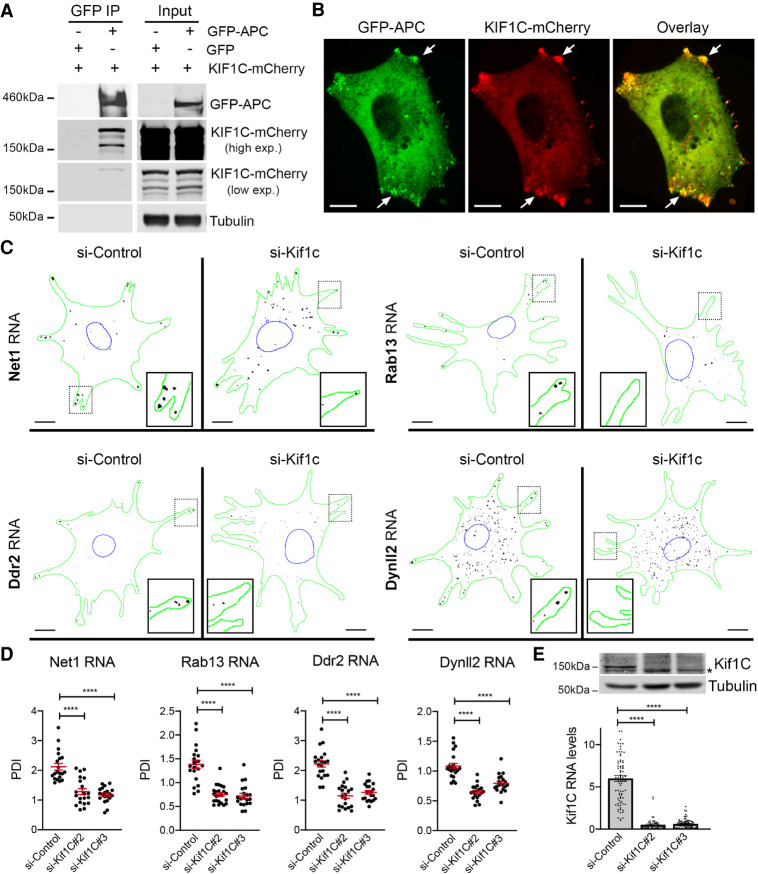
FIGURE 2.
KIF1C associates with APC and is required for the localization of APC-dependent mRNAs to cytoplasmic protrusions. (A) Coimmunoprecipitation of KIF1C with APC. GFP or GFP-APC were immunoprecipitated from cells also expressing KIF1C-mCherry and analyzed by western blot to detect the indicated proteins. Results are representative of three independent experiments. (B) Colocalization of GFP-APC and KIF1C-mCherry at cytoplasmic protrusions (arrows). Images are representative of multiple cells observed in two independent experiments. Scale bar: 10 microns. (C) Depletion of Kif1c prevents mRNA accumulation in protrusions. Images are micrographs of NIH/3T3 cells labeled by smFISH with probes against Net1, Rab13, Ddr2, and Dynll2 mRNAs, following treatment with siRNAs against Kif1C (panels si-Kif1c) or a control sequence (panel si-Control). Scale bars: 10 microns. Green: outline of the cells; blue: outline of the nuclei; black: smFISH signals. Insets represent magnifications of the boxed areas. (D) Quantification of mRNA localization of cells described in C. Graphs represent the intracellular distribution of the indicated mRNAs as measured by PDI index, with and without treatment of cells with the indicated siRNAs. Red bars represent the mean and 95% confidence interval. Points indicate individual cells observed in two independent experiments. (E) Detection of Kif1C protein (upper panels) or Kif1C RNA levels (lower graph) from cells treated with the indicated siRNAs. Stars in D and E are P-values: (****) P < 0.0001, (***) P < 0.001, estimated by analysis of variance with Bonferroni's multiple comparisons test.

