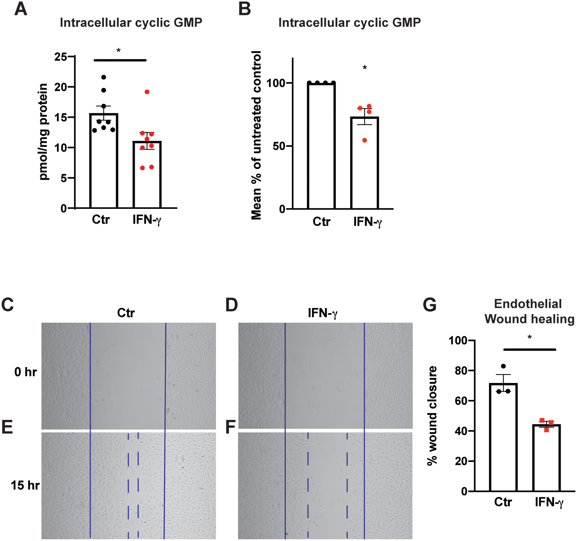
Figure 7. IFN-γ exposure depletes endothelial basal intracellular cGMP and impairs proliferation and migratory capacity.

A-B, Intracellular cGMP levels from HCAEC quantified using ELISA following 25 h of IFN-γ treatment at 50 ng/mL. Absolute intracellular cGMP measurements normalized to total protein/plate (pmol/mg protein) (A) and relative intracellular cGMP levels expressed as the percentages of those of untreated control groups (B). Mean ± SEM from 8 biological replicates from 4 independent experiments.
C-F, HCAEC proliferation and migration capacities were assessed by scratch assay after IFN-γ exposure for 30 h. Representative bright field images are shown at 0 h (C-D) and 15 h the scratch (E-F).
G, Percentages of wound closure or monolayer re-endothelialization following IFN-γ treatment were calculated by the following formula = [(open area at 0 h) - (open area at 15 h)]/(open area at 0 h) × 100. Mean ± SEM from 3 independent experiments.
Statistical significance was assessed using a two-tailed Student’s t-test (A,G) or one-sample t-test (B).
* p < 0.05
