Abstract
Background
Splanchnic vein thrombosis is a well-recognized local vascular complication of acute pancreatitis (AP), estimated to occur in approximately 15% of patients. While splanchnic vein recanalization occurs spontaneously in approximately one third of patients, severe complications such as bowel ischemia and liver failure have also been reported. At present, there is no consensus on whether patients presenting with AP-associated splanchnic vein thrombosis should receive therapeutic anticoagulation.
Methods
We searched multiple databases from inception through December 2020 to collect studies that compared the clinical outcomes of patients with AP and splanchnic vein thrombosis who received therapeutic anticoagulation (AC group) with those who did not (N-AC group). A meta-analysis was performed to calculate the relative risk (RR) of vessel recanalization, bleeding complications, collateral formation and death in the 2 groups.
Results
Seven studies with 8353 patients, 339 of whom had splanchnic vein thrombosis, were included in the final analysis. A total of 154 patients (45.4%) had acute severe pancreatitis. A significantly higher proportion of patients had vessel recanalization in the AC group: RR 1.6, 95% confidence interval 1.17-2.27; I2=0%; P=0.004. There was no difference between the 2 groups in the RR of bleeding complications, collateral formation and death.
Conclusions
Our analysis demonstrated that, among patients with AP-associated splanchnic vein thrombosis, therapeutic anticoagulation resulted in recanalization of the involved vessels without significantly increasing the risk of bleeding complications. There was no difference in the RR of death or the rates of collateral vessel formation during the follow up.
Keywords: Acute pancreatitis, splanchnic vein thrombosis, anticoagulation
Introduction
Acute pancreatitis (AP) is an inflammatory disorder of the pancreas associated with substantial morbidity and mortality. The global incidence of AP is 34 affected individuals per 100,000 person-years, and it has been increasing worldwide [1]. The worldwide obesity epidemic is also thought to be contributing to the overall increasing global incidence of AP [2]. While the morbidity and long-term sequelae of AP remain substantial, the associated mortality has seen a downward trend over the past decade, from 1.6% to 0.8%, in part due to improvements in timely and accurate diagnosis, as well as in the care of critically ill patients with AP [3]. Despite this, AP is currently one of the most common gastrointestinal disorders warranting hospitalization in the United States and accounts for $9.3 billion in healthcare costs annually [4].
Splanchnic vein thrombosis is a well-recognized local vascular complication of AP [5], estimated to occur in approximately 15% of patients [6]. Its etiology is thought to be due in part to the anatomic relationship of the large mesenteric vessels to the pancreas, the prothrombotic nature of the acute inflammatory reaction, along with contributions from systemic response to the injury, hypovolemia and fluid shifts [7]. Splanchnic vein thrombosis may involve thrombosis of the splenic vein (SpVT), portal vein (PVT) and superior mesenteric vein (SMVT), either separately or in combination, and is often detected incidentally on imaging performed to evaluate the symptoms and/or complications of AP. While splanchnic vein recanalization occurs spontaneously in approximately one third of patients [8,9], severe complications such as bowel ischemia and liver failure have also been reported [10]. Additionally, progressive splanchnic vein thrombosis can result in portal hypertension with resulting complications, including hemorrhage [11].
At present, there is no consensus on whether patients presenting with splanchnic vein thrombosis in the setting of AP should receive therapeutic anticoagulation. We conducted a systematic review of the published literature and performed a meta-analysis to assess the safety and clinical outcomes of anticoagulation in AP-associated splanchnic vein thrombosis.
Materials and methods
Search strategy
The relevant medical literature was searched by a medical librarian for studies reporting outcomes of anticoagulation in patients with splanchnic vein thrombosis in the setting of AP. The search strategy was created using a combination of keywords and standardized index terms. A systematic and detailed search was run in December 2020 in Ovid EBM Reviews, ClinicalTrials.gov, Ovid Embase (1974+), Ovid Medline (1946+ including Epub ahead of print, in-process & other non-indexed citations), Scopus (1970+), and Web of Science (1975+). Results were limited to English language publications only.
The full search strategy is available in Supplementary Appendix 1 (194.9KB, pdf) . As the included studies were observational in design, the MOOSE (Meta-analyses Of Observational Studies in Epidemiology) checklist was followed [12] and is provided as Supplementary Appendix 2 (194.9KB, pdf) . The PRISMA flowchart for study selection [13] was followed and is provided as Supplementary Fig. 1 (194.9KB, pdf) . The reference lists of evaluated studies were examined to identify other studies of interest.
Study selection
We included studies that compared the clinical outcomes of patients with AP and splanchnic vein thrombosis who received therapeutic anticoagulation with those who did not. Studies included were cohort and case-control studies that reported outcomes of both treatment approaches. Studies were included whether they were published as full manuscripts or conference abstracts, were performed in an inpatient or outpatient setting, irrespectively of follow-up time and country of origin, as long as they provided the appropriate data needed for the analysis.
Our exclusion criteria were as follows: 1) case series and case reports; 2) studies in which patients received prophylactic anticoagulation; 3) studies including patients with chronic pancreatitis; 4) studies performed in the pediatric population (age <18 years); and 4) studies not published in English. In cases of multiple publications from a single research group reporting on the same patient, same cohort and/or overlapping cohorts, data from the most recent and/or most appropriate comprehensive report were retained. The remaining studies were evaluated by 2 authors (SC, DR) based on the publication timing (most recent) and/or the sample size of the study (largest). In situations where a consensus could not be reached, overlapping studies were included in the final analysis and any potential effects were assessed in a sensitivity analysis of the pooled outcomes, leaving out one study at a time.
Data abstraction and quality assessment
Data on study-related outcomes from the individual studies were abstracted independently onto a standardized form by at least 2 authors (SC, SRK). Other authors (AP, BD, DR, NB) cross-verified the collected data for possible errors and 2 authors (SC, SRK) performed the quality scoring independently. We used the Newcastle-Ottawa scale to assess the quality of cohort studies [14]. This quality score consists of 8 questions, the details of which are provided in Supplementary Table 1 (194.9KB, pdf) .
Outcomes assessed
Patients were grouped based on whether they received anticoagulation (AC) or not (N-AC). The primary outcomes assessed were the relative risk (RR) of vessel recanalization, bleeding complications, collateral formation and death in patients who received anticoagulation compared with those who did not. The secondary outcomes measured were the pooled rates of vessel recanalization, bleeding complications, collateral formation and death in patients who received anticoagulation and in patients who did not receive anticoagulation.
Statistical analysis
We used meta-analysis techniques to calculate the pooled estimates in each case, following the methods suggested by DerSimonian and Laird and using the random-effects model. Results were expressed in terms of RR or mean difference (MD) along with relevant 95% confidence intervals (CI), when appropriate [15]. When the incidence of an outcome was zero in a study, a continuity correction of 0.5 was added to the number of incident cases before statistical analysis [16]. We assessed heterogeneity between study-specific estimates using the Cochran Q statistical test for heterogeneity, 95%CI, and the I2 statistics [16,17], in which values of <30%, 30-60%, 61-75%, and >75% were suggestive of low, moderate, substantial and considerable heterogeneity, respectively. We assessed publication bias, qualitatively, by visual inspection of funnel plot, and quantitatively, by the Egger test [18]. When publication bias was present, further statistics using the fail-safe N test and Duval and Tweedie’s “Trim and Fill” test was used to ascertain the impact of the bias [19]. Three levels of impact were reported, based on the concordance between the reported results and the actual estimate if there was no bias. The impact was reported as minimal if both versions were estimated to be same, modest if effect size changed substantially but the final finding would still remain the same, and severe if the basic final conclusion of the analysis was threatened by the bias [20]. A Knapp-Hartung 2-tailed P-value of <0.05 was considered statistically significant and the R2 value was calculated to study the goodness of fit. All analyses were performed using Comprehensive Meta-Analysis software, version 3 (BioStat, Englewood, NJ).
Results
Characteristics and quality of included studies
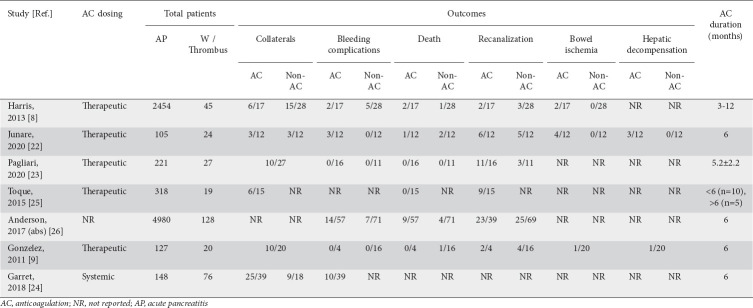
Seven studies with 8353 patients, 339 of whom had splanchnic vein thrombosis, were included in the final analysis. The severity of pancreatitis was defined as per the Atlanta Criteria [8,21], Revised Atlanta Classification [22-24] and Balthazar score [25]. Of 135 patients with splanchnic vein thrombosis reported in 5 studies [8,21-23,25], 78 (57.7%) were classified as having severe AP. In another study, all 148 of the included patients were classified as having moderately severe or severe AP [24].
Six of the included studies were retrospective in design [8,9,23-26], while one was a prospective multicenter study [22]. Only one of the included studies was published in abstract form but presented pertinent outcomes of interest [26]. Two studies were performed in the USA, 4 in Europe and 1 in India. Based on the Newcastle-Ottawa scoring system, all included studies were considered to be of high quality.
Search results and population characteristics
Our initial search strategy yielded 67 results. All search results were exported to Endnote, where 26 obvious duplicates were removed leaving 41 citations. A schematic diagram demonstrating our study selection is presented in Supplementary Fig. 1 (194.9KB, pdf) .
All studies reported information on thrombus location: 26 patients had isolated PVT, 194 had isolated SpVT and 5 patients had isolated SMVT. A vast majority of patients had a combination of vessels involved. Details of patient characteristics and demographics were available in 5 studies. A total of 172 males and 91 females were included in our analysis, with mean age ranging from 36.6-58 years. The follow-up period ranged from 3-24 months.
Imaging modalities utilized for diagnosis and follow up of splanchnic vein thrombosis
All studies used pancreas protocol contrast-enhanced computed tomography (CT), magnetic resonance imaging and/or color Doppler ultrasonography [8,9,22-25]. Splanchnic vein thrombosis was diagnosed when an actual thrombus was detected in the vein, or the vein appeared compressed, or was not visualized with presence of collaterals. Portal cavernoma was defined radiologically as the presence of large portoportal collaterals. Follow-up imaging, including contrast CT [21,23,25] and Doppler ultrasonography [8], was done to assess for recanalization of involved vessels.
Type and duration of anticoagulant agent used
Four studies provided information on the type of therapeutic anticoagulation used. In 2 studies, therapeutic low-molecular-weight heparin (LMWH) (1 mg/kg b.i.d.) or intravenous unfractionated heparin infusion (initial bolus of 80 U/kg, followed by an initial infusion rate of 18/kg/h), according to the standard nomogram, was given. This was followed by maintenance therapy with warfarin to keep the international normalized ratio (INR) between 2 and 3 [8,22]. Gonzelez et al utilized the same anticoagulation protocol, but in that study the INR range was kept between 1.8 and 2 [9].
In another study, a therapeutic dose of LMWH was administered (100 UI/kg b.i.d.) to in-patients at the time of diagnosis and patients were subsequently fully anticoagulated upon discharge with fondaparinux 7.5 mg/day, or vitamin K antagonist (warfarin) (target INR 2.0-3.0), or novel direct oral anticoagulants, [23]. Three studies did not provide information on the therapeutic modality used in the anticoagulated group [24-26]. Further details of patient characteristics and clinical outcomes are described in Tables 1 and 2.
Table 1.
Study population characteristics
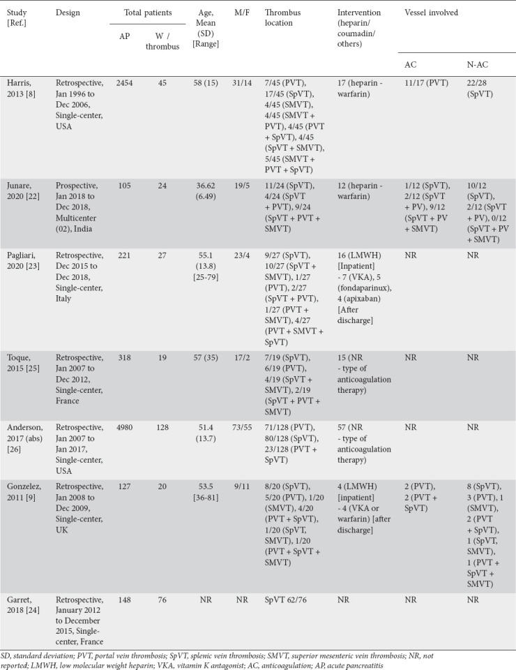
Table 2.
Study results and adverse events
Meta-analysis outcomes
Pooled RR and rates of vessel recanalization
The pooled rate of vessel recanalization at follow up in the AC group (6 studies, 53 of 103 patients) was 51.5%, 95%CI 35.5-67.1; I2 52%, while in the N-AC group (5 studies, 40 of 136 patients) it was 28.6%, 95%CI 18.6-41.3; I2=38%. The difference between the 2 was statistically significant: RR 1.6, 95%CI 1.17-2.27; I2=0%; P=0.004 (Fig. 1).
Figure 1.
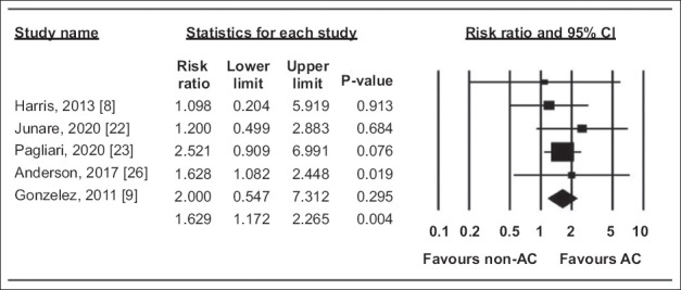
Forest plot, vessel recanalization CI, confidence interval, AC, anticoagulation
Pooled RR and rates of bleeding complications
The pooled rate of bleeding complications in the AC group was 21.2%, 95%CI 14-30.6; I2=0%, while in the N-AC group it was 11%, 95%CI 6.5-17.9; I2=0%. The difference between the 2 was not statistically significant: RR 1.95, 95%CI 0.98-3.88; I2=0%; P=0.06 (Fig. 2).
Figure 2.
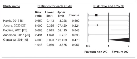
Forest plot, bleeding complications CI, confidence interval, AC, anticoagulation
Pooled RR and rates of collateral formation
The pooled rate of collaterals formation in the AC group was 43.3%, 95%CI 26.1-62.3; I2=61%, while in the N-AC group it was 46.2%, 95%CI 31.3-61.8; I2=26%. The difference between the 2 was not statistically significant: RR 1.24, 95%CI 0.75-2.05; I2=24%; P=0.4 (Fig. 3).
Figure 3.
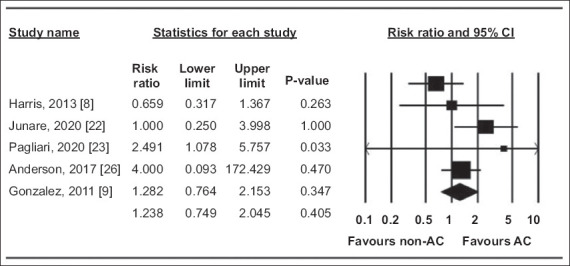
Forest plot, collateral formation CI, confidence interval, AC, anticoagulation
Pooled RR and rates of death
The pooled death rate in the AC group was 12.6%, 95%CI 7.5-20.4; I2=0%, while in the N-AC group it was 6.8%, 95%CI 3.5-12.8; I2=0%. The difference between the 2 was not statistically significant: RR 2.02, 95%CI 0.85-4.8; I2=0%; P=0.1 (Fig. 4).
Figure 4.
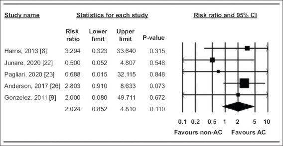
Forest plot, deaths CI, confidence interval, AC, anticoagulation
Validation of meta-analysis results
Sensitivity analysis
To assess whether any one study had a dominant effect on the meta-analysis, we excluded one study at a time and analyzed its effect on the main summary estimate. No one study had a dominant effect on our study outcomes.
Heterogeneity
We assessed dispersion of the calculated rates using the I2 percentage values, as reported in the meta-analysis outcomes section. While there was moderate to substantial heterogeneity in the pooled rates of vessel recanalization and collateral formation in both groups, the pooled RR ratios between the 2 groups had zero to low heterogeneity.
Publication bias
Publication bias could not be estimated as the number of studies included our final analysis was less than 10.
Discussion
Our analysis demonstrated that, among patients with AP found to have splanchnic vein thrombosis, therapeutic anticoagulation resulted in recanalization of the involved vessels without significantly increasing the risk of bleeding complications. We found no difference in the risk of death and rates of formation of collateral vessels at follow up between the AC and N-AC groups.
Splanchnic vein thrombosis is an increasingly recognized complication of AP. Its symptoms frequently overlap with those of AP and it is often diagnosed by imaging studies in the course of AP workup [27]. There is no consensus, however, with regard to anticoagulant treatment in these patients and whether it improves recanalization rates or overall clinical outcomes. Current society guidelines on AP do not specifically address this topic [28-30]. Timely intervention also needs to be considered, as splanchnic vein thrombosis may at times result in organ (bowel or hepatic) ischemia and necrosis if not recognized early [10,31]. Therefore, the risks and benefits of anticoagulation therapy in this subset of patients need to be thoroughly evaluated. Our results add to the current body of evidence on this challenging clinical scenario.
How does our study compare to the current published literature? A recent study by Zhou et al, including 273 patients with acute necrotizing pancreatitis, found that the application of timely systemic anticoagulation seems to reduce the incidence of splanchnic venous thrombosis and improves clinical outcomes without increasing the risk of hemorrhage. However, the percentage of patients developing recanalization or collateral circulation was not reported in this study [32]. Hajibandeh et al, in a recent Letter to the Editor, concluded that current evidence suggests that the routine use of AC in the management of pancreatitis-induced splanchnic venous thrombosis does not provide any benefit over no AC and may increase the risk of bleeding. However, only 252 patients were included in the analysis that was the subject of the letter [33]. A previous systematic review and meta-analysis of 16 studies evaluated the safety and efficacy of anticoagulation for splanchnic vein thrombosis in patients with AP. The authors noted a 14% rate of recanalization in patients receiving anticoagulation, compared to 11% among those who did not. Bleeding complications were observed in 16% of patients receiving anticoagulation compared to 5% who did not. However, the analysis also included 9 individual case reports and 2 case series, leading to significant heterogeneity in the results, precluding the authors from performing a meta-analysis [34]. Additionally, cohort studies in which prophylactic, rather than therapeutic dosage of anticoagulation was administered, were included [35]. In our analysis we only included studies in which patients received therapeutic or systemic anticoagulation and there was no or low heterogeneity in our results.
There are several strengths to our review. First, our search strategy was meticulous, with well-defined inclusion criteria, careful exclusion of redundant studies, inclusion of good quality studies with detailed extraction of data, and rigorous evaluation of study quality. Second, we included only those studies where the outcomes of patients who received therapeutic anticoagulation were directly compared to those who did not. This allowed us to perform a robust meta-analysis of clinical and safety outcomes. Third, in all of the studies included in our analysis, splanchnic venous thrombus was diagnosed when an actual thrombus was detected in the vein, or the vein appeared compressed, or was not visualized with presence of collaterals.
There are several limitations to this study, most of which are inherent to any meta-analysis. First and foremost, the type of anticoagulant used and duration of therapy varied widely among the studies. The vessels involved were not equally distributed among the AC and N-AC groups in the included studies. In one study, some patients received AC for less than 6 months and others for longer [25]. Second, one of the included studies was only published in abstract format [26] and in another study not all our outcomes of primary interest were presented [24]. Two studies reported rates of collateral formation for the entire cohort of patients with splanchnic venous thrombosis, not for AC and N-AC groups separately [23,9]. Limited literature suggests that the rate of spontaneous recanalization may be as high as 30% in patients with SpVT [36]. While 10-year recurrence-free survival is highest for isolated SpVT, this may not be true in patients with PVT and/or SMVT [37,38]. We were also unable to calculate the pooled outcomes of bowel ischemia and hepatic decompensation between the 2 groups, as this information was lacking in the studies. Third, 6 of the 7 studies included in our analysis were retrospective in design, while the severity classification criteria for AP varied across studies, which may have resulted in potential bias. Finally, a major concern for anticoagulation in splanchnic vein thrombosis is the risk of bleeding. While our analysis did not show a statistically significant difference in bleeding rates between the 2 groups, there was a trend towards more bleeding in patients receiving anticoagulation. Additionally, we did not report the etiology and severity of bleeding in our analysis, since this was not included in most studies. Further studies are warranted to stratify the risk of bleeding in patients with severe and non-severe AP receiving AC.
Despite these limitations, our study showed that the use of anticoagulation in patients with splanchnic vein thrombosis in the setting of AP resulted in higher rates of vessel recanalization, without an associated higher risk of bleeding complications. Our findings suggest that anticoagulation should be considered in AP patients with splanchnic vein thrombosis if there are no contraindications to anticoagulation.
Summary Box.
What is already known:
·Splanchnic vein thrombosis is a well-recognized local vascular complication of acute pancreatitis (AP) and is estimated to occur in approximately 15% of patients
·While splanchnic vein recanalization occurs spontaneously in approximately one third of patients, severe complications such as bowel ischemia and liver failure have also been reported
·At present, there is no consensus on whether patients presenting with splanchnic vein thrombosis in the setting of AP should receive therapeutic anticoagulation
What the new finding is:
·Based on this meta-analysis, in patients with AP found to have splanchnic vein thrombosis, therapeutic anticoagulation resulted in recanalization of the involved vessels without significantly increasing the risk of bleeding complications
Acknowledgment
The authors would like to thank Dana Gerberi, MLIS, Librarian, Mayo Clinic Libraries, for help with the systematic literature search
Biography
CHI Creighton University Medical Center, Omaha, Nebraska, USA; Harvard Medical School, Boston, Massachusetts, USA; University of Utah School of Medicine, Salt Lake City, Utah, USA; Brooklyn Hospital Center, Brooklyn, New York, USA; University of Minnesota & Minneapolis VA Health Care System, Minneapolis, Minnesota, USA; University of Nebraska Medical Center, Omaha, Nebraska, USA; Mayo Clinic, Rochester, Minnesota, USA; The Wright Center For Graduate Medical Education, Scranton, Philadelphia, USA; University of Arkansas for Medical Sciences, Little Rock, Arkansas, USA; University of Foggia, Foggia, Italy
Footnotes
Conflict of Interest: Douglas G. Adler is a consultant at Boston Scientific. All other authors report no conflict of interest
References
- 1.Petrov MS, Yadav D. Global epidemiology and holistic prevention of pancreatitis. Nat Rev Gastroenterol Hepatol. 2019;16:175–184. doi: 10.1038/s41575-018-0087-5. [DOI] [PMC free article] [PubMed] [Google Scholar]
- 2.NCD Risk Factor Collaboration (NCD-RisC) Worldwide trends in body-mass index, underweight, overweight, and obesity from 1975 to 2016:a pooled analysis of 2416 population-based measurement studies in 128·9 million children, adolescents, and adults. Lancet. 2017;390:2627–2642. doi: 10.1016/S0140-6736(17)32129-3. [DOI] [PMC free article] [PubMed] [Google Scholar]
- 3.Krishna SG, Kamboj AK, Hart PA, Hinton A, Conwell DL. The changing epidemiology of acute pancreatitis hospitalizations:a decade of trends and the impact of chronic pancreatitis. Pancreas. 2017;46:482–488. doi: 10.1097/MPA.0000000000000783. [DOI] [PMC free article] [PubMed] [Google Scholar]
- 4.Peery AF, Crockett SD, Murphy CC, et al. Burden and cost of gastrointestinal, liver, and pancreatic diseases in the United States:update 2018. Gastroenterology. 2019;156:254–272. doi: 10.1053/j.gastro.2018.08.063. [DOI] [PMC free article] [PubMed] [Google Scholar]
- 5.Rebours V, Boudaoud L, Vullierme MP, et al. Extrahepatic portal venous system thrombosis in recurrent acute and chronic alcoholic pancreatitis is caused by local inflammation and not thrombophilia. Am J Gastroenterol. 2012;107:1579–1585. doi: 10.1038/ajg.2012.231. [DOI] [PubMed] [Google Scholar]
- 6.Richards ER, Kabir SI, McNaught CE, MacFie J. Effect of thoracic epidural anaesthesia on splanchnic blood flow. Br J Surg. 2013;100:316–321. doi: 10.1002/bjs.8993. [DOI] [PubMed] [Google Scholar]
- 7.Ahmed SU, Rana SS, Ahluwalia J, et al. Role of thrombophilia in splanchnic venous thrombosis in acute pancreatitis. Ann Gastroenterol. 2018;31:371–378. doi: 10.20524/aog.2018.0242. [DOI] [PMC free article] [PubMed] [Google Scholar]
- 8.Harris S, Nadkarni NA, Naina HV, Vege SS. Splanchnic vein thrombosis in acute pancreatitis:a single-center experience. Pancreas. 2013;42:1251–1254. doi: 10.1097/MPA.0b013e3182968ff5. [DOI] [PubMed] [Google Scholar]
- 9.Gonzelez HJ, Sahay SJ, Samadi B, Davidson BR, Rahman SH. Splanchnic vein thrombosis in severe acute pancreatitis:a 2-year, single-institution experience. HPB (Oxford) 2011;13:860–864. doi: 10.1111/j.1477-2574.2011.00392.x. [DOI] [PMC free article] [PubMed] [Google Scholar]
- 10.Mallick IH, Winslet MC. Vascular complications of pancreatitis. JOP. 2004;5:328–337. [PubMed] [Google Scholar]
- 11.Gotto A, Lieberman M, Pochapin M. Gastric variceal bleeding due to pancreatitis-induced splenic vein thrombosis. BMJ Case Rep. 2014;2014:bcr2013201359. doi: 10.1136/bcr-2013-201359. [DOI] [PMC free article] [PubMed] [Google Scholar]
- 12.Stroup DF, Berlin JA, Morton SC, et al. Meta-analysis of observational studies in epidemiology:a proposal for reporting. Meta-analysis Of Observational Studies in Epidemiology (MOOSE) group. JAMA. 2000;283:2008–2012. doi: 10.1001/jama.283.15.2008. [DOI] [PubMed] [Google Scholar]
- 13.Moher D, Liberati A, Tetzlaff J, Altman DG PRISMA Group. Preferred reporting items for systematic reviews and meta-analyses:the PRISMA statement. Ann Intern Med. 2009;151:264–269. doi: 10.7326/0003-4819-151-4-200908180-00135. W64. [DOI] [PubMed] [Google Scholar]
- 14.Stang A. Critical evaluation of the Newcastle-Ottawa scale for the assessment of the quality of nonrandomized studies in meta-analyses. Eur J Epidemiol. 2010;25:603–605. doi: 10.1007/s10654-010-9491-z. [DOI] [PubMed] [Google Scholar]
- 15.DerSimonian R, Laird N. Meta-analysis in clinical trials. Control Clin Trials. 1986;7:177–188. doi: 10.1016/0197-2456(86)90046-2. [DOI] [PubMed] [Google Scholar]
- 16.Sutton AJ AK, Jones DR, et al. Methods for meta-analysis in medical research. John Wiley and Sons, Ltd; 2000. [Google Scholar]
- 17.Mohan BP, Adler DG. Heterogeneity in systematic review and meta-analysis:how to read between the numbers. Gastrointest Endosc. 2019;89:902–903. doi: 10.1016/j.gie.2018.10.036. [DOI] [PubMed] [Google Scholar]
- 18.Higgins JP, Thompson SG, Deeks JJ, Altman DG. Measuring inconsistency in meta-analyses. BMJ. 2003;327:557–560. doi: 10.1136/bmj.327.7414.557. [DOI] [PMC free article] [PubMed] [Google Scholar]
- 19.Duval S, Tweedie R. Trim and fill:A simple funnel-plot-based method of testing and adjusting for publication bias in meta-analysis. Biometrics. 2000;56:455–463. doi: 10.1111/j.0006-341x.2000.00455.x. [DOI] [PubMed] [Google Scholar]
- 20.Rothstein HR, Sutton AJ, Borenstein M. Publication bias in meta-analysis:prevention, assessment and adjustments. John Wiley &Sons, Ltd; 2006. [Google Scholar]
- 21.Gonzalez HD, Sahay SJ, John J, Davidson BR, Rahman SH. Splanchnic vein thrombosis in severe acute pancreatitis - is systemic anticoagulation indicated? Pancreatology. 2011;11:314. [Abstract] [Google Scholar]
- 22.Junare AR, Udgirkar S, Nair S, et al. Splanchnic venous thrombosis in acute pancreatitis:does anticoagulation affect outcome? Gastroenterology Res. 2020;13:25–31. doi: 10.14740/gr1223. [DOI] [PMC free article] [PubMed] [Google Scholar]
- 23.Pagliari D, Cianci R, Brizi MG, et al. Anticoagulant therapy in the treatment of splanchnic vein thrombosis associated to acute pancreatitis:a 3-year single-centre experience. Intern Emerg Med. 2020;15:1021–1029. doi: 10.1007/s11739-019-02271-5. [DOI] [PubMed] [Google Scholar]
- 24.Garret C, Péron M, Reignier J, et al. Risk factors and outcomes of infected pancreatic necrosis:Retrospective cohort of 148 patients admitted to the ICU for acute pancreatitis. United European Gastroenterol J. 2018;6:910–918. doi: 10.1177/2050640618764049. [DOI] [PMC free article] [PubMed] [Google Scholar]
- 25.Toqué L, Hamy A, Hamel JF, et al. Predictive factors of splanchnic vein thrombosis in acute pancreatitis:A 6-year single-center experience. J Dig Dis. 2015;16:734–740. doi: 10.1111/1751-2980.12298. [DOI] [PubMed] [Google Scholar]
- 26.Anderson W, Niccum B, Chitnavis M, Uppal D, Hays RA. Outcomes of anticoagulation for portal and/or splenic vein thrombosis in setting of acute pancreatitis. Am J Gastroenterol. 2017;112(S6) [Google Scholar]
- 27.Valla D. Splanchnic vein thrombosis. Semin Thromb Hemost. 2015;41:494–502. doi: 10.1055/s-0035-1550439. [DOI] [PubMed] [Google Scholar]
- 28.Crockett SD, Wani S, Gardner TB, Falck-Ytter Y, Barkun AN American Gastroenterological Association Institute Clinical Guidelines Committee. American Gastroenterological Association Institute Guideline on Initial Management of Acute Pancreatitis. Gastroenterology. 2018;154:1096–1101. doi: 10.1053/j.gastro.2018.01.032. [DOI] [PubMed] [Google Scholar]
- 29.Samarasekera E, Mahammed S, Carlisle S, Charnley R Guideline Committee. Pancreatitis:summary of NICE guidance. BMJ. 2018;362:k3443. doi: 10.1136/bmj.k3443. [DOI] [PubMed] [Google Scholar]
- 30.Leppäniemi A, Tolonen M, Tarasconi A, et al. Executive summary:WSES Guidelines for the management of severe acute pancreatitis. J Trauma Acute Care Surg. 2020;88:888–890. doi: 10.1097/TA.0000000000002691. [DOI] [PubMed] [Google Scholar]
- 31.Mendelson RM, Anderson J, Marshall M, Ramsay D. Vascular complications of pancreatitis. ANZ J Surg. 2005;75:1073–1079. doi: 10.1111/j.1445-2197.2005.03607.x. [DOI] [PubMed] [Google Scholar]
- 32.Zhou J, Zhang H, Mao W, et al. Efficacy and safety of early systemic anticoagulation for preventing splanchnic thrombosis in acute necrotizing pancreatitis. Pancreas. 2020;49:1220–1224. doi: 10.1097/MPA.0000000000001661. [DOI] [PubMed] [Google Scholar]
- 33.Hajibandeh S, Agrawal S, Irwin C, Obeidallah R, Subar D. Anticoagulation versus no anticoagulation for splanchnic venous thrombosis secondary to acute pancreatitis:do we really need to treat the incidental findings? Pancreas. 2020;49:e84–e85. doi: 10.1097/MPA.0000000000001644. [DOI] [PubMed] [Google Scholar]
- 34.Norton W, Lazaraviciute G, Ramsay G, Kreis I, Ahmed I, Bekheit M. Current practice of anticoagulant in the treatment of splanchnic vein thrombosis secondary to acute pancreatitis. Hepatobiliary Pancreat Dis Int. 2020;19:116–121. doi: 10.1016/j.hbpd.2019.12.007. [DOI] [PubMed] [Google Scholar]
- 35.Easler J, Muddana V, Furlan A, et al. Portosplenomesenteric venous thrombosis in patients with acute pancreatitis is associated with pancreatic necrosis and usually has a benign course. Clin Gastroenterol Hepatol. 2014;12:854–862. doi: 10.1016/j.cgh.2013.09.068. [DOI] [PubMed] [Google Scholar]
- 36.Nadkarni NA, Khanna S, Vege SS. Splanchnic venous thrombosis and pancreatitis. Pancreas. 2013;42:924–931. doi: 10.1097/MPA.0b013e318287cd3d. [DOI] [PubMed] [Google Scholar]
- 37.Thatipelli MR, McBane RD, Hodge DO, Wysokinski WE. Survival and recurrence in patients with splanchnic vein thromboses. Clin Gastroenterol Hepatol. 2010;8:200–205. doi: 10.1016/j.cgh.2009.09.019. [DOI] [PubMed] [Google Scholar]
- 38.Besselink MG. Splanchnic vein thrombosis complicating severe acute pancreatitis. HPB (Oxford) 2011;13:831–832. doi: 10.1111/j.1477-2574.2011.00411.x. [DOI] [PMC free article] [PubMed] [Google Scholar]
Associated Data
This section collects any data citations, data availability statements, or supplementary materials included in this article.


