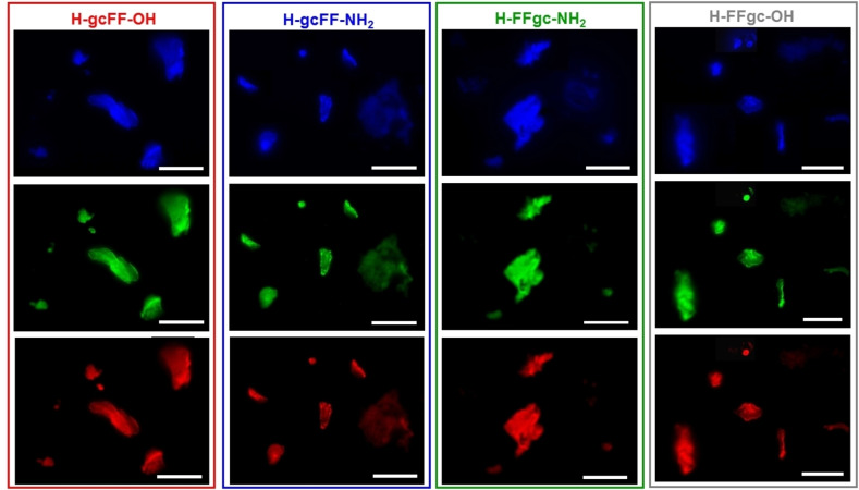
Figure 5.
Fluorescence microscopy images of self‐assembled PNA‐FF derivatives drop‐cast on glass slides and air‐dried at room temperature. Samples are obtained by drop‐casting 10 μL of each PNA‐FF solution at a concentration of 20.0 mg ⋅ mL−1. Images are obtained by exciting samples in the spectral region of: DAPI (4’,6‐diamidino‐2‐phenylindole; λexc=359 nm, λem=461 nm); GFP (Green fluorescent protein; λexc=488 nm, λem=507 nm) and Rho (Rhodamine; λexc=555 nm, λem=580 nm). The scale bar of all the images corresponds to 50 μm.

