Abstract
Objective:
To compare the skeletal, dentoalveolar, and soft tissue effects of the miniplate anchored Forsus Fatigue Resistant Device (FRD) and the conventional Forsus FRD in the treatment of Class II malocclusion.
Materials and Methods:
The study was carried out with 30 patients (10 girls, 20 boys). In the MA-Forsus group, 15 patients (2 girls, 13 boys) were treated with a miniplate anchored Forsus FRD for 9.40 ± 2.25 months. In the C-Forsus group, 15 patients (8 girls, 7 boys) were treated with a conventional Forsus FRD for 9.46 ± 0.81 months. A total of 16 measurements were calculated and statistically analyzed to find intragroup and intergroup differences.
Results:
Statistically significant differences were found between the groups in IMPA, SN/Occ, SN/GoGn, overjet, overbite, and Li-S measurements (P < .05). In the C-Forsus group, a substantial amount of lower incisor protrusion was observed, whereas retrusion was found in the MA-Forsus group (P < .001). The mandible rotated backward in the MA-Forsus group, whereas it remained unchanged in the C-Forsus group (P < .05). Reductions in overjet (P < .001) and overbite were greater in the C-Forsus group (P < .05).
Conclusion:
Stimulation of mandibular growth and inhibition of maxillary growth were achieved in both treatment groups. In the C-Forsus group, a substantial amount of lower incisor protrusion was observed, whereas retrusion of lower incisors was found in the MA-Forsus group. The MA-Forsus group was found to be more advantageous as it had no dentoalveolar side effects on mandibular dentition.
Keywords: Class II malocclusion, Forsus FRD, Fixed functional appliances, TAD, Miniplates
INTRODUCTION
The prevalence of Class II malocclusion was reported to be nearly 24% in an orthodontically referred population.1 Treatment of Class II malocclusion in a growing child usually involves stimulation of mandibular growth, inhibition of maxillary forward growth, or both. Fixed functional appliances have been used for decades in mandibular retrognathic patients. The appliances force the mandible forward, and by means of adaptational growth in the mandibular condyle and glenoid fossa remodeling, a significant increase in the mandibular effective length and a correction in facial convexity are attained. Fixed functional appliances have two major advantages: they are compliance-free alternatives to extraoral or intraoaral appliances, and they exert force full time. However, one major side effect of functional appliances is undesirable tooth movement in the anchor unit, which signifies anchorage loss. The protrusion of the lower incisors limits skeletal correction, and the results are more prone to relapse. To avoid this problem, temporary anchorage devices were introduced to the orthodontic community, and they are well accepted and used for stable anchorage units worldwide.
In 2001, the Forsus Fatigue Resistant Device (FRD; 3M Unitek Corp, Monrovia, Calif) was first introduced as a hybrid Herbst appliance.2 The latest modification, the Forsus FRD EZ2 appliance, is a semirigid telescoping system that consists of an EZ2 module and a pushing rod. It is attached to the maxillary molar headgear tube and mandibular archwire, creating a mesial force on the mandibular arch and a distal force on the maxillary arch.
In the literature, several studies have investigated the effects of a Forsus FRD used alone between dental arches3–10 or anchored with miniscrews11 or miniplates.12–14 However, to our knowledge, no study has compared the skeletal, dentoalveolar, and soft tissue effects of miniplate anchored Forsus FRDs and conventional Forsus FRDs in the treatment of Class II malocclusion.
The aim of this prospective study was to compare the skeletal, dentoalveolar and soft tissue effects of miniplate anchored Forsus FRDs and conventional Forsus FRDs in the treatment of Class II malocclusion.
Our null hypothesis was that there are no differences between the skeletal, dentoalveolar, and soft tissue effects of miniplate anchored Forsus FRDs and conventional Forsus FRDs in the treatment of Class II malocclusion.
MATERIALS AND METHODS
This study was approved by the ethics committee of Suleyman Demirel University, Faculty of Medicine. Written informed consent was obtained from patients and parents before evaluation.
Subjects meeting the following criteria were included in the study:
Healthy children with permanent dentition and in active growth stages
Angle Class II molar relationship
Convex profile characterized by mandibular deficiency
Minimum 7 mm overjet
Minimum crowding in the upper and lower dental arch
No previous orthodontic treatment
No systemic disease or craniofacial anomaly
Thirty-five patients who met the inclusion criteria were included in the study. However, three of them were excluded due to poor oral hygiene and frequent breakage of orthodontic bands and brackets, and two patients were excluded due to excess mobility of the miniplates. The study was carried out with 30 patients (10 girls, 20 boys). Because the treatment protocol involved surgical procedures in one group, a fully randomization procedure could not be performed. Fifteen patients (2 girls, 13 boys) who accepted surgical intervention were assigned to the MA-Forsus group, and treated with the Forsus FRD EZ2 attached to two miniplates for 9.40 ± 2.25 months. Fifteen patients (8 girls, 7 boys) who rejected surgical intervention were assigned to the C-Forsus group, and treated with the conventional Forsus FRD EZ2 for 9.46 ± 0.81 months. Mean chronological age was 12.77 ± 1.24 years for the MA-Forsus group and 13.26 ± 0.82 years for the C-Forsus group (Table 1).
Table 1.
Statistical Comparison of the Mean Chronologic Age of the Groups Before Treatment (T0), After the Leveling Stage (T1), and After the Fixed Functional Treatment Stage (T2)a
Treatment Protocol
In both groups, 0.018-inch preadjusted Roth brackets were used. In the MA-Forsus group, only teeth in the upper arch were bonded, while all maxillary and mandibular teeth, including second molars, were bonded in the C-Forsus group. The leveling stage started with 0.012-inch Ni-Ti archwires and continued until 0.016 × 0.022 inch stainless steel archwires were fitted. After the leveling phase, all maxillary teeth were ligated in a figure-8 pattern, and a 0.016 × 0.022 inch stainless steel archwire was inserted in the maxillary arch and cinched back to enforce anchorage.
In the C-Forsus group, Forsus FRDs were attached to the headgear tubes of the maxillary molar bands and to the mandibular archwire between the canine and first premolar brackets (Figure 1). In the MA-Forsus group, all patients underwent a surgical operation under local anesthesia for miniplate insertion. For the procedure, 10-mm-long horizontal incisions were made approximately 5 mm above the mucogingival junction in the anterior region of the mandible and mucoperiosteal flaps were raised. Two miniplates were bent and adjusted to the bone surface to achieve maximum bone contact. Next, 7-mm screws were inserted at the top of the plate and 9-mm screws in the lowest hole. A space of 1.5–2 mm was left between the plate arm and the mucosa to avoid irritation of the soft tissues. The incisions were closed with sutures and removed on postoperative day 7 (Figure 2). Forsus FRDs were attached to the headgear tubes of the maxillary molar bands and to the long arms of the miniplates (Figure 3).
Figure 1.
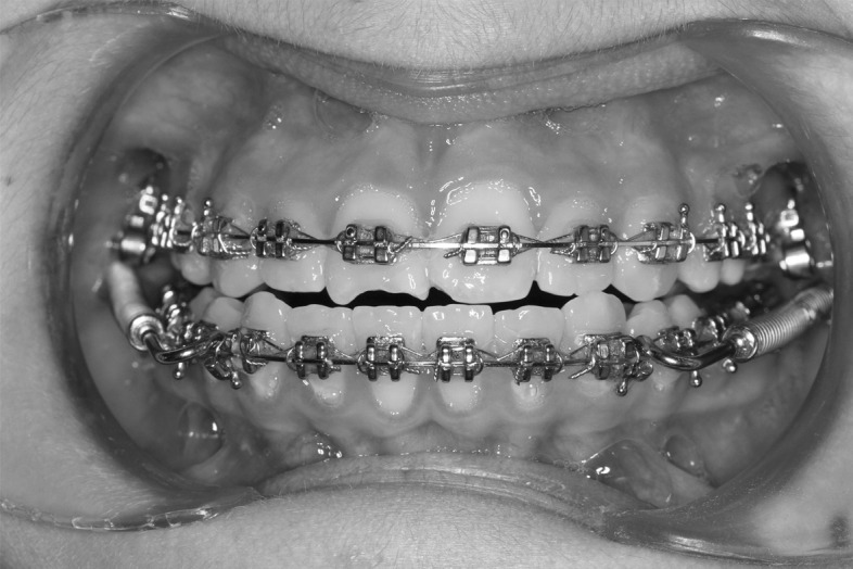
Application of Forsus Fatigue Resistant Devices to dental arches.
Figure 2.
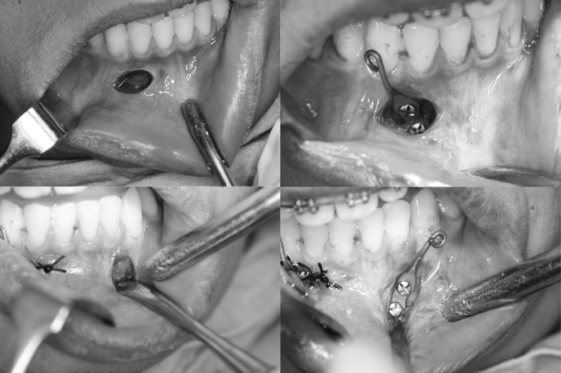
Surgical steps of miniplate insertion.
Figure 3.
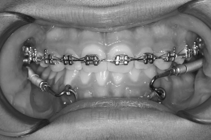
Application of Forsus Fatigue Resistant Devices to miniplates.
Patients were followed up every month and the appliances were checked. Forsus FRDs were reactivated with activation rings if needed. Treatment ended when a Class I molar relationship and successful elimination of the overjet were achieved.
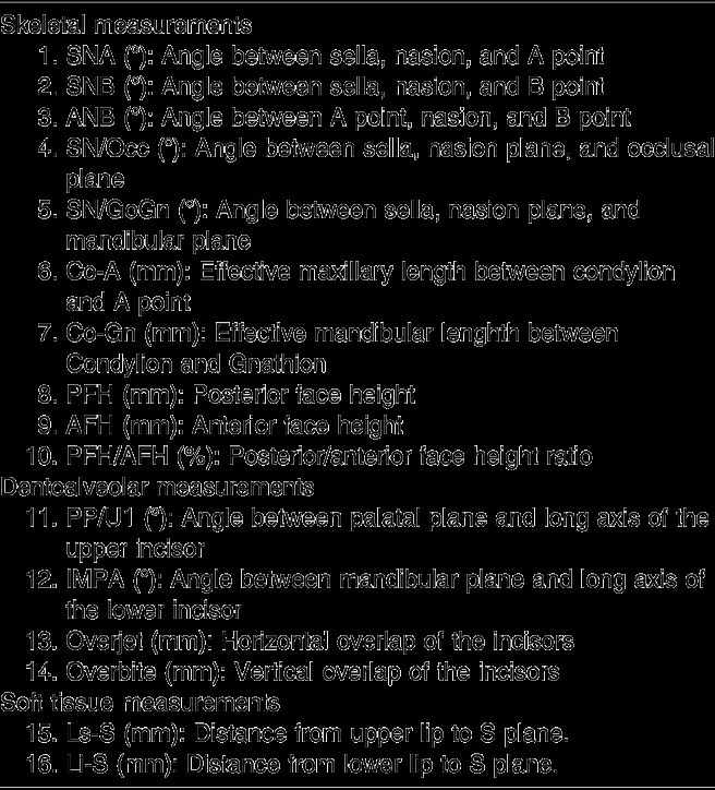
The study included 90 lateral cephalometric radiographs that were taken before treatment (T0), after the leveling stage (T1), and after the fixed functional treatment stage (T2). A total of 17 landmarks were identified on each cephalogram, and a total of 16 measurements (7 angular, 9 linear; Table 2) were performed with a Dolphin Imaging System (Dolphin Imaging and Management Solutions, Chatsworth, Calif)
Table 2.
Description of the Measurements
Statistical Analysis
For statistical analysis, SPSS version 18.0 for Windows (SPSS Inc, Chicago, Ill) was used. The Kolmogorov-Smirnov test was used to test the normalities of the data distributions. A Mann-Whitney U test was used to compare treatment changes according to gender, and no statistically significant difference was found. Therefore, gender differences between groups were ignored. An independent samples t-test was used for intergroup differences, and a paired samples t-test was used for intragroup differences.
To calculate method error and intraexaminer reliability, 20 randomly selected radiographs were retraced and remeasured, and a Cronbach's alpha test for reliability showed that the intraclass correlation was over 0.991.
The sample size of the study was calculated with G*Power version 3.1.9.2,15 based on a significance level of .05 and a power of 80% to detect a clinically significant difference of 2.0 mm for the effective mandibular length.
RESULTS
The success rate of the miniplates was 91% (29 of 32 miniplates), which was similar to that reported in the literature.12
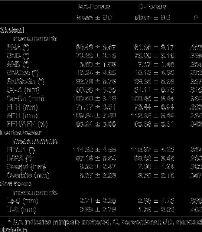
The results of the independent samples t-test revealed no statistically significant difference between the mean chronologic age of the groups (P > .05) (Table 1). Baseline comparison of the cephalometric variables of the groups was performed after the leveling stage (T1) to exclude leveling effects, and no significant differences were found between groups except overbite (P < .05), which was greater in the MA-Forsus group (Table 3). Intragroup and intergroup comparisons involved treatment changes between T1 and T2 (Table 4).
Table 3.
Comparison of the Groups After the Leveling Stage (T1)a
Table 4.
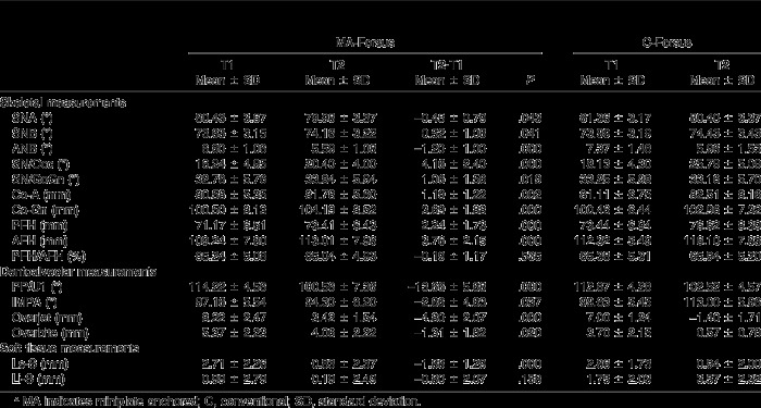
Intragroup and Intergroup Comparison of the Changes Between the Leveling Stage (T1) and the Fixed Functional Treatment Stage (T2)a
Table 4.
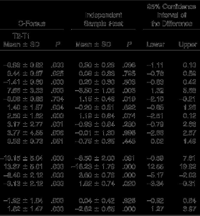
Extended
Intragroup Comparison
The results of the paired t-test are shown in Table 4. In the MA-Forsus group, statistically significant decreases in SNA (P < .05) and ANB angles (P < .001) and an increase in SNB angle (P < .05) were found. Retrusion of the upper (P < .001) and lower (P < .05) incisors were evident. Significant posterior rotation of the occlusal (P < .001) and mandibular planes (P < .05) was observed. Effective maxillary (P < .01) and mandibular (P < .001) lengths were increased. Increases in posterior and anterior face heights were statistically significant (P < .001). Overjet (P < .001) and overbite (P < .05) were decreased. The upper lip moved backward significantly (P < .001). The PFH/AFH ratio and lower lip position remained unchanged (P > .05).
In the C-Forsus group, statistically significant decreases in SNA and ANB angles (P < .001) and an increase in the SNB angle (P < .05) were found. The retrusion of upper (P < .001) and the protrusion of lower (P < .001) incisors were evident. Significant posterior rotation of the occlusal plane was found (P < .001), but the mandibular plane angle remained unchanged (P > .05). Effective maxillary (P < .01) and mandibular (P < .001) lengths were increased. Increases in posterior and anterior face heights were statistically significant (P < .01). Overjet and overbite were decreased (P < .001). Significant backward movement of the upper lip and forward movement of the lower lip were found (P < .001). The PFH/AFH ratio remained unchanged (P > .05).
Intergroup comparison
The results of the independent samples t-test are shown in Table 4. Statistically significant differences were found between the groups in IMPA, Occ-SN, SN/GoGn, overjet, overbite, and Li-S measurements (P < .05). In the C-Forsus group, a substantial amount of lower incisor protrusion was observed, while retrusion was found in the MA-Forsus group (P < .001). Posterior rotation of the occlusal plane was greater in the C-Forsus group (P < .01). The mandibular plane rotated backward in the MA-Forsus group, while it remained unchanged in the C-Forsus group (P < .05). Reductions in overjet (P < .001) and overbite (P < .05) were greater in the C-Forsus group. Significant protrusion of the lower lip was found in the C-Forsus group, while the lower lip in the MA-Forsus group remained unchanged (P < .001).
DISCUSSION
Fixed functional appliances have been used to correct skeletal Class II anomalies for decades, but their side effects on dentoalveolar structures limit the skeletal effect and correction.7,16 Studies have recently focused on eliminating these undesirable effects.12–14
The effects of the Forsus FRD appliance on maxillary growth were investigated by SNA and Co-A measurements. In both groups, a significant decrease was found in the SNA angle. This may be due to posteriorly directed forces acting on the maxilla. On the other hand, the significant increase in effective maxillary length (Co-A) may be due to adaptational growth in the mandibular condyle.16 Our results clearly showed that the conventional and miniplate anchored Forsus FRD both inhibited maxillary forward growth and had a headgear effect on the maxilla. Our results were in accordance with previous studies that have reported similar findings with Forsus appliances.3,6–9,12–14,16,17 In contrast, several studies have reported that the Forsus FRD had no significant effect on maxillary growth.2,5,10,11,18
The effects of the Forsus FRD on mandibular growth were investigated by SNB and Co-Gn measurements. In both groups, the anterior and downward force vector of the appliance significantly stimulated mandibular growth. The significant increase in effective mandibular length (Co-Gn) may have been due to adaptational growth in the mandibular condyle. Our results were in accordance with those of previous studies that reported stimulated mandibular growth with Forsus appliances.2,6,8,12–14,16–18 The increase in effective mandibular length in the MA-Forsus group was 3.69 mm, while it was 2.50 mm in the C-Forsus group. The difference between the groups was not statistically significant. However, the almost 50% greater increase in mandibular length by miniplate anchorage may be explained by a more stable anchorage unit and less anchorage loss. In contrast, several studies reported that the Forsus FRD little or no skeletal effect on mandibular growth.3,5,7,10,11
The rotational manner of the mandible under Forsus forces was investigated with the SN/GoGn angle. Posterior mandibular rotation was evident and significant in the MA-Forsus group, while the changes in the C-Forsus group were not significant. With miniplate anchorage, forward and downward forces of the Forsus appliance were directly transmitted to the anterior skeletal base of the mandible, causing greater and more significant posterior rotation. Moreover, the center of force application in the MA-Forsus group was located more downward vertically compared with the C-Forsus group. This might be another reason for increased mandibular rotation with skeletal anchorage. In accordance with our results, Unal et al.12 and Celikoglu et al.13 also reported significant mandibular posterior rotation with a skeletally anchored Forsus appliance. In contrast, Aslan et al.11 reported nonsignificant changes in the mandibular plane angle with miniscrew-supported and conventional Forsus appliances.
The effects of Forsus appliances on face heights were evaluated with posterior face height (S-Go), anterior face height (N-Me), and posterior/anterior face height ratio measurements. Significant increases in all measurements were found in both treatment groups. These increases may be due to the modified forward posture of the mandible by the appliances. The new forward position enhances condylar growth vertically and increases both posterior and anterior face height. Similar results were previously reported in the literature.11,12,16 However, Oztoprak et al.5 reported no significant change in face heights and explained this situation with the normal or horizontal growth patterns in their late adolescent patient sample.
In the present study, significant retrusion of the maxillary incisors was observed. This may be due to the posteriorly and upwardly directed forces acting on the maxillary molars. Distalization and intrusion of the maxillary molars can cause extrusion and retrusion of the maxillary incisors due to the heavy archwire connecting both segments. Our findings were almost completely in accordance with those of previous Forsus studies,2,3,5,6,8–13,16–18 indicating that extrusion and retrusion of the maxillary incisors are inevitable side effects of Forsus appliances.
One major disadvantage of fixed functional appliances is unwanted mesial tipping of the mandibular dentition and protrusion of the lower incisors. This situation causes early correction of the overjet and limits skeletal correction. In the present study, significant labial tipping of the lower incisors was found in the C-Forsus group. This finding was in accordance with findings of previous studies.2,3,5–11,16,18 However, in the MA-Forsus group, instead of protrusion, significant retrusion of the lower incisors was determined. This result was promising, as most unwanted complications of the fixed functional appliances were eliminated with this method. Moreover, in most Class II division 1 cases, lower incisors are already protrusive due to the dental compensatory mechanism and need to be retracted. Retrusion of the lower incisors with skeletal anchorage has also been reported in previous studies and case reports.12–14 Unal et al.12 and Celikoglu et al.13 explained this finding with the pressure of the maxillary incisors and the lower lip. In contrast, Aslan et al11 used Forsus with two miniscrews and reported that the protrusion of lower incisors was effectively minimized compared with the conventional Forsus, but it was not totally eliminated.
In the present study, significant retrusion of the upper lip was observed with both techniques, which was in accordance with previous studies.5,6,10–12 This was due to heavy distalizing forces acting on the upper arch and resultant retrusion of the upper incisors. The two techniques did not differ regarding changes in the upper lip. However, the lower lip was protruded in the C-Forsus group, while it remained almost unchanged in the MA-Forsus group. This difference was mainly due to changes in the position of the lower incisors, which were directly reflected on soft tissue. In accordance with our results, Oztoprak et al.5 reported no statistically significant changes in lower lip position with Forsus FRD. However, Unal et al.12 and Celikoglu et al.13 reported significant forward movement of the lower lip with skeletally anchored Forsus. This might be due to variations in soft tissue reference lines and measurements.
Although the results with miniplate anchorage were promising, the new technique has several disadvantages and limitations. First, at least two surgical operations are needed to insert and remove miniplates. Second, poor oral hygiene may cause severe inflammation and mobility around the miniplates. Third, increased costs of orthodontic treatment limit its usability.
CONCLUSIONS
Stimulation of mandibular growth and inhibition of maxillary growth were achieved in both treatment groups.
In the C-Forsus group, a substantial amount of lower incisor protrusion was observed, whereas retrusion of lower incisors was found in the MA-Forsus group.
Overjet reduction was greater with the conventional Forsus FRD due to significant amounts of lower incisor protrusion.
The miniplate anchored Forsus FRD was found to be more advantageous as it had no dentoalveolar side effects on the mandibular dentition.
ACKNOWLEDGMENTS
We wish to thank Suleyman Demirel University's Scientific Research Coordination Unit for funding this project (Grant no: 3426-D2-13) and Dr. Hikmet Orhan from the Department of Biostatistics, Faculty of Medicine of Suleyman Demirel University, for his help with statistical analyses.
REFERENCES
- 1.Sayin MO, Turkkahraman H. Malocclusion and crowding in an orthodontically referred Turkish population. Angle Orthod. 2004;74:635–639. doi: 10.1043/0003-3219(2004)074<0635:MACIAO>2.0.CO;2. [DOI] [PubMed] [Google Scholar]
- 2.Heinig N, Goz G. Clinical application and effects of the Forsus spring. A study of a new Herbst hybrid. J Orofac Orthop. 2001;62:436–450. doi: 10.1007/s00056-001-0053-6. [DOI] [PubMed] [Google Scholar]
- 3.Cacciatore G, Ghislanzoni LT, Alvetro L, Giuntini V, Franchi L. Treatment and posttreatment effects induced by the Forsus appliance: a controlled clinical study. Angle Orthod. 2014;84:1010–1017. doi: 10.2319/112613-867.1. [DOI] [PMC free article] [PubMed] [Google Scholar]
- 4.Adusumilli SP, Sudhakar P, Mummidi B, et al. Biomechanical and clinical considerations in correcting skeletal class II malocclusion with Forsus. J Contemp Dent Pract. 2012;13:918–924. doi: 10.5005/jp-journals-10024-1254. [DOI] [PubMed] [Google Scholar]
- 5.Oztoprak MO, Nalbantgil D, Uyanlar A, Arun T. A cephalometric comparative study of class II correction with Sabbagh Universal Spring (SUS(2)) and Forsus FRD appliances. Eur J Dent. 2012;6:302–310. [PMC free article] [PubMed] [Google Scholar]
- 6.Franchi L, Alvetro L, Giuntini V, Masucci C, Defraia E, Baccetti T. Effectiveness of comprehensive fixed appliance treatment used with the Forsus Fatigue Resistant Device in Class II patients. Angle Orthod. 2011;81:678–683. doi: 10.2319/102710-629.1. [DOI] [PMC free article] [PubMed] [Google Scholar]
- 7.Jones G, Buschang PH, Kim KB, Oliver DR. Class II non-extraction patients treated with the Forsus Fatigue Resistant Device versus intermaxillary elastics. Angle Orthod. 2008;78:332–338. doi: 10.2319/030607-115.1. [DOI] [PubMed] [Google Scholar]
- 8.Heinrichs DA, Shammaa I, Martin C, Razmus T, Gunel E, Ngan P. Treatment effects of a fixed intermaxillary device to correct class II malocclusions in growing patients. Prog Orthod. 2014;15:45. doi: 10.1186/s40510-014-0045-x. [DOI] [PMC free article] [PubMed] [Google Scholar]
- 9.Giuntini V, Vangelisti A, Masucci C, Defraia E, McNamara JA, Jr, Franchi L. Treatment effects produced by the Twin-block appliance vs the Forsus Fatigue Resistant Device in growing Class II patients. Angle Orthod. 2015;85:784–789. doi: 10.2319/090514-624.1. [DOI] [PMC free article] [PubMed] [Google Scholar]
- 10.Gunay EA, Arun T, Nalbantgil D. Evaluation of the immediate dentofacial changes in late adolescent patients treated with the Forsus(TM) FRD. Eur J Dent. 2011;5:423–432. [PMC free article] [PubMed] [Google Scholar]
- 11.Aslan BI, Kucukkaraca E, Turkoz C, Dincer M. Treatment effects of the Forsus Fatigue Resistant Device used with miniscrew anchorage. Angle Orthod. 2014;84:76–87. doi: 10.2319/032613-240.1. [DOI] [PMC free article] [PubMed] [Google Scholar]
- 12.Unal T, Celikoglu M, Candirli C. Evaluation of the effects of skeletal anchoraged Forsus FRD using miniplates inserted on mandibular symphysis: a new approach for the treatment of Class II malocclusion. Angle Orthod. 2015;85:413–419. doi: 10.2319/051314-345.1. [DOI] [PMC free article] [PubMed] [Google Scholar]
- 13.Celikoglu M, Buyuk SK, Ekizer A, Unal T. Treatment effects of skeletally anchored Forsus FRD EZ and Herbst appliances: a retrospective clinical study. Angle Orthod doi: 10.2319/040315-225.1. . In press. [DOI] [PMC free article] [PubMed] [Google Scholar]
- 14.Celikoglu M, Unal T, Bayram M, Candirli C. Treatment of a skeletal Class II malocclusion using fixed functional appliance with miniplate anchorage. Eur J Dent. 2014;8:276–280. doi: 10.4103/1305-7456.130637. [DOI] [PMC free article] [PubMed] [Google Scholar]
- 15.Faul F, Erdfelder E, Lang AG, Buchner A. G*Power 3: a flexible statistical power analysis program for the social, behavioral, and biomedical sciences. Behav Res Methods. 2007;39:175–191. doi: 10.3758/bf03193146. [DOI] [PubMed] [Google Scholar]
- 16.Karacay S, Akin E, Olmez H, Gurton AU, Sagdic D. Forsus Nitinol Flat Spring and Jasper Jumper corrections of Class II division 1 malocclusions. Angle Orthod. 2006;76:666–672. doi: 10.1043/0003-3219(2006)076[0666:FNFSAJ]2.0.CO;2. [DOI] [PubMed] [Google Scholar]
- 17.Bilgic F, Basaran G, Hamamci O. Comparison of Forsus FRD EZ and Andresen activator in the treatment of class II, division 1 malocclusions. Clin Oral Investig. 2015;19:445–451. doi: 10.1007/s00784-014-1237-y. [DOI] [PubMed] [Google Scholar]
- 18.Aras A, Ada E, Saracoglu H, Gezer NS, Aras I. Comparison of treatments with the Forsus fatigue resistant device in relation to skeletal maturity: a cephalometric and magnetic resonance imaging study. Am J Orthod Dentofacial Orthop. 2011;140:616–625. doi: 10.1016/j.ajodo.2010.12.018. [DOI] [PubMed] [Google Scholar]


