Abstract
Purpose:
The aim of this study was to carry out blue light photography of fluorescein-stained corneas using a novel smartphone attachment.
Methods:
A smartphone attachment known as the cobalt blue light unit (C-BLU) was developed. It can filter out all wavelengths of light except the blue light emerging from the flashlight of a smartphone. A pilot study was carried out wherein the images captured with the C-BLU system were compared with slit-lamp photographs of the same patients. This setup was then used to photo document fluorescein-stained corneas in various clinical settings assembled at point-of-care.
Results:
Many pathologies of the fluorescein-stained cornea were captured using the C-BLU filter. It was used effectively in various settings (remote eye camps, intensive care units (ICU), pediatric group, corneal trauma triaging, etc.). C-BLU was assembled and used by optometrists and ophthalmology residents. The images captured were used for documenting, assisting in the treatment, and also for telecommunication of the patients’ findings.
Conclusion:
C-BLU is a low-cost pocket-size filter which is easy to use with a modern smartphone without any technical expertise needed to obtain a clear image of fluorescein-stained pathological corneas.
Keywords: Blue light, cornea, imaging, smartphone, telemedicine
Fluorescein staining is an integral part of the examination of the ocular surface, allowing adequate documentation of ocular surface lesions.[1] In 1882, Pfluger first identified discontinuity in the corneal epithelium by using sodium fluorescein.[2] Traditionally, the cobalt blue filters mounted on the commercially available photo slit lamps are used to take photographs of fluorescein-stained corneas. Portable slit lamps with similar filters are available but are expensive and not of widespread use in developing countries.[3]
While examining infants and critically ill patients in the intensive care unit (ICU), it is almost impossible to photo document the stained cornea of these patients effectively. Conducting eye camps is also an important part of Ophthalmology services in developing countries. Slit-lamp facility and photography may not be available always in such situations, and hence it is difficult to document the corneal fluorescein stain findings. There is a paucity of affordable devices to exclusively document the fluorescein-stained corneal images in these situations.
Modern smartphones are already being commonly used by ophthalmologists to capture images of the anterior segment of the eye.[4,5,6,7,8,9] We repurposed the flashlight of smartphones as the light source and developed the cobalt blue light unit (C-BLU) filter. We report the development of our first-generation, compact, user-friendly, smartphone attachment and some clinical photographs obtained with it.
Innovation
Specifications
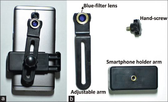
C-BLU is made of polycarbonate and has three integral components. The first and most critical component is the blue filter lens, which is aligned in front of the flashlight of the smartphone. The second component is the adjustable arm that houses the blue filter lens and measures 11.5 cm × 2.5 cm × 1.5 cm. This arm helps in aligning the blue lens in front of the flashlight appropriately. The final component is the expandable smartphone holder arm, and it facilitates the attachment of smartphones to the assembly. It has dimensions of 7.0 cm × 3.5 cm × 2.5 cm in the contracted state and its holder ranges from 5.8 to 10.5 cm, and can house most modern smartphones. The inside of the holder is lined with a soft rubber foam to ensure that the phone is safe and protected in its grip. A hand-screw helps in fixing and titrating the position of the adjustable arm relative to the smartphone holder arm. The minimum dimension of the entire assembled C-BLU is 11.5 cm × 4.0 cm × 3.5 cm and can easily fit into one’s pocket. C-BLU is compatible with both Android and Apple smartphones, yielding comparable results. When C-BLU is combined with any modern smartphone camera, a portable blue light photography unit is attained [Fig. 1]. Approval has been obtained on 20/03/2019.
Figure 1.

(a) Smartphone with C-BLU filter mounted—the blue lens is aligned in front of its flashlight. (b) The various components of C-BLU filter that can be assembled with ease
Technique
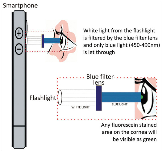
Keeping in mind that a majority of people in the developing world are android phone users, we have used an android device—OnePlus 3T (OnePlus Technology Co., Ltd., Shenzhen, Guangdong, China) for developing our proof of concept [Video Clip 1]. The patient is seated or asked to lie supine in a dimly lit room. The traditional staining method of the cornea after wetting with 1% sodium fluorescein strip is followed here as well.[10] C-BLU filter is mounted onto the smartphone and the blue lens aligned in front of the flashlight using the hand-screw. The basic principle of C-BLU is that the blue lens ensures the passage of blue light with a wavelength in the range 450–490 nm while filtering out other wavelengths when the flashlight is switched on [Fig. 2]. After adequate alignment is achieved, the native camera app is opened, and the camera or video mode is enabled. Blue light is emitted when the flashlight turns on during the capture process or can be enabled continuously if in video mode. If a video has been captured, the best frame can be isolated later into a photograph. Most of the latest smartphones have optical zoom and image stabilization, enabling superior quality C-BLU images.
Figure 2.

Optics of the cobalt blue light unit (C-BLU) filter system
Methods
The C-BLU system was used first used in a pilot case with a known diagnosis of a corneal pathology and the images were compared with slit-lamp photographs. The images captured by both modalities were compared by a single reviewer in terms of their ability to detect and describe the staining pattern seen in different corneal pathologies, and also to arrive at a diagnosis of the staining pattern. After establishing the effectiveness of the C-BLU system, it was then used in various settings to deliver point-of-care identification of various corneal lesions. It was used in remote eye camps, in assessing the response of corneal ulcers to treatment, in ICU, in the emergency area for triaging corneal trauma, in a case of corneal ulcer, and also in a pediatric patient.
Results
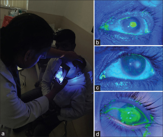
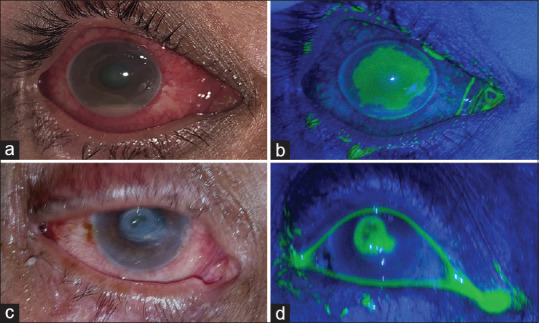
Varied pathologies of the fluorescein-stained cornea were captured using the C-BLU filter [Fig. 3a]. A patient with a central corneal ulcer secondary to trauma with the vegetative matter was documented with C-BLU during a remote eye camp [Fig. 3b]. C-BLU filter is also helpful in assessing the response of corneal ulcers to treatment. Another patient of Sjogren syndrome, with superficial punctate staining of a cornea due to chronic dry eye, was captured using C-BLU [Fig. 3c]. The cornea of a 1-year-old child with xerophthalmia secondary to vitamin A deficiency was photo-documented with the C-BLU system [Fig. 3d]. Corneal photographs under diffuse illumination prior to fluorescein stain application were captured followed by staining and capturing with C-BLU system [Fig. 4]. A patient with a deep central ulcer and descemetocele was also photographed using C-BLU which showed the characteristic “green donut” appearance of descemetocele with a ring of stain-positive stroma surrounding a dark center [Fig. 4d]. C-BLU was also effectively used in the emergency area to document fluorescein stain findings in corneal trauma, chemical burns, and abrasions. Since most modern smartphones have an efficient built-in camera, when combined with C-BLU, they were able to capture clear images of the fluorescein-stained cornea to allow triaging and screening during remote eye camps as well.
Figure 3.

(a) Fluorescein-stained cornea of a child being photographed using a cobalt blue light unit (C-BLU) filter mounted on a smartphone. (b) Still images captured using C-BLU technique during remote eye camp that demonstrate stain-positive central corneal ulcer. (c) Diffuse corneal staining due to chronic dry eye. (d) A 1-year-old child presented with xerophthalmia which was documented using C-BLU
Figure 4.

(a) Large central corneal ulcer with hypopyon seen under diffuse illumination. (b) The same ulcer as in (a) photographed using the C-BLU filter system after staining with fluorescein. (c) A deep central corneal ulcer with descemetocele under diffuse illumination. (d) C-BLU system used to capture fluorescein-stained ulcer seen in (c)—the characteristic “green donut” appearance of descemetocele with a ring of stain-positive stroma surrounding a dark center can be seen
Discussion
The C-BLU filter is an effective and cheap method to capture fluorescein-stained findings of the cornea. Although modern smartphones can capture good-quality ocular images, none has a blue filter to capture images of fluorescein-stained images of the cornea.[4,5,6,7] Even though there are a number of adapters available for anterior segment photography, most do not possess an external light source or blue filter.[8,9,11,12,13] The C-BLU filter was developed keeping three salient things in mind: easy to assemble, easy to operate, and easy on the pocket. The cost of production of a C-BLU filter is less than $20 including all the raw materials.
C-BLU filter has performed exceptionally well, reproducing good-quality usable images. C-BLU was used in ICU settings where it is difficult to document perforations. In remote eye camps, corneal pathological staining can be identified without a slit lamp, and hence, aiding in telecommunication. This filter can be used both in rural and remote areas with ease; images can be transferred easily for an expert opinion if needed. C-BLU assisted in documenting disease progression or regression, and also in explaining the disease to patients and their families.
In most situations where the more expensive slit lamp is not available, we can use the C-BLU filter as a cheaper alternative for corneal fluorescein stain photography. This can be assembled by an optometrist, ophthalmologist, intensivist, or even a health care worker during eye camps. This can be implemented with much ease in various health care facilities without any modification of existing protocols. This is a point-of-care device that enables us to diagnose and prognosticate various corneal stain pathologies.
A minor limitation of this device is reduced field of view as the flashlight and the camera module are inherently placed in near each other within certain smartphones. So, we are now working on refining the design to address this limitation.
The field of teleophthalmology is showing promising developments with many advancements and the quick relay through the Internet makes such technology much accessible to the vast majority. We envisage that this simple device can easily be utilized by any health care worker to capture the corneal images at a remote place and can become an effective tool in telemedicine.
Conclusion
C-BLU is a pocket-size filter that is low-cost and is easy to use with a modern smartphone without any technical expertise needed to obtain a clear image of the fluorescein-stained cornea. This will open a whole array of opportunities in teleophthalmology for photo documentation and monitoring at an affordable level in settings where imaging cannot be done otherwise.
Financial support and sponsorship
Nil.
Conflicts of interest
There are no conflicts of interest.
Video Available on: www.ijo.in
References
- 1.Campbell FW, Boyd TAS. Use of sodium fluorescein in assessing the rate of healing in corneal ulcers. Br J Ophthalmol. 1950;34:545–9. doi: 10.1136/bjo.34.9.545. [DOI] [PMC free article] [PubMed] [Google Scholar]
- 2.Kim J. The use of vital dyes in corneal disease. Curr Opin Ophthalmol. 2000;11:241–7. doi: 10.1097/00055735-200008000-00005. [DOI] [PubMed] [Google Scholar]
- 3.Hand-Held Portable Slit Lamp from Reichert. Review of Ophthalmology. [Last accessed on 2020 May 26]. Available from: https://www.reviewofophthalmology.com/article/hand-held-portable-slit-lamp-from-reichert .
- 4.Chhablani J, Kaja S, Shah VA. Smartphones in ophthalmology. Indian J Ophthalmol. 2012;60:127–31. doi: 10.4103/0301-4738.94054. [DOI] [PMC free article] [PubMed] [Google Scholar]
- 5.Lord RK, Shah VA, San Filippo AN, Krishna R. Novel uses of smartphones in ophthalmology. Ophthalmology. 2010;117:1274–1274.e3. doi: 10.1016/j.ophtha.2010.01.001. [DOI] [PubMed] [Google Scholar]
- 6.Bastawrous A, Cheeseman RC, Kumar A. iPhones for eye surgeons. Eye (Lond) 2012;26:343–54. doi: 10.1038/eye.2012.6. [DOI] [PMC free article] [PubMed] [Google Scholar]
- 7.Chandrakanth P, Nallamuthu P. Anterior segment photography with intraocular lens. Indian J Ophthalmol. 2019;67:1690–1. doi: 10.4103/ijo.IJO_52_19. [DOI] [PMC free article] [PubMed] [Google Scholar]
- 8.Akkara JD, Kuriakose A. How-to guide for smartphone slit-lamp imaging. Kerala J Ophthalmol. 2019;31:64–71. [Google Scholar]
- 9.Akkara JD, Kuriakose A. Commentary:The glued intraocular lens smartphone microscope. Indian J Ophthalmol. 2019;67:1692. doi: 10.4103/ijo.IJO_986_19. [DOI] [PMC free article] [PubMed] [Google Scholar]
- 10.Holland MC. Fluorescein staining of the cornea. JAMA. 1964;188:81. doi: 10.1001/jama.1964.03060270087025. [DOI] [PubMed] [Google Scholar]
- 11.Teichman JC, Sher JH, Ahmed IIK. From iPhone to eyePhone:A technique for photodocumentation. Can J Ophthalmol. 2011;46:284–6. doi: 10.1016/j.jcjo.2011.05.016. [DOI] [PubMed] [Google Scholar]
- 12.Ludwig CA, Murthy SI, Pappuru RR, Jais A, Myung DJ, Chang RT. A novel smartphone ophthalmic imaging adapter:User feasibility studies in Hyderabad, India. Indian J Ophthalmol. 2016;64:191–200. doi: 10.4103/0301-4738.181742. [DOI] [PMC free article] [PubMed] [Google Scholar]
- 13.Chakrabarti R. Application of mobile technology in ophthalmology to meet the demands of low-resource settings. J Mob Technol Med. 2012;1:1–3. [Google Scholar]
Associated Data
This section collects any data citations, data availability statements, or supplementary materials included in this article.


