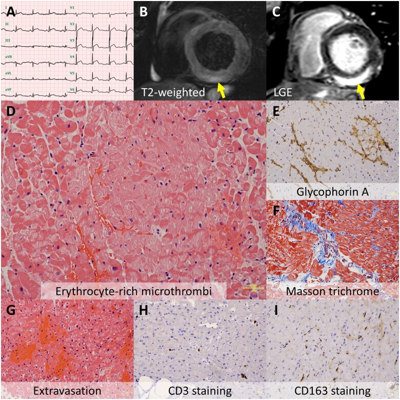
A 37-year-old man was admitted to the emergency room with acute chest pain 19 days after his first dose of mRNA-1273 SARS-CoV-2 vaccination (Moderna). He was afebrile and his Abbott ID NOW COVID-19 point-of-care test returned negative. Electrocardiography showed diffuse ST-segment elevation (Panel A); echocardiography showed subtle wall motion abnormality in the left ventricle; and his troponin T, creatinine kinase, and C-reactive protein levels were raised at 1660 ng/L, 1200 U/L, and 5.76 mg/dL, respectively. Urgent coronary angiography revealed no coronary abnormalities. Cardiovascular magnetic resonance demonstrated T2-weighted hyperintense (Panel B, arrow) and gadolinium-delayed hyperenhancement (Panel C, arrow; Supplementary material online, Videos S1 and S2) in the subepicardial myocardium of the left ventricle, indicating acute myocarditis. Right ventricular endomyocardial biopsy revealed that erythrocyte-rich microthrombi occluded capillary vessels (Panels D and E) accompanied by extravasation of erythrocytes without inflammatory cell infiltration in the myocardium (Panels F–I), thereby precluding the pathological diagnosis of myocarditis. The levels of D-dimer and haptoglobin were within the normal range. Serological testing ruled out systemic virus infections. He was discharged without any complications on Day 7 and had no symptoms after discharge. Although myocardial injury has been described as a rare adverse reaction of SARS-CoV-2 vaccination, caution should be exercised in individuals presenting with chest pain after the vaccination. The underlying mechanisms of COVID-19 vaccine-related myocardial injury remain to be elucidated; myocardial microthrombi without inflammatory cell infiltration may be proposed as a possible explanation. Our case highlights that histological examination is important to clarify the mechanism of COVID-19 vaccine-related myocardial injury.
Supplementary material is available at European Heart Journal online.
Funding: This work was supported in part by JSPS KAKENHI (grant number 19K17189 to T.A.), Fukuda Foundation for Medical Technology (to T.A.), the Akiyama Life Science Foundation (to T.A.), and Grants-in-Aid for Regional R&D Proposal-Based Program from Northern Advancement Center for Science & Technology of Hokkaido Japan (to T.A.). The authors thank Daishi Nakayama RT, Takashi Kato RT, Yuji Hiroshima RT, and Tamaki Kudo RT for their technical support.
Conflict of interest: The authors have submitted their declaration which can be found in the article Supplementary Material online.
Supplementary Material
Contributor Information
Tadao Aikawa, Department of Cardiology, Hokkaido Cardiovascular Hospital, 1-30, Minami-27, Nishi-13, Chuo-ku, Sapporo 064-8622, Japan; Department of Radiology, Jichi Medical University Saitama Medical Center, 1-847 Amanuma-cho, Omiya-ku, Saitama 330-8503, Japan.
Jiro Ogino, Department of Pathology, JR Sapporo Hospital, Kita-3, Higashi-1, Sapporo 060-0033, Japan .
Yuichi Kita, Department of Radiology, Hokkaido Cardiovascular Hospital, 1-30, Minami-27, Nishi-13, Chuo-ku, Sapporo 064-8622, Japan.
Naohiro Funayama, Department of Cardiology, Hokkaido Cardiovascular Hospital, 1-30, Minami-27, Nishi-13, Chuo-ku, Sapporo 064-8622, Japan.
Associated Data
This section collects any data citations, data availability statements, or supplementary materials included in this article.


