Abstract
Objective:
To investigate the relationship between a micropulse vibration device and pain perception during orthodontic treatment.
Materials and Methods:
This study was a parallel group, randomized clinical trial. A total of 58 patients meeting eligibility criteria were assigned using block allocation to one of two groups: an experimental group using the vibration device or a control group (n = 29 for each group). Patients used the device for 20 minutes daily. Patients rated pain intensity on a visual analog scale at appropriate intervals during the weeks after the separator or archwire appointment. Data were analyzed using repeated measures analysis of variance at α = .05.
Results:
During the 4-month test period, significant differences between the micropulse vibration device group and the control group for overall pain (P = .002) and biting pain (P = .003) were identified. The authors observed that perceived pain was highest at the beginning of the month, following archwire adjustment.
Conclusion:
The micropulse vibration device significantly lowered the pain scores for overall pain and biting pain during the 4-month study period.
Keywords: Pain, Vibration, Orthodontics
INTRODUCTION
Pain is a common side effect of orthodontic treatment. It is a complex phenomenon involving multiple variants and is influenced by factors such as age, gender, individual pain threshold, and amount of force applied.1 In orthodontics, a mechanical stimulus is introduced by placing fixed appliances on the teeth resulting in tooth movement. To achieve this movement, forces are applied to the dentoalveolar complex resulting in inflammation or ischemia to the periodontal ligament (PDL) with subsequent release of histamine, bradykinin, prostaglandins, substance P, and serotonin.2 These mediators stimulate local nerve endings and send pain signals to the brain.
Many methods have been used to alleviate pain arising from orthodontic origins. Nonsteroidal anti-inflammatory drugs (NSAIDS) are the most common method employed for pain relief. NSAIDS block the formation of arachidonic acid in the production cycle of prostaglandin, which would lead to pain.3 Other methods include low-level laser therapy,4 acupuncture,5 transcutaneous electrical nerve stimulation,6 vibratory stimulation of the PDL,7 viscoelastic bite wafers8, and even chewing gum.9 These methods can relieve compression of the PDL and restore normal vascular and lymphatic circulation to eliminate edema and inflammation, reducing pain.10
The focus of this study is vibratory stimulation to reduce pain. Previous studies have demonstrated that vibration effectively reduces pain originating from teeth or the surrounding tissues.11 Vibration may help relieve compression of the PDL, promoting normal circulation to prevent the proliferation of inflammatory by-products. Another possibility is the "gate control" theory, which suggests that pain can be reduced by simultaneous activation of nerve fibers that conduct non-noxious stimuli.12
AcceleDent® (OrthoAccel Technologies Inc, Bellaire, TX) is patented as a “vibrating orthodontic remodeling device” (U.S. Department of Commerce's United States Patent and Trademark Office, 2013). It is a U.S. Food and Drug Administration–approved, Class II medical device designed for faster orthodontic treatment.13 The manufacturer states that the device applies cyclic forces to the dentition for the safe acceleration of the bone remodeling process to complement conventional orthodontic treatment. In a series of rabbit experiments, Mao14 demonstrated that cyclical forces applied at 2 N with frequencies of 0.2 and 1 Hz for 20 minutes daily, in conjunction with typical static orthodontic forces 24 hours per day, induced increased cranial growth, sutural separation, and proliferation of osteoblast-like cells. The primary purpose for using AcceleDent® is to decrease overall orthodontic treatment time.
There are reports from clinicians who have noticed pain reduction as an additional benefit for those patients using AcceleDent®. The purpose of this clinical study was to investigate the relationship between micropulse vibration therapy for pain relief compared to a control group of no pain therapy during the first 4 months of orthodontic tooth movement and adjustment and to investigate whether age and gender were significant factors in the perception of orthodontic pain.
MATERIALS AND METHODS
This parallel group, randomized clinical trial was approved by the local institutional review board (IRB) and was determined to be no greater than minimal risk. Informed consent was obtained from each patient in the study, and a medical monitor was assigned to the study. The power/sample size calculation was based on Student’s t-test and compared the overall change from baseline between the two groups. It was determined that a sample size of 30 participants per group would be sufficient to answer the research question at the 95% confidence interval. Other data were compared graphically and, if indicated, statistically over the various time points.
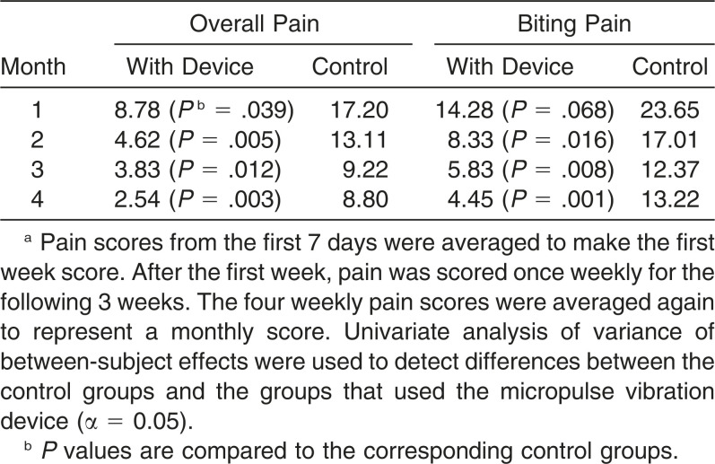
The study was carried out during a 4-month period with adjustments and wire changes made at the beginning of each month. Pain data from a visual analog scale (VAS) were collected from each patient daily on the first 7 days of each month after the adjustment appointment. These scores were averaged to determine the first week pain score. After the first week, pain was scored once weekly for the remainder of the month. The four weekly pain scores were averaged to represent a monthly score for comparisons. Each patient had a data set of 40 pain scores total: seven data points for the first week plus one data point for the following 3 weeks multiplied by 4 months. Age and gender were recorded as subvariables.
A total of 70 participants (35 per treatment group) were selected from patients who presented for initial orthodontic treatment. Participants were selected based on the following inclusion criteria: healthy child (aged 10 years and older) and adult patients approved for comprehensive orthodontic treatment. For adolescent patients meeting the requirements for inclusion, the parent or guardian and child were informed of the research study. Adult patients willing to participate in the study were enrolled; adolescent patients willing to participate were enrolled only with parent or guardian permission. Participants were excluded from recruitment if they currently had any pre-existing pain conditions or if they were not able to comply with the restriction on using any analgesic drugs during the course of the study.
Participants were assigned to comparison groups using a block allocation sequence. This sequence was concealed from the investigators. Participants were randomized in blocks of 10 with five patients being allocated to each arm of the trial until all 70 patients were randomized. For participant allocation, a computer-generated list of random numbers was used. The randomization sequence was created using Stata 9.0 statistical software (StataCorp, College Station, Tex) with a 1:1 allocation using a random block size of 10. A designated individual (not part of the investigative team) performed the allocation. All participants were given an informed consent form that was approved by the IRB. Only the primary investigator (PI) and associate investigators with approved human subjects training were allowed to obtain informed consent. For patients younger than 18 years of age, both parental or legal guardian consent and patient assent were required. All participants were given routine posttreatment instructions and asked to complete a pain scale survey at appropriate intervals during the weeks after the separator or archwire appointment. The pain VAS was in the format of a multipage booklet that contained a series of 10-cm horizontal scales on which the patient marked the degree of discomfort (none to worst pain imaginable) at the indicated time periods. The patients were instructed to make a mark on a new scale sheet at each time interval to record the perceived severity of pain in two categories: chewing/biting and overall pain. Patients using the AcceleDent® Aura micropulse vibration device were instructed to mark the pain scales within 1 hour after using the device. Patients had archwires placed and adjusted each month. Incidence and severity of pain were recorded by the patient after the separator or archwire placement appointment daily for the first 7 days and then weekly for the remainder of the month for 4 months. The PI monitored compliance regarding usage of the device via the integrated Universal Serial Bus (USB) interface of the AcceleDent® Aura.
Patients in both groups were directed not to take any pain medication or analgesics, including over-the-counter (OTC) medications and topical ointments. If “rescue” medication was needed for pain control of any kind, the patient was instructed to indicate on the pain scale survey the date, time, dosage, reason, and specific rescue medication taken. The patient remained in the study if rescue medication was not used on the day of or the day after adjustment and if the patient did not take more than one dose of medication.
The hands-free micropulse vibration device was used following the manufacturer’s recommendations. The primary components of the device are the activator and mouthpiece. The activator is battery powered and delivers gentle micropulses (0.25 N at 30 Hz). It includes a USB interface for downloading usage history. The rigid inner frame of the device is made of Makrolon 2458, a polycarbonate material. The occlusal surface is Versaflex® CL 2250 (Vista Technologies, Stillwater, MN), a soft, thermoplastic elastomer that is commonly used in pacifiers and teethers. Biting on the mouthpiece activated the device, and the vibration was transferred to the teeth. Patients assigned to the experimental group were instructed to use the device for 20 minutes daily beginning on the day separators were placed and continuing daily for the first 4 months of leveling and aligning.
Participants were continuously reminded to complete their VAS and record pain scores and whether they were taking rescue medications. At the end of the 4-month trial, patients returned the pain scale data to the PI. Collected data were kept in a locked cabinet in a room with restricted card-badge access. The VAS was selected as the measurement tool because it has been validated and used extensively in randomized trials15,16 and has shown good construct validity in comparison with other pain measures.17 The pain VAS is a continuous scale composed of a horizontal line, usually 10 cm (100 mm) in length, anchored by two verbal descriptors, one for each symptom extreme. For pain intensity, the scale is most commonly anchored by “no pain” (score of 0) and “pain as bad as it could be” or “worst imaginable pain” (score of 100 on the 100-mm scale). Data were collected from participants who had no exclusion criteria (n = 29 for both groups) and were analyzed to see if differences existed among the groups using the vibratory device and the control groups and also between subgroups for gender and age. Repeated-measures analysis of variance (ANOVA) was used to detect differences among groups at α = .05.
RESULTS
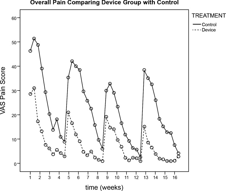
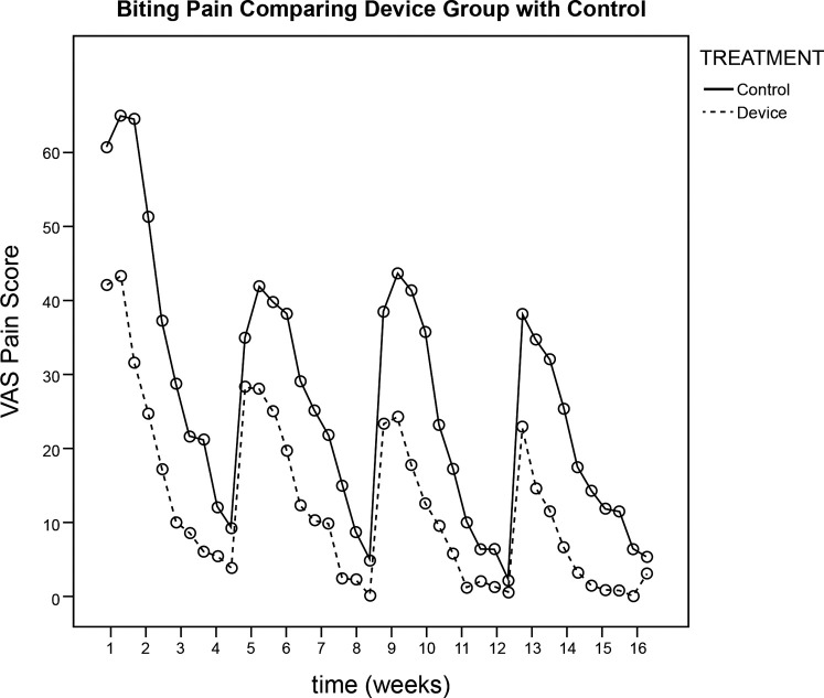
Average monthly pain scores were numerically generated from the patients’ VAS information and are reported in Table 1. In each group, 29 of 35 participants (83%) remained in the study after the 4-month trial. Six patients from each group were excluded from the study. Four of six patients from the device groups used a quantity of rescue medication that was considered excessive, mostly for nondental pain. The other two patients were noncompliant with their pain diary. In the control group, three patients used rescue medication too often (headache, body pain) and three others were noncompliant with respect to the pain diary. Usage compliance was verified electronically by the USB interface in the device. During the 4-month test period, repeated-measures ANOVA detected significant differences between the micropulse vibration device group and the control group for overall pain (P = .002) and for biting pain (P = .003) at α = .05. The authors also observed from graphical data that perceived pain was highest at the beginning of the month, following archwire adjustment. Graphical representation of average overall pain scores for the device and control groups during the 4-month study period is shown in Figure 1. Graphical representation of average biting pain for the device and control groups during the 4-month study period is depicted in Figure 2. Stratified analysis was used for gender and age; however, the study was not powered adequately to look at subgroup differences. No harms or unintended effects were noticed. All participants who were given a device reported that they were in less pain when using the device.
Table 1.
Mean Pain Scores Numerically Generated From Patient Visual Analog Scale Information (0–100)a
Figure 1.
Visual analog scale pain scores for overall pain.
Figure 2.
Visual analog scale pain scores for biting pain.
DISCUSSION
Researchers attribute initial and delayed pain responses following orthodontic treatment to compression and hyperalgesia of the periodontal ligament, respectively.18 The periodontal ligament becomes sensitive to released substances such as histamine, bradykinin, prostaglandins, and serotonins.19 These pain mediators are found in high levels when a pain response occurs. Given that pain is a subjective experience, it is difficult to assess and few in vivo studies have measured and quantified it. In our study, pain quickly increased and peaked at approximately 24 hours postinitial archwire or separator insertion. Figures 1 and 2 illustrate the differences in pain perception during the 4-month study period for device and control groups for overall and biting pain, respectively. Pain scores were higher throughout the course of treatment for biting pain. This agrees with the current concept that sustained PDL pressure from orthodontic adjustment decreases blood flow and recruitment of the pain-producing substances over time.19 Therefore, increasing blood flow to the PDL by vibratory stimulation at regular intervals may be effective in reducing the perception of orthodontic pain.
The lack of a placebo group is a limitation of the study. We cannot dismiss the possibility that a placebo effect from the device may have influenced the results. A sham device was not used in the current study for several reasons. First, possible skewed results could have occurred because a bite plate could essentially function as a bite wafer and, second, a sham device could be interpreted as misleading or deceptive to the patient. Murdock et al.20 found that plastic bite wafers chewed by the patient were as effective as OTC pain medications after initial archwire placement. Hwang et al.21 found that pain relief occurred in 56% of patients after using a bite wafer; however, the other 44% of the patients reported increased discomfort. In contrast, Otasevic et al.22 found that their bite wafer group reported more pain than the group that avoided masticatory activity. Because of possible unwanted treatment effects of bite wafers on pain reporting, the authors chose not to use a sham device that may have a bite wafer effect.
A VAS was used in the current study for pain assessment. This scaled survey gives the respondent the freedom to choose the exact intensity of the pain, and it is a reliable and sensitive method of measuring pain and the effect of pain-reducing methods. Test–retest reliability for the VAS has been shown to be very good.17 In the absence of a gold standard for pain, criterion validity unfortunately cannot be reliably evaluated. For construct validity, the VAS has been shown to be highly correlated with a five-point verbal descriptive scale (“nil,” “mild,” “moderate,” “severe,” and “very severe”) and a numeric rating scale (with response options from “no pain” to “unbearable pain”).16
Table 1 compares the pain data for the study groups and indicates whether there were significant differences by month. The data reveal that for all groups except one there were significantly lower pain scores for the device groups when compared with the control groups for overall pain and biting pain. Only the first month data set for biting pain was not statistically different (P = .068). However, when repeated-measures ANOVA was performed on all 4 months of data, significantly lower pain scores were recorded for overall pain and biting pain when the device was used. Gender and age data were collected, but there was not a sufficient number of participants to make statistical conclusions.
Several authors have found that vibration diminishes pain responses.23,24 However, at least one study demonstrated no pain relief with the use of a vibratory device.25 Vibration therapy in this randomized clinical trial resulted in significantly lower perceived pain and less OTC medication use. Therefore, it may be a safer, more effective means of postorthodontic adjustment pain control. More study is needed in this area of vibratory stimulus and pain modulation.
CONCLUSION
Based on the parameters of this randomized clinical trial, the use of a micropulse vibration device significantly reduced the perception of overall and biting pain in patients undergoing orthodontic treatment.
Future studies on micropulse vibration are needed. The use of a device without any occlusal extension could be used as a placebo.
REFERENCES
- 1.Scheurer PA, Firestone AR, Bürgin WB. Perception of pain as a result of orthodontic treatment with fixed appliances. Eur J Orthod. 1996;18:349–357. doi: 10.1093/ejo/18.4.349. [DOI] [PubMed] [Google Scholar]
- 2.Lökken P, Skoglund LA, Skjelbred P. Anti-inflammatory efficacy of treatments with aspirin and acetaminophen. Pain. 1995;60:231–233. doi: 10.1016/0304-3959(94)00198-N. [DOI] [PubMed] [Google Scholar]
- 3.Ngan P, Wilson S, Shanfeld J, Amini H. The effect of ibuprofen on the level of discomfort in patients undergoing orthodontic treatment. Am J Orthod Dentofacial Orthop. 1994;106:88–95. doi: 10.1016/S0889-5406(94)70025-7. [DOI] [PubMed] [Google Scholar]
- 4.Tortamano A, Lenzi DC, Haddad AC, Bottino MC, Dominguez GC, Vigorito JW. Low-level laser therapy for pain caused by placement of the first orthodontic archwire: a randomized clinical trial. Am J Orthod Dentofacial Orthop. 2009;136:662–667. doi: 10.1016/j.ajodo.2008.06.028. [DOI] [PubMed] [Google Scholar]
- 5.Vachiramon A, Wang WC. Acupuncture and acupressure techniques for reducing orthodontic post-adjustment pain. J Contemp Dent Pract. 2005;6:163–167. [PubMed] [Google Scholar]
- 6.Roth PM, Thrash WJ. Effect of transcutaneous electrical nerve stimulation for controlling pain associated with orthodontic tooth movement. Am J Orthod Dentofacial Orthop. 1986;90:132–138. doi: 10.1016/0889-5406(86)90045-4. [DOI] [PubMed] [Google Scholar]
- 7.Marie SS, Powers M, Sheridan JJ. Vibratory stimulation as a method of reducing pain after orthodontic appliance adjustment. J Clin Orthod. 2003;37:205–208. [PubMed] [Google Scholar]
- 8.Farzanegan F, Zebarjad SM, Alizadeh S, Ahrari F. Pain reduction after initial archwire placement in orthodontic patients: a randomized clinical trial. Am J Orthod Dentofacial Orthop. 2012;141:169–173. doi: 10.1016/j.ajodo.2011.06.042. [DOI] [PubMed] [Google Scholar]
- 9.Proffit WR, Fields HW. Occlusal forces in normal and long-face children. J Dent Res. 1983;62:571–574. doi: 10.1177/00220345830620051301. [DOI] [PubMed] [Google Scholar]
- 10.Furstman L, Bernick S. Clinical considerations of the periodontium. Am J Orthod. 1972;61:138–155. doi: 10.1016/0002-9416(72)90092-9. [DOI] [PubMed] [Google Scholar]
- 11.Ottoson D, Ekblom A, Hansson P. Vibratory stimulation for the relief of pain of dental origin. Pain. 1981;10:37–45. doi: 10.1016/0304-3959(81)90043-9. [DOI] [PubMed] [Google Scholar]
- 12.Melzack R, Wall PD. Pain mechanisms: a new theory. Science. 1965;150:971–979. doi: 10.1126/science.150.3699.971. [DOI] [PubMed] [Google Scholar]
- 13.Lowe MK. Vibrating orthodontic remodeling device. US Patent 8,939,762 filed Aug 22, 2013, issued Jan 27, 2015. [Google Scholar]
- 14.Mao JJ, Nah HD. Growth and development: hereditary and mechanical modulations. Am J Orthod Dentofacial Orthop. 2004;125:676–689. doi: 10.1016/j.ajodo.2003.08.024. [DOI] [PubMed] [Google Scholar]
- 15.Conti PC, dos Santos CN, Kogawa EM, de Castro Ferreira Conti AC, de Araujo Cdos R. The treatment of painful temporomandibular joint clicking with oral splints: a randomized clinical trial. J Am Dent Assoc. 2006;137:1108–1114. doi: 10.14219/jada.archive.2006.0349. [DOI] [PubMed] [Google Scholar]
- 16.Ferraz MB, Quaresma MR, Aquino LR, Atra E, Tugwell P, Goldsmith CH. Reliability of pain scales in the assessment of literate and illiterate patients with rheumatoid arthritis. J Rheumatol. 1990;17:1022–1024. [PubMed] [Google Scholar]
- 17.Joyce CR, Zutshi DW, Hrubes VF, Mason RM. Comparison of fixed interval and visual analogue scales for rating chronic pain. Eur J Clin Pharmacol. 1975;8:415–420. doi: 10.1007/BF00562315. [DOI] [PubMed] [Google Scholar]
- 18.Burstone CJ. Rationale of the segmented arch. Am J Orthod. 1962;48:805–822. doi: 10.1016/0002-9416(62)90001-5. [DOI] [PubMed] [Google Scholar]
- 19.Polat O, Karaman AI. Pain control during fixed orthodontic appliance therapy. Angle Orthod. 2005;75:214–219. doi: 10.1043/0003-3219(2005)075<0210:PCDFOA>2.0.CO;2. [DOI] [PubMed] [Google Scholar]
- 20.Murdock S, Phillips C, Khondker Z, Hershey HG. Treatment of pain after initial archwire placement: a noninferiority randomized clinical trial comparing over-the-counter analgesics and bite-wafer use. Am J Orthod Dentofacial Orthop. 2010;137:316–323. doi: 10.1016/j.ajodo.2008.12.021. [DOI] [PubMed] [Google Scholar]
- 21.Hwang JY, Tee CH, Huang AT, Taft L. Effectiveness of thera-bite wafers in reducing pain. J Clin Orthod. 1994;28:291–292. [PubMed] [Google Scholar]
- 22.Otasevic M, Naini FB, Gill DS, Lee RT. Prospective randomized clinical trial comparing the effects of a masticatory bite wafer and avoidance of hard food on pain associated with initial orthodontic tooth movement. Am J Orthod Dentofacial Orthop. 2006;130:6.e9–e15. doi: 10.1016/j.ajodo.2005.11.033. [DOI] [PubMed] [Google Scholar]
- 23.Lundeberg T, Nordemar R, Ottoson D. Pain alleviation by vibratory stimulation. Pain. 1984;20:25–44. doi: 10.1016/0304-3959(84)90808-X. [DOI] [PubMed] [Google Scholar]
- 24.Roy EA, Hollins M, Maixner W. Reduction of TMD pain by high-frequency vibration: a spatial and temporal analysis. Pain. 2003;101:267–274. doi: 10.1016/S0304-3959(02)00332-9. [DOI] [PubMed] [Google Scholar]
- 25.Miles P, Smith H, Weyant R, Rinchuse DJ. The effects of a vibrational appliance on tooth movement and patient discomfort: a prospective randomized clinical trial. Aust Orthod J. 2012;28:213–218. [PubMed] [Google Scholar]


