Abstract
Objective:
To evaluate the effect of using low-level laser therapy (LLLT) to control pain and discomfort during orthodontic treatment.
Materials and Methods:
A randomized, split-mouth clinical trial was conducted with 30 volunteers in need of orthodontic treatment, of both genders, aged between 18 and 40 years, who were randomly divided into two groups. One hemiarch was considered the exposed group (EG) and the other, the placebo group (PG). Both groups had elastic separators placed mesially and distally to the first molars of the two hemiarches at different times. The EG received an AIGaAs diode LLLT (810 nm, 100 mW, 2J/cm2) application for 15 seconds per point (interdental papilla at the mesial, distal, and near the root apex) immediately after separator placement on the maxillary right side. The PG also had elastics placed around the maxillary right molars, but received only simulated LLLT application. The elastics were left in place for 5 days, and after a waiting period of 1 week, they were inserted on the left side in both groups; however, the order of laser application was changed. While the separator remained in place, the patient marked his degree of perceived discomfort on a Visual Analog Scale (VAS) at 5 minutes (T0), 24 hours (T1), and 120 hours (T2), after LLLT application.
Results:
A statistically significant difference was observed (P < .005) in reducing discomfort in the exposed group compared with the placebo group. This reduction of discomfort in the EG was observed at all time intervals.
Conclusions:
A sincle AIGaAs diode LLLT application may be indicated for the control or reduction of pain in the early stages of orthodontic treatment.
Keywords: Orthodontics, Pain, Low level laser therapy, Clinical trial
INTRODUCTION
The correction of malocclusion during orthodontic treatment, especially in the early stages, results in the patient's experiencing some degree of pain.1–7 The pain mechanism in orthodontic treatment is a result of compression forces, consequently leading to ischemia, inflammation, and edema in the periodontal tissues.4,8
In patients who experience a higher degree of pain, the orthodontist may recommend the use of pharmacological agents or nonpharmacological methods for pain relief, considering their pain sensitivity threshold or reported emotional condition. As a nonpharmacological method, low-level laser therapy (LLLT) has recently been used. It has analgesic properties and anti-inflammatory effects7,9–11 through increasing the local blood flow by reduction of prostaglandin levels E2 and inhibition of cycloxygenase-2.10–12
Some studies have already investigated the action of LLLT in pain reduction during orthodontic movement13–15 and the placing of elastic separators.7,16–20 Some researchers have pointed out that the use of LLLT reduced the risk of incidence of pain by 24% compared with control or placebo groups.8 However, standardization of the type and use of LLLT needs to be established. The clinical results and efficacy of LLLT in reducing orthodontic pain are directly related to the type of laser, wavelength, energy density (J/cm2), time of application per point, and frequency.8,21
The risk of bias of studies included in the systematic reviews8,21 indicate that the use of LLLT for the reduction of pain or discomfort, at any stage of orthodontic treatment, presents limited clinical evidence. The aim of developing this study was to evaluate the effect of using LLLT on pain experienced by patients undergoing elastic separation of the first molars during the early stages of orthodontic treatment, following the recommendations of the statement declaration.22
MATERIALS AND METHODS
This randomized, split-mouth clinical trial was approved by the Ethics Committee on Research with Human Beings of the Universidade Luterana do Brasil (ULBRA).
Patients of both sexes, aged between 18 and 40 years, were selected from a private orthodontic clinic. All patients in need of orthodontic treatment were invited to participate in the study and were selected according to the following inclusion criteria: patients with healthy molars, presence of proximal contacts around the maxillary first permanent molars on both sides, and without periodontal disease. The latter was based on indexes of visible plaque, gingival bleeding, bleeding on probing, periodontal probing depth, and clinical attachment loss. Absence of periapical pathology was verified by periapical radiographs from the initial records. Exclusion criteria were as follows: excessive tooth crowding (>3 mm) as measured by misalignment of teeth on the study models according to the Dental Aesthetic Index, any systemic disease that would contraindicate the use of LLLT (eg, malignant neoplasias), metabolic diseases (eg, diabetes), chronic pain, neurological or psychiatric disorders, and use of medications (antibiotics, corticosteroids, bisphosphonates, analgesics, anti-inflammatory agents, or contraceptives) taken up to 1 month before the selection exam. This information was answered by the patients in an anamnestic questionnaire.
Exams and all research procedures were performed at the same clinic, and volunteering patients who agreed to participate and sign an informed consent form were included in the study.
After complying with these requirementss, all selected patients were randomly divided into two groups (n = 30) with respect to the right and left first molars in each hemiarch in order to eliminate intersubject variability. One hemiarch was designated the exposed group (EG) and the other, the placebo group (PG). In the EG, effective applications of LLLT were performed; the PG received simulated laser applications (Figure 1).
Figure 1.
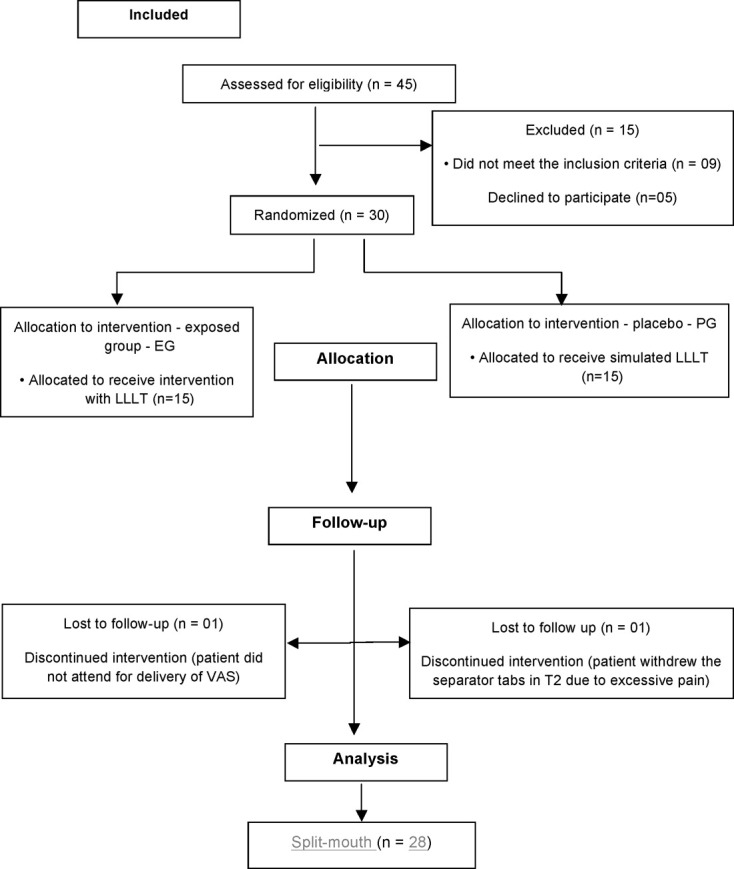
Flowchart representing strategies and follow-up of the study.
To randomize the groups for comparison of placebo with intervention (simulation vs laser application), each patient took an envelope that had either the letter A (EG) or B (PG) printed on it. Patients who took the A envelope had the separators placed on the right side and the laser treatment applied. The elastics were left in place for 5 days. After 1 week, the same patient had the separators placed on the left side and received only the simulated laser application. For patients who took the letter B (PG) the same methodology was used, only the order of laser application was changed, that is, the LLLT was applied on the left side and the simulated application on the right side. Patients were blinded as to which group the letter or intervention belonged. A single operator performed all the clinical procedures, and was blinded to the groups and objectives of the study. Data collection and analysis was performed by a single investigator, who was also blinded to the study groups.
Half-millimeter-thick elastic separators (American Orthodontics, Sheboygan, Wisc) that had been previously selected and measured by electronic caliper (Mitutoyo America, Aurora, Ill) were inserted at the mesial and distal of the right and left first molars with the aid of two pieces of dental floss. After placement of the separators, the LLLT AIGaAs diode was used; the area of the spot tip of this tool was 0.028cm2. Laser irradiation was performed in continuous wave mode in accordance with the protocol of the Photon Lase Plus unit (DMC, São Carlos, São Paulo, Brazil): 810 nm (infrared) wave length, 100mW output power, 2J/cm2 energy density per point (6J total dose per tooth), with a single spot application in the immediate region corresponding to the buccal surfaces of the tooth at three points. One point was aimed at the interdental papilla from the mesial direction, one point was aimed from the distal, and another near the apex of the root. For the PG, the laser unit was switched off; however, the sound signal was maintained in order to be aware of the application time, thereby maintaining blinding of the operator and participants as to allocation of the group. The laser was applied to each group for the same amount of time (15 seconds per point), corresponding to a total of 45 seconds per tooth.
The volunteers were instructed to quantify the discomfort or pain by means of a VAS, noting the intensity on a scale of zero to 10 according to the participant' s self-perception. This evaluation was performed after separator placement at the following times: 5 minutes (T0), 24 hours (T1), and 120 hours (T2). The marking the patient made on the VAS was measured using a digital caliper (Mitutoyo America) and recorded in millimeters (100 mm).
After data collection, 6.67% of the volunteers (n = 2) had to be excluded from the analysis (Figure 1): One participant from the EG, who used the analgesic, removed the separators before the deadline (T2) due to excessive pain. Another (PG) did not attend the appointment for delivery of the form (VAS). For these reasons, the final sample consisted of 28 patients.
Statistical Analysis
All data were tabulated and analyzed using the v.18.0 statistical software program (SPSS Inc, Chicago, Ill). Categorical variables were described as frequencies and percentages and compared by using Fisher's exact test. Quantitative variables with normal distribution were described by the mean and standard deviation and compared between groups by Student's t test for independent samples. To verify the distribution of pain in the groups, the Kolmogorov Smirnov test was used. Variables with asymmetrical distribution were described by the median and interquartile interval. Comparisons between the hemiarches and groups were performed using the Wilcoxon test. Comparisons between the different times were performed by the Friedman test, and differences by the Wilcoxon test. Bonferroni adjustment modified by Finner was used to adjust P values. The maximum level of significance of 5% was adopted.
RESULTS
Of the 28 participants included in the data collection and analysis, 14 belonged to the EG and 14 to the PG. In the EG, five were men (35.7%) and nine were women (64.3%), while in the PG, eight were males (57.1%) and six, females (42.9%). As regards gender, there was no statistically significant difference between groups (P = .449).
The mean (SD) age of the EG was 24.9 years (± 7) and of the PG, 22.8 years (± 5.3). In the two groups, there was no statistically significant difference between ages (P = .378).
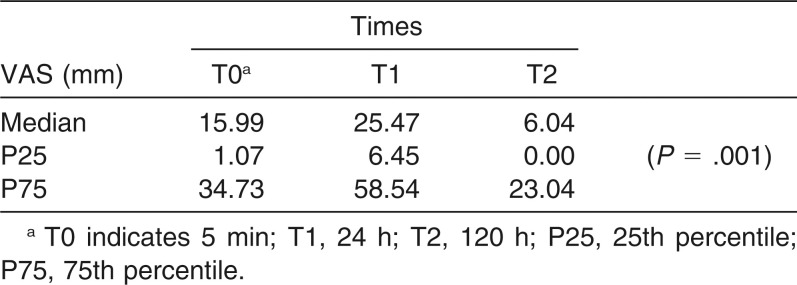
In Table 1, comparisons between the different evaluation time intervals in all the hemiarches allocated to the placebo group can be verified. A statistically significant difference between the times (P = .001) was verified with regard to pain reduction.
Table 1.
Comparison of Hemiarches of the Placebo Group (PG) with the Time Intervals of Evaluation in Pain Perception (VAS)
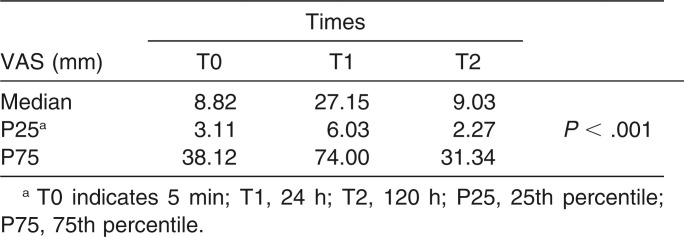
When the hemiarches allocated to the EG were compared with the various times of evaluation (Table 2), a statistically significant difference was verified between the times (P < .001), with this difference being located between times T0 and T1 (P = .030) and between T1 and T2 (P = .003).
Table 2.
Comparison of Hemiarches of the Exposed Group (EG) with the Different Time Intervals of Evaluation in Pain Perception (VAS)
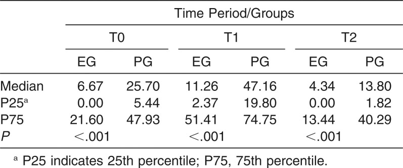
When the EG was compared with the PG, there was a statistically significant difference between groups at all time intervals (P < .001). The EG exhibited reduced pain at time intervals T0, T1, and T2 (P < .001) (Table 3).
Table 3.
Comparison of Hemiarches of the Exposed Group (EG) and Placebo Group (PG) with the Different Time Intervals of Evaluating Pain Perception (VAS)
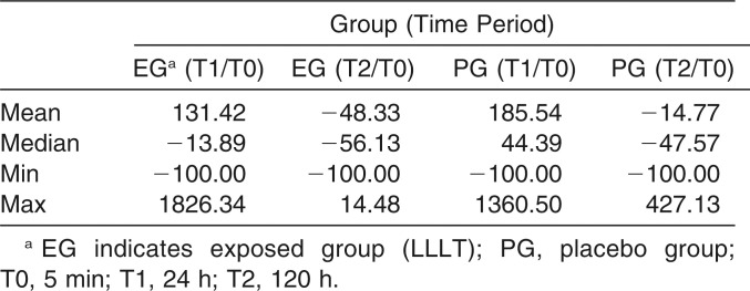
In Table 4, using the Wilcoxon test and Bonferroni adjustment modified by Finner, it can be seen that after 24 hours (T1), there was a decrease of pain in 13.89% of the EG, while in the PG there was a 44.39% increase.
Table 4.
Difference Between Initial Time (T0) and Other Time Intervals of Evaluating Pain Reduction in the Groups (Exposed and Placebo)
DISCUSSION
In our study, it was observed that pain increased 24 hours after insertion of the separators, and in both the exposed and placebo groups, pain regressed over time, showing uniformity of the groups and providing greater reliability of the posttherapy results.20 Similar studies have reported that pain is usually highest during the first 24 hours after application of orthodontic force. The frequency decreases to baseline levels in up to 7 days.23–26
However, the goal in this study was to verify the positive effect of LLLT in reducing pain in the EG at all times evaluated compared with the PG. Another important fact was that in the first 24 hours after application of LLLT, a 13.89% reduction in pain was promoted, while in the PG, there was a 44.39% increase in pain during the same period.
Compared with other studies, we observed a significant reduction in pain levels when LLLT was applied.20,27,27 However, there was a large variation in the methodologies used, as well as the presence of methodological bias risk, as reported in a systematic review with meta-analysis. Therefore, it is difficult to compare the results obtained and described in the various studies.8,21
According to Li et al.,21 who considered the results of published studies, the use of LLLT cannot yet be considered a standard treatment for orthodontic pain, because the various commercial laser systems differ both in technical specifications and in methods of application, as well as in the study designs, which are limited and have risk of bias. Furthermore, studies should be analyzed separately with regard to the origin of pain, whether it is caused by orthodontic movement or by the use of separators.
If we consider only clinical trials that used elastic separators and the use of LLLT with a AIGaAs diode and the same technical and application parameters for pain reduction, we could compare our results with those of the study of Eslamian et al.,20 who verified that LLLT (810 nm) was effective during the first 3 days after placement of the separators and made a substantial reduction in pain after the fifth day (120 h). Similarly, in the present study, LLLT (810 nm) was effective in pain reduction from the first 24 hours up to the fifth day (120 hours) after separator placement.
The LLLT at a wavelength of 810 nm used in the study showed an analgesic action in all patients. Other studies have confirmed this effect using powers ranging from 650 nm to 910 nm, with an average of 830 nm.8,13,19,29,30
With respect to the dose and wavelength, more profound penetration has been shown to occur with infrared radiation at 810 nm, with a possible effect on both cortical and alveolar bone tissue, and it was more effective than laser at wavelengths between 620 nm and 670 nm.18
Differently from other studies,16–20 pain control was obtained with only a single application of GaAlAs diode LLLT immediately after separator placement, with a total time of 45 seconds per tooth. This method was effective mainly after the first 24 hours, considering the peak of pain, which showed a significant difference compared with the placebo group.
As for the design, the study was conducted as a randomized, split-mouth clinical trial, preventing interindividual biological variation in pain perception of the participants.13,30 It must be taken in account that perception of pain intensity is variable for each individual. This bias occurs when the difference of the means are compared between the groups, as verified in other studies.18,19 Moreover, use of this design associated with masking of the participants may have reduced the Hawthorne effect.21
With reference to the means of inducing pain, patients can experience acute pain immediately after placement of separators or express “medium pain” for 1–2 days. LLLT seemed to be effective in delaying pain onset, shortening pain duration, and reducing average pain intensity.8
One of the limitations of these studies is that they do not specify the origin of the subjects14 or they use a very specific population, such as dental students,7,19 so that the results do not present sufficient variety in pain response. Therefore, the conclusions obtained from these samples may not reflect the same results obtainable in the general population requiring orthodontic treatment, thereby increasing the risk of bias21 and reducing the reliability and external validity of the studies. Another limitation observed in previous studies is sample size calculation. It should be determined based on the pain outcome size, but some studies do not justify that and use sample size as described in previous articles.14,20
The VAS used in this work, although subjective, is a widely used and reliable method to quantify pain levels over a period of time when one expects to observe a large variability between individuals.6,31
Other studies have evaluated the use of LLLT in reducing pain during the active stages of conventional treatment in which all the teeth have been banded, bracketed, and wired.13,15,15 While this methodology assesses the routine of conventional treatment, it may be biased with many variations with regard to different types of malocclusion and range of motion. In the present study, the technique of separating the teeth followed by immediate application of LLLT, as recommended in other randomized, controlled, and and in quasirandomized trials,29,32 was done to facilitate comparisons of localized pain. Nevertheless, differences in methods were noted, since some investigators used the first molars and others chose to use the first premolars of their volunteers.7,32
CONCLUSIONS
There was a statistically significant reduction in pain in the exposed side of patients receiving a single application of AlGaAs diode LLLT (810nm) compared with the placebo or control side at all time intervals evaluated.
The use of a single AlGaAs diode LLLT (810nm) is suggested as an effective therapeutic method to control or reduce pain in the early stages of orthodontic treatment.
REFERENCES
- 1.Kvam E, Bondevik O, Gjerdet NR. Traumatic ulcers and pain in adults during orthodontic treatment. Community Dent Oral Epidemiol. 1989;17:154–157. doi: 10.1111/j.1600-0528.1989.tb00012.x. [DOI] [PubMed] [Google Scholar]
- 2.Bergius M, Kiliaridis S, Berggren U. Pain in orthodontics. A review and discussion of the literature. J Orofac Orthop. 2000;61:125–137. doi: 10.1007/BF01300354. [DOI] [PubMed] [Google Scholar]
- 3.Polat O, Karaman AI. Pain control during fixed orthodontic appliance therapy. Angle Orthod. 2005;75:214–219. doi: 10.1043/0003-3219(2005)075<0210:PCDFOA>2.0.CO;2. [DOI] [PubMed] [Google Scholar]
- 4.Krishnan V. Orthodontic pain: from causes to management—a review. Eur J Orthod. 2007;29:170–179. doi: 10.1093/ejo/cjl081. [DOI] [PubMed] [Google Scholar]
- 5.Polat O. Pain and discomfort after orthodontic appointments. Semin Orthod. 2007;13:292–300. [Google Scholar]
- 6.Youssef M, Ashkar S, Hamade E, Gutknecht N, Lampert F, Mir M. The effect of low-level laser therapy during orthodontic movement: a preliminary study. Lasers Med Sci. 2008;23:27–33. doi: 10.1007/s10103-007-0449-7. [DOI] [PubMed] [Google Scholar]
- 7.Lim HM, Lew KK, Tay DK. A clinical investigation of the efficacy of low level laser therapy in reducing orthodontic postadjustment pain. Am J Orthod Dentofacial Orthop. 1995;108:614–622. doi: 10.1016/s0889-5406(95)70007-2. [DOI] [PubMed] [Google Scholar]
- 8.He WL, Li CJ, Liu ZP, et al. Efficacy of low-level laser therapy in the management of orthodontic pain: a systematic review and meta-analysis. Lasers Med Sci. 2013;28:1581–1589. doi: 10.1007/s10103-012-1196-y. [DOI] [PubMed] [Google Scholar]
- 9.Holmberg PF, Zaror SC, Fabres SR, Sandoval VP. Use the laser therapy in orthodonthic pain control. Rev Clin Periodoncia Implantol Rehabil Oral. 2011;4:114–116. [Google Scholar]
- 10.Pallotta RC, Bjordal JM, Frigo L, et al. Infrared (810-nm) low-level laser therapy on rat experimental knee inflammation. Lasers Med Sci. 2012;27:71–78. doi: 10.1007/s10103-011-0906-1. [DOI] [PMC free article] [PubMed] [Google Scholar]
- 11.Silveira PC, Silva LA, Freitas TP, Latini A, Pinho RA. Effects of low-power laser irradiation (LPLI) at different wavelengths and doses on oxidative stress and fibrogenesis parameters in an animal model of wound healing. Lasers Med Sci. 2011;26:125–131. doi: 10.1007/s10103-010-0839-0. [DOI] [PubMed] [Google Scholar]
- 12.Sakurai Y, Yamaguchi M, Abiko Y. Inhibitory effect of low-level laser irradiation on LPS-stimulated prostaglandin E2 production and cyclooxygenase-2 in human gingival fibroblasts. Eur J Oral Sci. 2000;108:29–34. doi: 10.1034/j.1600-0722.2000.00783.x. [DOI] [PubMed] [Google Scholar]
- 13.Doshi-Mehta G, Bhad-Patil WA. Efficacy of low-intensity laser therapy in reducing treatment time and orthodontic pain: a clinical investigation. Am J Orthod Dentofacial Orthop. 2012;141:289–297. doi: 10.1016/j.ajodo.2011.09.009. [DOI] [PubMed] [Google Scholar]
- 14.Abtahi SM, Mousavi SA, Shafaee H, Tanbakuchi B. Effect of low-level laser therapy on dental pain induced by separator force in orthodontic treatment. Dent Res J (Isfahan) 2013;10:647–651. [PMC free article] [PubMed] [Google Scholar]
- 15.Domínguez A, Velásquez SA. Effect of low-level laser therapy on pain following activation of orthodontic final archwires: a randomized controlled clinical trial. Photomed Laser Surg. 2013;31:36–40. doi: 10.1089/pho.2012.3360. [DOI] [PubMed] [Google Scholar]
- 16.Esper MA, Nicolau RA, Arisawa EA. The effect of two phototherapy protocols on pain control in orthodontic procedure—a preliminary clinical study. Lasers Med Sci. 2011;26:657–663. doi: 10.1007/s10103-011-0938-6. [DOI] [PubMed] [Google Scholar]
- 17.Kim WT, Bayome M, Park JB, Park JH, Baek SH, Kook YA. Effect of frequent laser irradiation on orthodontic pain. A single-blind randomized clinical trial. Angle Orthod. 2013;83:611–616. doi: 10.2319/082012-665.1. [DOI] [PMC free article] [PubMed] [Google Scholar]
- 18.Nóbrega C, da Silva EM, de Macedo CR. Low-level laser therapy for treatment of pain associated with orthodontic elastomeric separator placement: a placebo-controlled randomized double-blind clinical trial. Photomed Laser Surg. 2013;31:10–16. doi: 10.1089/pho.2012.3338. [DOI] [PubMed] [Google Scholar]
- 19.Marini I, Bartolucci ML, Bortolotti F, Innocenti G, Gatto MR, Alessandri Bonetti G. The effect of diode superpulsed low-level laser therapy on experimental orthodontic pain caused by elastomeric separators: a randomized controlled clinical trial. Lasers Med Sci. 2015;30:35–41. doi: 10.1007/s10103-013-1345-y. [DOI] [PubMed] [Google Scholar]
- 20.Eslamian L, Borzabadi-Farahani A, Hassanzadeh-Azhiri A, Badiee MR, Fekrazad R. The effect of 810-nm low-level laser therapy on pain caused by orthodontic elastomeric separators. Lasers Med Sci. 2014;29:559–564. doi: 10.1007/s10103-012-1258-1. [DOI] [PubMed] [Google Scholar]
- 21.Li FJ, Zhang JY, Zeng XT, Guo Y. Low-level laser therapy for orthodontic pain: a systematic review. Lasers Med Sci. 2014 doi: 10.1007/s10103-014-1661-x. [Epub ahead of print] [DOI] [PubMed] [Google Scholar]
- 22.CONSORT. http://www.consort-statement.org/consort-statement/
- 23.Scheurer PA, Firestone AR, Burgin WB. Perception of pain as a result of orthodontic treatment with fixed appliances. Eur J Orthod. 1996;18:349–357. doi: 10.1093/ejo/18.4.349. [DOI] [PubMed] [Google Scholar]
- 24.Fernandes LM, Ogaard B, Skoglund L. Pain and discomfort experienced after placement of a conventional or a superelastic NiTi aligning archwire. A randomized clinical trial. J Orofac Orthop. 1998;59:331–339. doi: 10.1007/BF01299769. [DOI] [PubMed] [Google Scholar]
- 25.Grieve WG, III, Johnson GK, Moore RN, Reinhardt RA, DuBois LM. Prostaglandin E (PGE) and interleukin-1 beta (IL-1 beta) levels in gingival crevicular fluid during human orthodontic tooth movement. Am J Orthod Dentofacial Orthop. 1994;105:369–374. doi: 10.1016/s0889-5406(94)70131-8. [DOI] [PubMed] [Google Scholar]
- 26.Chow RT, David MA, Armati PJ. 830 nm laser irradiation induces varicosity formation, reduces mitochondrial membrane potential and blocks fast axonal flow in small and medium diameter rat dorsal root ganglion neurons: implications for the analgesic effects of 830 nm laser. J Peripher Nerv Syst. 2007;12:28–39. doi: 10.1111/j.1529-8027.2007.00114.x. [DOI] [PubMed] [Google Scholar]
- 27.Jones M, Chan C. The pain and discomfort experienced during orthodontic treatment: a randomized controlled clinical trial of two initial aligning arch wires. Am J Orthod Dentofacial Orthop. 1992;102:373–381. doi: 10.1016/0889-5406(92)70054-e. [DOI] [PubMed] [Google Scholar]
- 28.Artes-Ribas M, Arnabat-Dominguez J, Puigdollers A. Analgesic effect of a low-level laser therapy (830 nm) in early orthodontic treatment. Lasers Med Sci. 2013;28:335–341. doi: 10.1007/s10103-012-1135-y. [DOI] [PubMed] [Google Scholar]
- 29.Kawasaki K, Shimizu N. Effects of low-energy laser irradiation on bone remodeling during experimental tooth movement in rats. Lasers Surg Med. 2000;26:282–291. doi: 10.1002/(sici)1096-9101(2000)26:3<282::aid-lsm6>3.0.co;2-x. [DOI] [PubMed] [Google Scholar]
- 30.Shirani AM, Gutknecht N, Taghizadeh M, Mir M. Low-level laser therapy and myofacial pain dysfunction syndrome: a randomized controlled clinical trial. Lasers Med Sci. 2009;24:715–720. doi: 10.1007/s10103-008-0624-5. [DOI] [PubMed] [Google Scholar]
- 31.Sriwatanakul K, Kelvie W, Lasagna L, Calimlim JF, Weis OF, Mehta G. Studies with different types of visual analog scales for measurement of pain. Clin Pharmacol Ther. 1983;34:234–239. doi: 10.1038/clpt.1983.159. [DOI] [PubMed] [Google Scholar]
- 32.Fujiyama K, Deguchi T, Murakami T, Fujii A, Kushima K, Takano-Yamamoto T. Clinical effect of CO2 laser in reducing pain in orthodontics. Angle Orthod. 2008;78:299–303. doi: 10.2319/033007-153.1. [DOI] [PubMed] [Google Scholar]


