Abstract
Objective:
To compare volumetric root resorption after rapid maxillary expansion (RME) between tooth-borne and tissue-borne appliances using CBCT. Repair in resorption cavities after 6 months of fixed retention was also compared.
Materials and Methods:
A sample of 33 subjects were randomly divided into two groups: Hyrax (n = 16) and Haas (n = 17). CBCT scans were taken 6 months before expansion, immediately after expansion, and 6 months after fixed retention. Mimics Innovation V 16.0 software was used for segmentation and volumetric measurement of 198 teeth. Bland-Altman plots, independent samples t test, repeated measures analysis of variance, and the Friedman test were used for statistical analysis.
Results:
Differences in root resorption after RME and repair after retention were not significant between the hyrax and Haas appliances or between male and female. Significant differences were found between preexpansion and postexpansion root volumes in the first premolars and molars—even in unattached second premolars. When the percentage of root volume loss is considered, no significant difference was found between the first premolar, second premolar, and first molar. Volumetric changes after 6 months of retention were not statistically significant.
Conclusions:
More resorption was observed in the Hyrax expander group. But it was not statistically significant. Repair was observed after 6 months of retention. Heavy RME forces affected premolars and molar similarly.
Keywords: Root resorption, CBCT, Segmentation
INTRODUCTION
Rapid maxillary expansion (RME) is used to correct maxillary skeletal transverse deficiency. Expansion occurs when the force applied to the teeth and maxilla exceeds the limits needed for tooth movement.1 The applied force causes widening and gradual opening of the midpalatal suture, bending of the alveolar processes, and dental tipping.
Although RME has several clinical benefits, there are some complications that may occur throughout the expansion. These are technical problems such as loosening or breakage of the expander and biological problems such as pain,2 nonopening of the midpalatal suture,3 dental tipping,4 root resorption of the anchor teeth,5 gingival recession,6 widening of the nasal root and nasal bridge,7 swelling and reddening,8 and occurrence of nasal septum asymmetries.8
Root resorption occurs because of individual biologic and genetic predisposition and the effect of mechanical factors.9,10 When considering root resorption in orthodontics, we must realize that resorption cavities can arise anywhere along the root surface without decreasing root length.11
Several studies have evaluated orthodontic and biologic factors related to root resorption using 2-D periapical radiographs,12 digital radiographs,13 panoramic radiographs,14 and lateral cephalometric radiographs.15 Three-dimensional methods such as SEM, serial sectioning, and micro CT have several advantages; however, the need to extract teeth before evaluation and data loss during tooth preparation and high radiation exposure with micro CT are disadvantages of the methods.11 After CBCT was introduced, several studies were conducted on tooth segmentation and volumetric measurements.16–18 It is possible to detect and measure root resorption with a lower radiation dose and without the need to extract teeth. Only two studies have evaluated volumetrically with CBCT the amount of root resorption occurring during orthodontic treatment.5,16
The aim of this study was to evaluate root resorption in the first premolar (P1), second premolar (P2), and first molar (M1) after RME and to compare the amount of resorption between tooth-borne (hyrax) and tooth-and-tissue–borne (Haas) expanders. Repair in resorption cavities after 6 months of fixed retention was also compared.
MATERIALS AND METHODS
This study was approved by the ethical committee of the School of Medicine, Ege University in Izmir, Turkey. Informed consent was obtained from the parents of all patients who agreed to participate in the study. There was a palatal acrylic support in the Haas-type expander that covered the screw and extensions.
Subjects
The study sample consisted of 33 patients with a mean age of 12.8 years (16 girls, 17 boys) randomly divided into two groups: hyrax (16 patients; mean age, 12.9 ± 0.9 years; range, 11.3–14.1 years) and Haas (17 patients; mean age, 12.8 ± 0.9 years; range, 11.1–13.9 years). The skeletal age of each patient was determined by the hand-wrist radiograph, and patients who did not exceed MP3Cap were included in the study sample.
The criteria for selection of the study groups were as follows: skeletal maxillary constriction with unilateral or bilateral posterior crossbite; completion of permanent dentition; no apical lesions, root canal treatment, or coronal restoration in the P1s, P2s, or M1s. The subjects were selected consecutively according to male and female distribution in the sample groups.
Methods
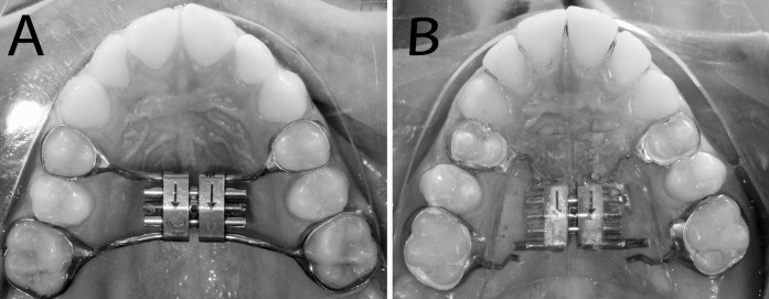
Hyrax and Haas expanders consist of an expansion screw and stainless steel extensions welded on the bands of P1 and M1. No additional buccal or lingual bars were used to attach the P2s (Figures 1A, B).
Figure 1.
(A) Tooth-borne expander (Hyrax-type) and (B) tooth-tissue–borne expander (Haas-type).
All expanders were activated 0.5 mm per day until the palatal cusps of the maxillary molars were in contact with the buccal cusps of the mandibular first molars. We used a digital caliper to measure the exact amount of expansion. All patients wore mandibular anterior bite planes to eliminate premature contacts during active expansion.
A Kodak 9000 3-D extraoral imaging system (Kodak Dental Systems, Carestream Health, Rochester, NY) was used at 70 kV and 10 mA, a scan time of 10.8 seconds, and a field of view of 5 × 3.75 cm2. Voxel dimension was 0.2 mm. CBCT images were obtained 6 months before RME (T0), immediately after RME (T1), and 6 months after expansion (T2) without expanders in place to prevent metal artifacts. A total of 198 teeth (96 in the hyrax group; 102 in the Haas group) were segmented, and volumetric measurements were done (Table 1).
Table 1.
Distribution of First Premolars, Second Premolars, and First Molars in Hyrax and Haas Expanders
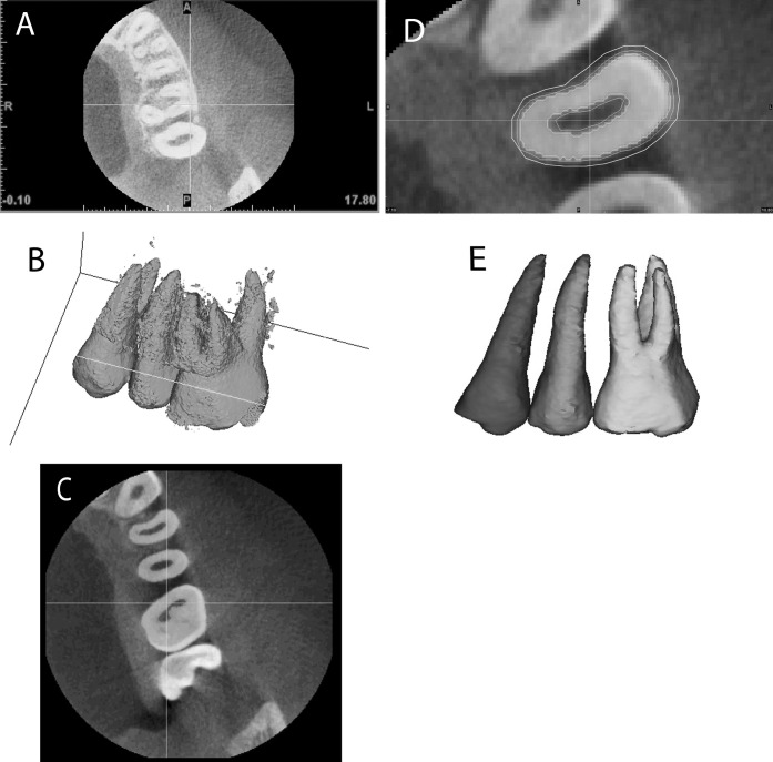
Segmentation was done with Mimics software V 16.0 (Materialise NV, Leuven, Belgium) randomly by the same researcher, and all CBCT images were blinded. The segmentation process included four steps: selecting the threshold, editing the mask in 3-D, editing the mask in 2-D, and reconstructing in 3-D (Figure 2). Threshold values were set individually with regard to each tooth. The same Hounsfield units were used for each tooth before expansion, after expansion, and after 6 months of retention.18
Figure 2.
Segmentation and volumetric measurement: (A) Selecting the optimal threshold; (B) Edit mask in 3-D; (C) Edit mask in 2-D; (D, E) 3-D reconstruction.
Tooth volume loss and its percentage were measured. The percentage of root volume loss was calculated by subtracting the postexpansion root volume from the preexpansion volume. Then the difference was divided by the preexpansion volume and multiplied by 100.
Statistical Analysis
All analyses were performed with SPSS software (version 19, SPSS Inc, Chicago, Ill). CBCT images of 20 teeth were randomly selected 1 month later. Segmentation and volumetric measurements were repeated and intraobserver repeatability was evaluated using Bland-Altman plots. The normality test of Shapiro-Wilks and Levene’s variance homogeneity test were conducted. The data were found normally distributed. Independent samples t test was used to compare mean ages of the study groups, expansion amounts (mm), and days between expander groups. For P1, P2, and M1, volumetric changes in T0, T1, and T2 periods and comparison of volumetric changes between Hyrax and Haas appliances and right and left sides were analyzed using repeated-measure analysis of variance. When a statistical difference was detected, a Bonferroni post-hoc test was performed to determine which groups caused the difference. Percentage of volume loss was compared between P1, P2, and M1 within each expander group with the Freidman test. A statistical significance (alpha) level of .05 was used for all statistical analyses. Root resorption between the left and right posterior segments was not statistically significant (P = .537), so these two data were pooled.
RESULTS
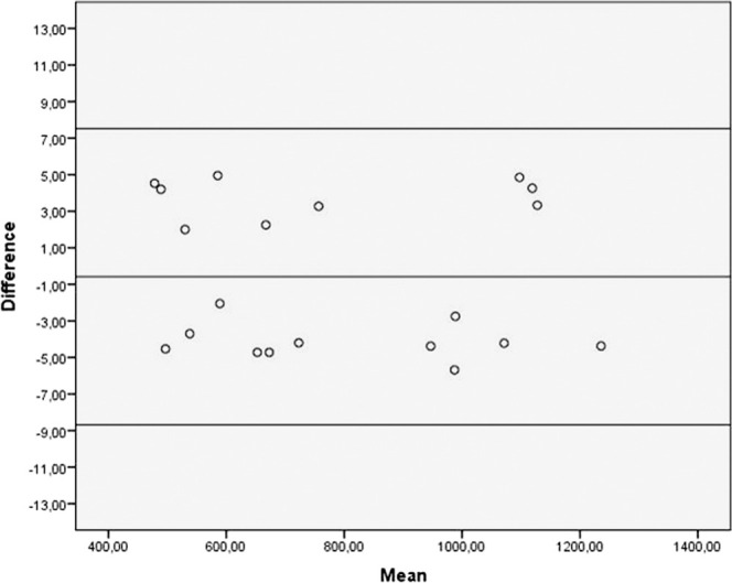
The Bland-Altman plot shows the 95% limits of agreement (upper and lower lines), estimated by mean difference ± 1.96 standard deviation (SD) of the differences (Figure 3). The differences were not significant for mean ages, expansion days, or expansion amounts between expanders (Table 2).
Figure 3.

Bland-Altman plots to observe intraobserver repeatability. Upper and lower lines show the agreement. A range of agreement was defined as mean bias ± 1.96 SD.
Table 2.
Means, Standard Deviations, and Ranges of Age, Appliance Expansion, Duration of Activation, and Comparison Between Hyrax and Haas Expanders
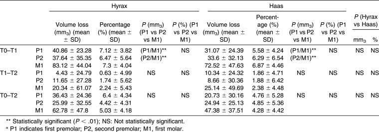
Volume loss in P1, P2, and M1 was significant between the T0 and T1 periods for both the hyrax and the Haas expanders (P < .001) (Table 3). When the premolars and first molar were compared with each other, the highest mean volume loss was found in M1 (Hyrax, 83.12 mm3; Haas, 72.52 mm3). However, no significant difference was found when the percentage of root volume loss was considered (P > .05) (Table 4).
Table 3.
Descriptive Statistics and Statistical Significance of Volumetric Change in P1a, P2, and M1 Between T0 (Pretreatment), T1 (Posttreatment), and T2 (After Retention) Time Points
Table 4.
Comparison of Amount and Percentage of Volumetric Change in P1a, P2, and M1 Between Each Other and Between Hyrax and Haas Expanders
Amount of root resorption in P1 (Hyrax, 40.86 mm3; Haas, 31.07 mm3), P2 (Hyrax 37.64 mm3; Haas, 33.6 mm3), and M1 (Hyrax: 83.12 mm3; Haas, 72.52 mm3) after RME was higher in the hyrax group, but the difference was not statistically significant (P = .095; P = .576; P = .424, respectively) (Table 4).
After 6 months’ retention, repair was observed in P1, P2, and M1, but it was not statistically significant (P > .05) for either expander. The amount of repair was between 4.43 mm3 and 20.34 mm3 in the hyrax group and 8.66 mm3 to 25.14 mm3 in the Haas group. The difference between P1, P2, and M1 was not significant (P > .05) (Table 4).
DISCUSSION
Two-dimensional imaging techniques are insufficient in quantitative evaluation of resorption cavities because of lack of information in the third dimension.11 Therefore, it is necessary to use 3-D methods to assess the amount of root resorption. So far, the effect of RME on root resorption has been evaluated with histological and SEM studies. However, it is possible to detect and measure root resorption in any tooth three-dimensionally without extracting teeth by using CBCT with a lower radiation dose. Many studies have concluded that volumetric measurement of teeth using CBCT is a reliable, reproducible, and valid method.16–18 As of today, there has been only one study evaluating root resorption after RME with CBCT in vivo.5
The volumetric data set comprises isotropic structures, known as voxels, each representing a specific degree of X-ray absorption.19 When the CBCT images were compared with micro CT and laser scanning, the authors found that volumetric measurements made with CBCT were all similar for voxel sizes up to 200 µm.17–18 Liedke et al.20 recommended a minimal voxel resolution of 0.3 mm for the detection of external root resorption. In this study, segmentation and volumetric measurements were done on 0.2 mm CBCT images.
Different conclusions have been made about the orthopedic effects of Haas and hyrax expanders.21–24 As for root resorption, Do acrylic pads decrease it? Recently, Weisheimer et al.22 found that the hyrax expander had more of an orthopedic effect than did the Haas immediately after RME. Further, appliance designs that incorporate an acrylic interface with the teeth are not stiff enough to achieve an orthopedic effect compared with those constructed solely of soldered stainless steel wire.23 In our study, the amount of volume loss after RME was significant for P1, P2, and M1. But this volumetric loss was not statistically significant between the hyrax and Haas expanders. It has been thought that the acrylic pads in contact with the palatal vault distribute the forces between the tooth and the palatal vault.24 Our result showed that, regardless of their skeletal effects, high expansion forces in the Haas expander had similar effects despite the acrylic pads. Erverdi et al.25 found that the Haas and cast cap splint device caused root resorption similar to that in our study. On the other hand, Odenrick et al.26 stated that hyrax expanders caused much more root resorption than did the Haas expander, as distinct from our study. Although we found no statistically significant difference between two expanders, the difference was 10.6 mm3 for M1, 9.79 mm3 for P1, and 4.04 mm3 for P2. The question is, What difference between the two appliances is clinically significant? We need studies evaluating root resorption volumetrically so we can correlate the results with the clinical findings to make more reliable comparisons between different treatment techniques using CBCT and segmentation.
Erverdi et al.,25 Odenrick et al.,26 Barber and Sims,27 Langford,28 and Langford and Sims29 found that statistically significant root resorption occurred after RME in extracted first premolars. We found statistically significant resorption in P1. In this study the amount of resorption in our first premolars was more than Baysal et al.5 had evaluated root resorption volumetrically with CBCT after RME. In our opinion, the difference was due to including the cervical region in our volumetric assessment.
Several researchers have focused on how orthopedic and orthodontic effects differed from anterior to posterior regions of the maxilla.22,30 Garrett et al.30 found that the orthopedic effect decreases from anterior to posterior and that alveolar bending and dental tipping increase at the same time. Besides the interlocking of the palatine bone with the pterygoid plates of the sphenoid bone,31 the direction of the expansion force23 is the cause of the V-shaped opening of the suture. In light of these contributions, the question may arise: Is it possible for different amounts of root resorption to occur from anterior to posterior in the dental arch? According to the results of this study, although the orthopedic effect decreases rearward in the maxilla, the percentages of mean root resorption were not significantly different between P1, P2, and M1.
Although it was not statistically significant, we found repair in resorption cavities between 4.43 mm3 and 25.14 mm3 including P1, P2, and M1. Volumetric repair was not the same as volumetric loss for these three. Also, the difference between T0 and T2 volumes was still statistically significant (Table 3), which can be explained as follows: Relapse forces after active expansion and cellular activation of the repair process cannot be examined with CBCT. Resistance of the median suture and circummaxillary sutures, tooth movement, and resistance of the surrounding soft tissues after RME constituted the sum of relapse forces, which were resisted by the tooth segments that were stabilized using the expander itself as rigid fixation. It has been reported that the cementum reparative process occurs when the orthodontic force is stopped or is decreased below a certain level.32 Researchers have briefly pointed out that repair increases with time histologically but active resorption may be seen after longer fixed retention periods.25,27,28 Thus, during the retention period in this study, resorption continued—or at least repair did not occur—in the resorption cavities for a time because of the relapse forces mentioned earlier. In addition, when the density of the repaired cementum approached root density, the outcome could be segmented using tomography.33 So, the results after 6 months of retention in our study were the sum of active resorption cavities, partial repair or stable resorption cavities, and advanced repair cavities.
New imaging techniques are needed, as distinct from X-ray, to regularly evaluate root resorption during orthodontic treatment. Advancements in imaging modalities and related software may facilitate the segmentation process and increase the sensitivity of the technique.
Limitations
We took first the CBCT records 6 months before active expansion for ethical reasons. It is noteworthy that the results of this study were obtained from images having a voxel resolution of 200 µm. Wang et al.34 evaluated the accuracy of volumetric measurement of simulated root resorption cavities with CBCT using a 125-µm voxel size and concluded that CBCT was not sufficient for detecting cavities smaller than 1.07 mm3 because of manual segmentation. Thus, in our study, small volumetric changes might not be discerned, causing an underestimation of volumetric changes.
CONCLUSIONS
Although not statistically significant, more resorption was observed in the hyrax expander group. Following RME treatment, significant root volume loss was observed in both the hyrax and the Haas appliances.
Both the hyrax and Haas appliances affected the unattached P2 in a manner similar to that of the attached P1 and M1.
The amounts of resorption and repair were not significantly different between P1, P2, and M1.
Repair was observed in resorption cavities after 6 months of retention.
REFERENCES
- 1.Ghoneima A, Abdel-Fattah E, Hartsfield J, El-Bedwehi A, Kamel A, Kula K. Effects of rapid maxillary expansion on the cranial and circummaxillary sutures. Am J Orthod Dentofacial Orthop. 2011;140:510–519. doi: 10.1016/j.ajodo.2010.10.024. [DOI] [PMC free article] [PubMed] [Google Scholar]
- 2.Needleman HL, Hoang CD, Allred E, et al. Reports of pain by children undergoing rapid palatal expansion. Pediatr Dent. 2000;22:221–226. [PubMed] [Google Scholar]
- 3.Jafari A, Shetty KS, Kumar M. Study of stress distribution and displacement of various craniofacial structures following application of transverse orthopedic forces—a three-dimensional FEM study. Angle Orthod. 2003;73:12–20. doi: 10.1043/0003-3219(2003)073<0012:SOSDAD>2.0.CO;2. [DOI] [PubMed] [Google Scholar]
- 4.Adkins MD, Nanda RS, Currier GF. Arch perimeter changes on rapid palatal expansion. Am J Orthod Dentofacial Orthop. 1990;97:194–199. doi: 10.1016/S0889-5406(05)80051-4. [DOI] [PubMed] [Google Scholar]
- 5.Baysal A, Karadede I, Hekimoglu S, et al. Evaluation of root resorption following rapid maxillary expansion using cone-beam computed tomography. Angle Orthod. 2012;82:488–494. doi: 10.2319/060411-367.1. [DOI] [PMC free article] [PubMed] [Google Scholar]
- 6.Greenbaum K, Zachrisson BU. The effect of palatal expansion therapy on the periodontal supporting tissues. Am J Orthod. 1982;81:12–21. doi: 10.1016/0002-9416(82)90283-4. [DOI] [PubMed] [Google Scholar]
- 7.Cross DL, McDonald JP. Effects of rapid maxillary expansion on skeletal, dental, and nasal structures: a postero-anterior cephalometric study. Eur J Orthod. 2000;22:519–528. doi: 10.1093/ejo/22.5.519. [DOI] [PubMed] [Google Scholar]
- 8.Schuster G, Borel-Scherf I, Schopf PM. Frequency of and complications in the use of RPE appliances—results of a survey in the federal state of Hesse, Germany. J Orofac Orthop. 2005;66:148–161. doi: 10.1007/s00056-005-0431-6. [DOI] [PubMed] [Google Scholar]
- 9.Weltman B, Vig KW, Fields HW, Shanker S, Kaizar EE. Root resorption associated with orthodontic tooth movement: a systematic review. Am J Orthod Dentofacial Orthop. 2010;137:462–476. doi: 10.1016/j.ajodo.2009.06.021. [DOI] [PubMed] [Google Scholar]
- 10.Zahrowski J, Jeske A. Apical root resorption is associated with comprehensive orthodontic treatment but not clearly dependent on prior tooth characteristics or orthodontic techniques. J Am Dent Assoc. 2011;142:66–68. doi: 10.14219/jada.archive.2011.0030. [DOI] [PubMed] [Google Scholar]
- 11.Chan EKM, Darendeliler MA. Exploring the third dimension in root resorption. Orthod Craniofac Res. 2004;7:64–70. doi: 10.1111/j.1601-6343.2004.00280.x. [DOI] [PubMed] [Google Scholar]
- 12.Picanço GV, Freitas KM, Cançado RH, Valarelli FP, Picanço PR, Feijão CP. Predisposing factors to severe external root resorption associated to orthodontic treatment. Dental Press J Orthod. 2013;18:110–120. doi: 10.1590/s2176-94512013000100022. [DOI] [PubMed] [Google Scholar]
- 13.Westphalen VP, Moraes IG, Westphalen FH. Efficacy of conventional and digital radiographic imaging methods for diagnosis of simulated external root resorption. J Appl Oral Sci. 2004;12:108–112. doi: 10.1590/s1678-77572004000200005. [DOI] [PubMed] [Google Scholar]
- 14.Sameshima GT, Asgarifar KO. Assessment of root resorption and root shape: periapical vs panoramic films. Angle Orthod. 2001;71:185–189. doi: 10.1043/0003-3219(2001)071<0185:AORRAR>2.0.CO;2. [DOI] [PubMed] [Google Scholar]
- 15.Leach HA, Ireland AJ, Whaites EJ. Radiographic diagnosis of root resorption in relation to orthodontics. Brit Dent J. 1989;190:16–22. doi: 10.1038/sj.bdj.4800870. [DOI] [PubMed] [Google Scholar]
- 16.Li W, Chen F, Zhang F, et al. Volumetric measurement of root resorption following molar mini-screw implant intrusion using cone beam computed tomography. PLoS One. 2013;8:e60962. doi: 10.1371/journal.pone.0060962. [DOI] [PMC free article] [PubMed] [Google Scholar]
- 17.Ye N, Jian F, Lai W. Dentomaxillofac Radiol. 2013;42: 2013;0070; Effect of voxel size and partial volume effect on accuracy of tooth volumetric measurements with cone beam CT. [DOI] [PMC free article] [PubMed] [Google Scholar]
- 18.Wang Y, He S, Yu L, Li J, Chen S. Accuracy of volumetric measurement of teeth in vivo based on cone beam computer tomography. Orthod Craniofac Res. 2011;14:206–212. doi: 10.1111/j.1601-6343.2011.01525.x. [DOI] [PubMed] [Google Scholar]
- 19.Hatcher DC. Operational principles for cone-beam computed tomography. J Am Dent Assoc. 2010;141:3S–6S. doi: 10.14219/jada.archive.2010.0359. [DOI] [PubMed] [Google Scholar]
- 20.Liedke GS, da Silveira HE, da Silveira HL, Dutra V, de Figueiredo JA. Influence of voxel size in the diagnostic ability of cone beam tomography to evaluate simulated external root resorption. J Endod. 2009;35:233–235. doi: 10.1016/j.joen.2008.11.005. [DOI] [PubMed] [Google Scholar]
- 21.Oliveira NL, Da Silveira AC, Kusnoto B, Viana G. Three-dimensional assessment of morphologic changes of the maxilla: a comparison of 2 kinds of palatal expanders. Am J Orthod Dentofacial Orthop. 2004;126:354–362. doi: 10.1016/j.ajodo.2003.07.008. [DOI] [PubMed] [Google Scholar]
- 22.Weissheimer A, de Menezes LM, Mezomo M, Dias DM, De Lima EM, Rizzatto SM. Immediate effects of rapid maxillary expansion with Haas-type and hyrax-type expanders: a randomized clinical trial. Am J Orthod Dentofacial Orthop. 2011;140:366–376. doi: 10.1016/j.ajodo.2010.07.025. [DOI] [PubMed] [Google Scholar]
- 23.Braun S, Bottrel JA, Lee KG, Lunazzi JJ, Legan HL. The biomechanics of rapid maxillary sutural expansion. Am J Orthod Dentofacial Orthop. 2000;118:257–261. doi: 10.1067/mod.2000.108254. [DOI] [PubMed] [Google Scholar]
- 24.Haas AJ. The treatment of maxillary deficiency by opening the midpalatal suture. Angle Orthod. 1965;35:200–217. doi: 10.1043/0003-3219(1965)035<0200:TTOMDB>2.0.CO;2. [DOI] [PubMed] [Google Scholar]
- 25.Garrett BJ, Caruso JM, Rungcharassaeng K, Farrage JR, Kim JS, Taylor GD. Skeletal effects to the maxilla after rapid maxillary expansion assessed with cone–beam computed tomography. Am J Orthod Dentofacial Orthop. 2008;134:8–9. doi: 10.1016/j.ajodo.2008.06.004. [DOI] [PubMed] [Google Scholar]
- 26.Bishara SE, Staley RN. Maxillary expansion: Clinical implications. Am J Orthod Dentofacial Orthop. 1987;91:3–14. doi: 10.1016/0889-5406(87)90202-2. [DOI] [PubMed] [Google Scholar]
- 27.Ramanathan C, Hofman Z. Root resorption in relation to orthodontic tooth movement. Acta Medica. 2006;49:91–95. [PubMed] [Google Scholar]
- 28.Erverdi N, Okar I, Kucukkeles N, Arbak S. A comparison of two different rapid palatal expansion techniques from the point of root resorption. Am J Orthod Dentofac Orthop. 1994;106:47–51. doi: 10.1016/S0889-5406(94)70020-6. [DOI] [PubMed] [Google Scholar]
- 29.Odenrick L, Karlander EL, Pierce A, Kretschmar U. Surface resorption following two forms of rapid maxillary expansion. Eur J Orthod. 1991;13:264–270. doi: 10.1093/ejo/13.4.264. [DOI] [PubMed] [Google Scholar]
- 30.Barber AF, Sims MR. Rapid Maxillary expansion and external root resorption in man: a scanning electron microscope study. Am J Orthod. 1981;79:630–652. doi: 10.1016/0002-9416(81)90356-0. [DOI] [PubMed] [Google Scholar]
- 31.Langford, SR Root resorption extremes resulting from clinical RME. Am J Orthod. 1982;81:371–377. doi: 10.1016/0002-9416(82)90074-4. [DOI] [PubMed] [Google Scholar]
- 32.Langford SR, Sims MR. Root surface resorption, repair and periodontal attachment following rapid maxillary expansion in man. Am J Orthod. 1982;81:108–115. doi: 10.1016/0002-9416(82)90034-3. [DOI] [PubMed] [Google Scholar]
- 33.Cheng LL, Türk T, Elekdağ-Türk S, Jones AS, Petocz P, Darendeliler MA. Am J Orthod Dentofacial Orthop. Repair of root resorption 4 and 8 weeks after the application of continuous light and heavy forces for 4 weeks: a microcomputed-tomography study; Physical properties of root cementum: Part 13. 2009;136. 320 e1–e10. [DOI] [PubMed] [Google Scholar]
- 34.Wang Y, He S, Guo Y, Wang S, Chen S. Accuracy of volumetric measurement of simulated root resorption lacunas based on cone beam computed tomography. Orthod Craniofac Res. 2013;16:169–176. doi: 10.1111/ocr.12016. [DOI] [PubMed] [Google Scholar]


