Abstract
Cyclic GMP-AMP (cGAMP) synthase (cGAS) is a cytosolic DNA sensor and innate immune response initiator. Binding with exogenous or endogenous nucleic acids, cGAS activates its downstream adaptor, stimulator of interferon genes (STING). STING then triggers protective immune to enable the elimination of the pathogens and the clearance of cancerous cells. Apparently, aberrantly activated by self-DNA, cGAS/STING pathway is threatening to cause autoimmune and inflammatory diseases. The effects of cGAS/STING in defenses against infection and autoimmune diseases have been well studied, still it is worthwhile to discuss the roles of cGAS/STING pathway beyond the “classical” realm of innate immunity. Recent studies have revealed its involvement in non-canonical inflammasome formation, calcium hemostasis regulation, endoplasmic reticulum (ER) stress response, perception of leaking mitochondrial DNA (mtDNA), autophagy induction, cellular senescence and senescence-associated secretory phenotype (SASP) production, providing an exciting area for future exploration. Previous studies generally focused on the function of cGAS/STING pathway in cytoplasm and immune response. In this review, we summarize the latest research of this pathway on the regulation of other physiological process and STING independent reactions to DNA in micronuclei and nuclei. Together, these studies provide a new perspective of cGAS/STING pathway in human diseases.
Keywords: cGAS, STING, DNA sensor, Immune, Nuclei, Micronuclei, Tumor, Senescence
Introduction
Human body has a complicated defensive system against foreign pathogens, senescent and cancerous cells to maintain internal homeostasis. In this process, correctly detecting aberrant molecules is the first and foremost step, where two main immunity strategies – the adaptive immune system and the innate immune system – play indispensable roles. Adaptive immunity is performed by lymphocytes which are highly specific to a particular pathogen and provide long-lasting protection [1]. Unlike the adaptive immune system, the innate immune system is the first line of defense that respond to pathogens in a non-specific and generic way [2]. Extracellular pathogens are sensed and removed after binding to transmembrane receptors such as Toll-like receptors (TLRs), RIG-I-like receptors (RLRs) and NOD-like receptors (NLRs). When pathogens gain access into the cell or cell carcinogenesis happens due to harmful intrinsic damage, accumulated cytosolic DNA would function as a danger sign [3].
Cytosolic DNA delivers a signal of threat to innate immune system. Cytosolic DNA appears when certain pathogens infect cells or cellular genome is unstable. A number of mechanisms are involved in maintaining DNA level below the danger-signal threshold to prevent unnecessary waste of cellular energy. For example, the deoxyribonuclease (DNase) system. There are many types of DNases located in different subcellular sites. DNases located in the extracellular space (such as DNase I), endosomes (such as DNase II) and the cytoplasm (such as three prime repair exonuclease 1 (TREX1, also known as DNase III)) are all responsible for disposing mis-localized DNA [4]. But in some pathological conditions, abnormally distributed or accumulated DNA activates the self-defense mechanisms of cells by binding to DNA sensors. For example, in bacterial infection, unmethylated CpG DNA is recognized by TLR9 in endolysosomal compartment [5]. Absent in melanoma 2 (AIM2) detects DNA in the cytoplasm and activates the inflammasome pathway in response to exogenous and endogenous DNA challenge [6]. In some cases, RNA polymerase III also acts as a DNA sensor [7].
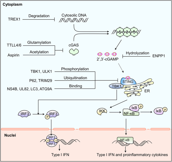
cGAS is a kind of cytosolic DNA sensor which initiates innate immune (Fig.1). Notably, cGAS and its downstream regulators form a major DNA-sensing mechanism, sensing foreign and self-DNA in the cytoplasm and sometimes in the micronucleus and nuclei as well [8]. The canonical cGAS/STING pathway starts with the activation of cGAS. cGAS is activated by accumulated cytosolic DNA and produces cGAMP as a second messenger. cGAMP then activates STING, an ER resident transmembrane protein [9]. Activated by cGAMP, STING will be transferred from ER to the Golgi body via the ER-Golgi intermediate compartment (ERGIC) and recruits TANK-binding kinase 1 (TBK1) [10]. Combined with STING, TBK1 will be delivered to lysosomal compartments to catalyze the phosphorylation of interferon regulatory factor 3 (IRF3) [11, 12]. Phosphorylated IRF3 will be dimerized and translocated into the nucleus to stimulate the expression of IFN-I and IFN-stimulated genes (ISGs). In parallel, STING also activates inhibitor of nuclear factor kappa-B (NF-κB) kinase (IKK). IKK phosphorylates and deactivates the inhibitor of NF-κB (IκB). NF-kB then is released from IκB and enters the nucleus, where it functions together with IRF3 and other transcription factors to induce the expression of interferons and inflammatory cytokines such as TNF, interleukin (IL)-1b and IL-6 [13]. STING may also directly bind to the cytosolic DNA but the pathological background is not fully elaborated [14]. Recent evidence has shown that the primordial function of STING is related to autophagy [15], as is proved in Zika virus infection of the Drosophila brain [16].
Fig. 1.
cGAS provoking pro-inflammatory reaction and its regulations. cGAS leads to a pro-inflammatory reaction through the classic cGAS-STING-IFN1 axis. And this axis can be regulated in different levels: dsDNA, cGAS, cGAMP, STING and downstream regulators
In addition to the classic cGAS/STING-IFN axis (Fig. 1), study has revealed that cGAS interacted with Beclin-1 to trigger autophagy, which would reduce cGAMP production independent of STING activation [17]. Similarly, activation of STING could be achieved in a cGAS-independent manner. Noncanonical STING signaling in response to etoposide-induced DNA damage could be activated by DNA-repair proteins ataxia telangiectasia mutated (ATM), poly-ADP ribose polymerase 1 (PARP1) and DNA binding protein interferon-γ-inducible factor 16 (IFI16) mediated NF-κB signaling in keratinocytes [18]. Similar regulatory processes can also be seen in some specific tumor models, such as in HCT116 colorectal carcinomas [19].
As a classic pathway, components of cGAS pathway, up and down stream regulatory factors and roles in autoimmune diseases and tumor immunity have been fully described and summarized [8, 20, 21]. In this review, we only briefly introduce these aspects as background information. Emphasis will be placed on the latest research on position-dependent cGAS function and non-immune physiological regulatory processes such as cellular senescence, programmed cell death (PCD), mitophagy Ca2+ homeostasis and ER stress and the different roles of cGAS/STING pathway in some human diseases.
cGAS interacting with different sources of DNA
The structure of cGAS
Like other proteins, cGAS has a C terminus (160–522) and a N-terminus (1 ~ 159). C terminus contains a nucleotidyltransferase domain and two DNA binding sites [22, 23] which assist cGAS dimerizes and binds to the sugar-phosphate backbone of double strand DNA (dsDNA) in the form of 2:2 [24, 25]. Single-stranded DNA (ssDNA) generated from reverse transcription weakly activates cGAS [8]. Specifically, unpaired guanosines are the necessary DNA structures for the activation of cGAS, suggesting that cGAS has the ability to recognize specific DNA sequence under certain circumstances. For example, cGAS can be activated effectively by the short (12- to 20-bp) human immunodeficiency virus type 1 (HIV-1) Y-form DNA in a sequence-dependent manner [26]. After binding to DNA, its nucleotidyltransferase domain transfers adenosine 5′-triphosphate (ATP) and guanosine 5′-triphosphate (GTP) to cGAMP and activates downstream pathways [27, 28].
For a long time, the functional significance of cGAS N-terminus (1 ~ 159) remains unclear [23]. However, researchers have recently taken a small step closer toward this. Du and Chen reported that N-terminus contributes to cGAS and DNA liquid droplets formation in physiological buffer and human cell lines, and therefore promotes the activation of cGAMP production [29]. N-terminus is also important for the subcellular localization of cGAS [30]. In the absence of DNA, N-terminus helps cGAS bind to the PI (4,5) P2 at the cell plasma membrane which reduces the sensitivity of cGAS to self-DNA. When the interaction between N-terminus and the plasma membrane disappears, cGAS will translocate into the cytoplasm and nucleus [30]. C-terminal domain 161 ~ 212 is crucial for cytoplasmic retention. While two nuclear localization sequences (NLS): N-terminal NLS1 (21 ~ 51) and C-terminal NLS2 (295 ~ 305) are needed for nuclear translocation [31].
cGAS and cytosolic DNA
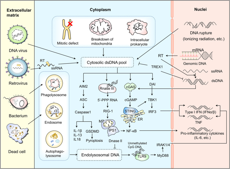
The main sources of cytoplasmic DNA are as follows (Fig. 2) [32, 33]: (1) Intracellular pathogens infection, such as DNA viruses, retroviruses and intracellular prokaryotes; (2) Reactivation of endogenous retroviral sequences which codes a catalytically active retro transcriptase; (3) Imbalanced control of endogenous DNA such as mitochondrial breakdown, mitotic defects and DNA rupture; (4) Impaired ability to clear exogenous DNA; (5) Importing extracellular DNA-containing exosomes and/or micropinocytosis. Crystal structures of human cGAS and DNA-bound cGAS shows that cGAS is activated when two cGAS molecules and two dsDNA molecules compose together to form a ladder-like structure [34]. Bacterial and mitochondrial nucleoid proteins HU, mitochondrial transcription factor A (TFAM) and high-mobility group box 1 protein (HMGB1) support the recognition of dsDNA by elongating DNA sensing time via inducing the formation of U-turns and bends in DNA [35]. cGAS modified by other molecules also links tightly with the activity of dsDNA induced immune responses [36–39]. For example, polyglutamylases such as tubulin tyrosine ligase-like family member 6 (TTLL6) catalyzes the polyglutamylation of cGAS and hinders its binding with DNA, which can be canceled and reversed by the cytosolic carboxypeptidase 6 (CCP 6); Monoglutamylases such as TTLL4 catalyzes the monoglutamylation of cGAS and impedes the synthase of GAMP, which can be removed and recovered by CCP 5 [36]; Tripartite motif 56 (TRIM 56) E3 ubiquitin ligase monoubiquitinates cGAS at the Lysine 335 (K335), facilitating the binding of DNA and the synthesis of cGAMP by increasing the dimerization of cGAS [37]; The small ubiquitin-like modifier (SUMO) SUMOylates cGAS at K335, K372, and K382, restraining the following reactions, which can be reserved by sentrin/SUMO-specific protease 7 (SENP7) [38]; And also, Recently, both in vivo and in vitro studies have shown that GTPase-activating protein SH3 domain-binding protein 1 (G3BP1) binds to cGAS directly and helps cGAS bind with dsDNA by forming the large G3BP1-cGAS complexes [39].
Fig. 2.
Source and function of cytosolic DNA. The cytosolic dsDNA pool is composed of DNA originating from retroviruses, DNA viruses, mitotic defects, mitochondria, intracellular prokaryotes and DNA rupture debris from the nuclei. In the cytosol, these DNA are either degraded by DNase III and TREX1 or sensed by DNA sensors. Once sensed by cytosolic sensors including AIM2, RNaseIII, cGAS and DAI, a cascade of reactions are triggered. Downstream signal molecules regulate the expression of immune-related genes, eventually leading to DNA clearance or cell death. In addition, DNA also comes from dead cells and bacteria. Membrane vesicles are formed by endocytosis and DNA is transported into the cell. DNase II is localized in lysosomes and digests DNA from pathogens and dead cells that end up in this cellular compartment. TLR9 directly binds with the remaining unmethylated CpG DNA and triggers downstream immune activation, inducing the expression of inflammation-related genes
Cytoplasmic DNA-induced cell death and immune response are self-defense against harmful substances from the internal and external environment. Drugs using DNA to induce immunity against tumors and infectious diseases are under developing and testing [40].
cGAS and micronuclear DNA
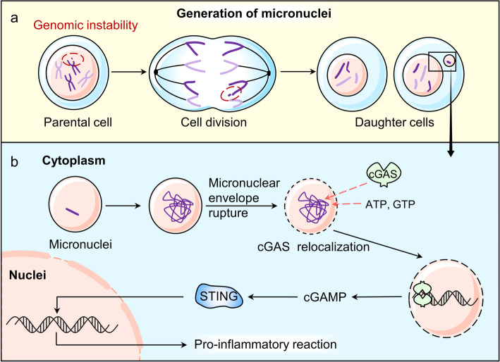
Micronuclei is a cytoplasmic compartment which is composed of a membrane envelope and chromatin in it. Generally, micronuclei is regarded as an accurate indicator of genomic instability [41] (Fig. 3). It is formed when mitosis process encounters with mis-segregations of a whole or a part of a chromosome, accompanied with chromatin bridging and chromosomes/chromatin formation lagging behind [42, 43].
Fig. 3.
The generation of micronuclei and cGAS’s role in micronuclei. a. Micronuclei are generated when the genome is unstable during cell division, often associated with an abnormal nucleus in a daughter cell. b. After micronuclei form, the micronuclear envelope ruptures irreversibly. Then, cGAS enters into the micronuclei, binding to the chromatin and facilitating the downstream proinflammatory signals
Some evidence shows that cGAS binds with chromosomes during mitosis [44]. But cGAS/STING pathway remains inactive, probably due to the tight-compacted structure of chromosomes [45]. It is noteworthy that cGAS will dissociate from chromosomes when mitosis is done. However, when micronuclei forms, a high level of cGAS shows up in the micronuclei during the interphase [46]. Then micronuclear cGAS mediates the downstream process [10, 22, 46] in a timely and cell-cycle dependent manner [47]. Under such circumstances, micronuclei acts as a reservoir of immunostimulatory DNA, which may function as a compensative cell cycle checkpoint [48, 49].
Activation of cGAS by micronuclear DNA requires the entry of cGAS into micronuclei. Micronuclei forms when lagging chromosome separates from the primary nucleus and has its own membrane [41]. Then, the micronuclear envelope shatters irreversibly which occurs at a random phase. That means it is not confined to a specific phase but sensitive to DNA damage [44]. This failure of membrane integrity is associated with the reduction of lamin B1 [41, 47], which may reverse by nuclear β-dystroglycan (β-DG) [50]. As a consequence, the cytosolic cGAS gains the access to micronuclei and thereby binds to chromatin and initiates downstream proinflammatory responses [47].
As we discussed above, the formation of micronuclei is an important mark of genomic instability. And genomic instability caused by the activation of multipolar mitotic spindles [50], the production of DNA-damage agents [51], centrosome abnormalities [52], telomeres dysfunctions [53–55] andp53/p21 mutation [56] are hallmarks of cancer and cellular senescence. The disruption of the micronuclei caused by aberrant accumulation of the endosomal sorting complex required for transport-III (ESCRT-III) complex or the mutant prelamin A (progerin) accentuates DNA damage and enhances pro-inflammatory responses via cGAS/STING pathway [57–59]. The formation of micronuclei has a dual function. On the one hand, abnormal accumulation of micronuclei is related to cancer and aging. On the other hand, the micronuclear dsDNA activates cGAS-mediated immune response, which is an important innate immune surveillance mechanism for the clearance of cancer cells and senescent cells.
cGAS and nuclear DNA
cGAS interacting with endogenous DNA in nuclei
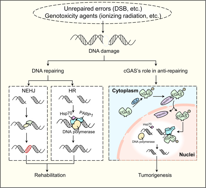
Micronuclear cGAS works as a supervisor. When unstable genetic material appears, cGAS will initiate its downstream pro-inflammatory responses, linking the genome instability with innate immune. However, in some cases, cGAS enters into nuclei and inhibits DNA repair when DNA damage happens, and therefore playing a tumorigenic role adversely [31] (Fig. 4).
Fig. 4.
cGAS interacting with endogenous DNA in nuclei. Naturally, DNA damage caused by unrepaired errors or genotoxic agents can be corrected by two basic DNA DSB repair mechanisms: NHEJ and HR. However, in some circumstances, when DNA is damaged, cGAS can be phosphorylated by BLK and then translocated into the nucleus with the help of importin-α. In nucleus, the phosphorylated cGAS interacts indirectly with PARP1, impeding HR and thus promoting tumorigenesis
DNA damage arises more frequently when exposed to genotoxic therapies such as chemotherapies [60]. Among all forms of damage, the double-stranded breaks (DSBs) of DNA strains are the severest type [61]. These damages can be rescued by two basic DNA DSB repair ways: homologous recombination (HR) and nonhomologous end joining (NHEJ) [62–64]. However, studies have confirmed that this repair process can be interrupted by cGAS via binding with chromosomes and initiates immune response [31]. The underlying mechanism and significance of cGAS binding with chromosomes have not been fully elucidated. One thing we can confirm is that it is the NLS-mediated entry of cGAS rather than the low-level physiological accumulation of cGAS that activates innate immune [65]. More in-depth research is needed to better understand this process.
cGAS interacting with exogenous DNA in nuclei
Viruses live a highly parasitic live. They use host’s resources and organelles as production materials and workshops and own genetic material as templates to proliferate. In response, mammals have a set of complicated mechanisms to detect and kill those viruses. At the same time, viruses never stop attempting to elude and defect the surveillance system of the host.
The innate immune system of human body reacts immediately in response to virus infections. Pattern recognition receptors (PRRs) recognize the conservative pathogen-associated molecular patterns (PAMPs) or host damage associated molecular patterns (DAMPs) [66], followed by cascade signaling reactions. In this process, cGAS plays a pivotal role in detecting viruses and activating dendritic cells (DCs) and macrophages [67, 68]. The recognition process often starts in the cytoplasm, sometimes in nucleus (Fig. 5).
Fig. 5.
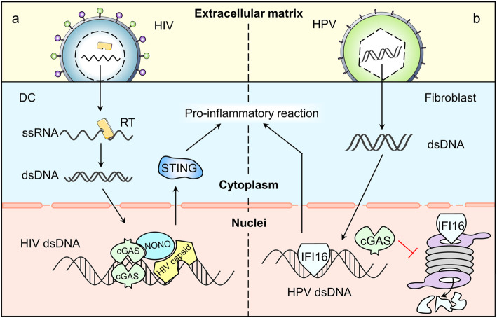
cGAS interacting with exogenous DNA in nuclei. a. HIV’s ssRNA enters into the DC and is synthesized into HIV dsDNA in the cytoplasm. Then, the dsDNA is admitted into the nuclei, where capsid protein binds directly to NONO, promoting cGAS’s recognition of HIV dsDNA. b. HPV’s dsDNA is transmitted into the fibroblast and then enters into the nuclei. The nuclear cGAS prolongs the half-life of IFI16 (another DNA sensor) by promoting the degradation of proteasomes
HIV infection is a typical example of nuclear recognition process. Earlier studies show that cGAS is vital for the recognition of retro-transcriptional synthetic HIV dsDNA by immune cells both inside and outside the cell nuclei [26]. To active cGAS, Non-POU domain-containing octamer-binding protein (NONO) first binds to capsid protein DCs nucleus. Then, NONO interacts with cGAS to form a complex which in turn promotes the recognition of HIV-2 DNA and the initiation of nuclear cGAS induced STING activation [69].
cGAS also participates in the recognition of nuclear DNA through an indirect, STING independent way. This is achieved by helping maintain the stability of other DNA sensors such as IFI16 [70]. When infected by human papillomavirus (HPV), normal human fibroblasts will develop a certain mechanism to facilitate IFI16 in binding directly with HPV DNA in the nucleus [70]. At the same time, the existence of cGAS prolongs the half-life of IFI16, may by promoting the degradation of proteasome [70]. Though more research is needed to confirm the role of cGAS in this process, we may able to develop cGAS as an immune enhancer to support our body in defending virus.
Regulation of cGAS/STING pathway
Regulation of cGAS/STING pathway has been thoroughly discussed elsewhere [8]. Here we only give a brief summary and add some new findings (Fig. 1). The regulation is complex and multidimensional, mainly from the following aspects:
Degradation of cytosolic dsDNA. To avoid the cellular disorder triggered by cytosolic DNA, TREX1 degrade mis-localized DNA and maintain the balance of homeostasis and inflammation response [4].
Regulation of cGAS. Transcriptional, epigenetic regulations and post-translational modifications are all involved. Besides these aspects discussed in the referent paper, chemical modifications also participate in regulating the activity of cGAS. For instance, Aspirin robustly inhibits cGAS activation and cGAS-mediated IFN production by directly acetylating the cGAS at K384 and/or K394 and K414 [4].
Regulation of cGAMP. Activity and location of cGAMP are controlled by ecto-nucleotide pyrophosphatase/ phosphodiesterase 1 (ENPP1) and intercellular transmission respectively [71]. In vivo study is still limited but the in vitro study shows that overexpression of ENPP1 significantly lowers cGAMP level and reduces production of IFN-β and NF-κB in porcine cells infected with pseudorabies virus (PRV) [72].
Modification of STING. Post-translational modifications, trafficking degradation and binding affinities with cGAMP are involved. Phosphorylation by serine/threonine UNC-51-like kinase (ULK1) and TBK1 and ubiquitination by ubiquitin-binding protein p62 lead to the degradation of STING [73, 74]. Interestingly, the function of p62 is dependent on TBK1 and IRF3, which indicates negative feedback on the attenuation of signaling [73]. Moreover, the tyrosine-protein phosphatase nonreceptor type (PTPN) 1 and 2 dephosphorylate STING at Y245 which promotes its 20S proteasomal degradation [75]. Additionally, DNA virus infection triggered the ubiquitination of STING by upregulating TRIM29 [76].
In addition to modification, regulators inhibit STING by directly binding to STING. Such as autophagy proteins, including microtubule-associated protein 1 light chain 3 (LC3) and autophagy-related protein 9a (ATG9A) [77]. Pathogenic proteins, such as Hepatitis C virus non-structural 4B (NS4B) protein and human cytomegalovirus (HCMV) tegument protein UL82, directly interact with STING to reduce STING activity [78, 79]. Meanwhile, DNA tumor viruses, such as HPV18 and human adenoviruses 5 (hAd5), inhibit the activation of cGAS/STING pathway by producing oncoprotein binding STING [68]. Moreover, the Ca2+ sensor stromal interaction molecule 1 (STIM1) binds to STING and elongates STING retention with ER, consequently hindering the following cascade [80].
Roles of the cGAS/STING pathway in physiological regulatory processes
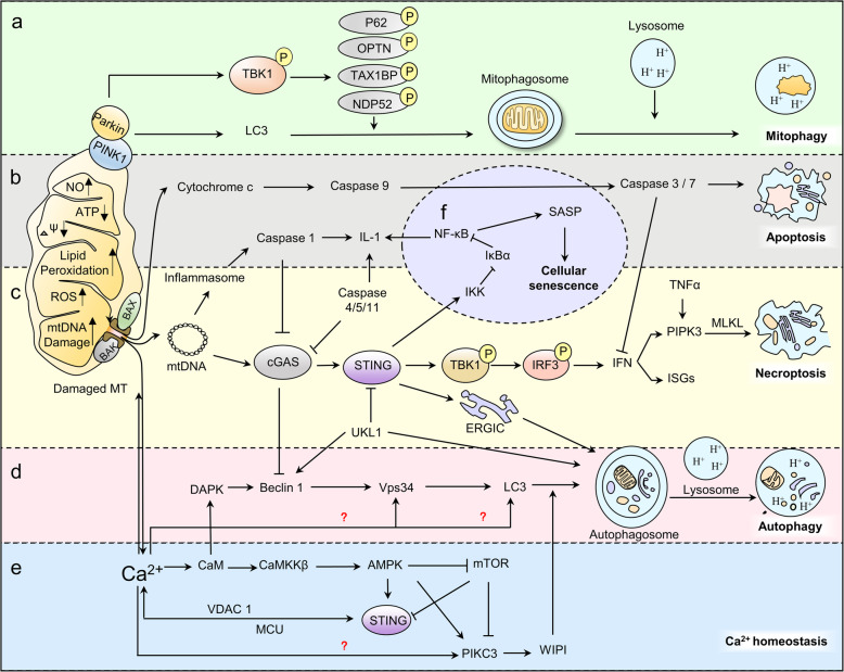
A bulk of studies have exhaustively summarized the role of cGAS/STING pathway in regulating antipathogenic and antitumor responses and the adaptive changes of cancerous cells and pathogens to escape cGAS supervision [8, 81–84]. As we explore and know more about this pathway, we are able to find more in other aspects. Therefore, here we focus on other newly found and also important aspects, including its role in nuclei, senescence, mitochondrial dysfunction, ER stress and Ca2+ homeostasis (Fig. 6).
Fig. 6.
Regulatory role of cGAS–STING pathway in physiological processes. cGAS/STING participates in regulating mitophagy (a), apoptosis (b), necroptosis (c), autophagy (d), cytoplasmic Ca2+ homeostasis (e), production of SASP and cellular senescence (f)
Cellular senescence
Cellular senescence is first proposed by Hayflick and Moorhead in 1961, which is defined as irreversible cell-cycle arrest that occurs when cells experience potentially oncogenic stress [85]. According to different incentives, such as telomere shortening, certain oncogenes and chemotherapeutic drugs or ionizing radiation, senescence can be subdivided into replicative senescence, oncogene-induced senescence (OIS) and therapy-induced senescence (TIS) respectively [86]. The p53/p21 and p16INK4a/pRB pathways are responsible for senescence related growth arrest [87]. Senescent cells, though fail to initiate DNA replication, remain metabolically active and secret sorts of proteins, including proteases, various growth factors, cytokines and chemokines with proinflammatory properties. Collectively, these secretions are termed as SASP that have complex effects on cell behaviors, especially in aging and tumorigenesis [87, 88]. As we mentioned before, mtDNA and micronuclei are two sources of cytoplasmic dsDNAs. Considering that mitochondrial dysfunction and genomic instability are typical features of aging [89], it is not difficult to link cGAS/STING with cellular senescence.
In senescent cells, several factors contribute to the accumulation of cytoplasmic DNA and the activation of cGAS: (1) Loss of the nuclear lamina protein Lamin B [90]. Decreased Lamin B is a hallmark of senescence which leads to collapse of the nuclear envelope, triggering release of chromatin fragments from the nucleus to the cytosol, termed as cytoplasmic chromatin fragments (CCF) [91]. (2) Leakage of mtDNA. Accumulated oxidative damage to mitochondrial membrane proteins and lipids leads to increased membrane permeability. Membrane break up results in the leakage of mtDNA to cytoplasm [92]. (3) Downregulation of TREX1 [93]. TREX1 is responsible for degrading the double-stranded and single-stranded DNA in the cytoplasm to prevent accumulation of DNA. (4) Upregulated long-interspersed element-1 (LINE-1, also known as L1) [94]. LINE-1 is a retro-transposable element which transcribes mRNA to cDNA and causes cytoplasmatic DNA accumulation. (5) Increased MUS81protein [95]. MUS81 is a structure-specific endonuclease that resolves inter-strand DNA structures such as stalled replication forks and Holliday junctions. MUS81is engaged in changing nuclear DNA into cytoplasmic forms which causes elevated cytoplasmic DNA [96].
Binding with accumulated self-derived DNA fragments in senescent cells, cGAS then activates downstream expression of NF-κB and triggers SASP production in senescent cells [86]. Studies find that suppressing either cGAS, STING or NF-κB in mouse and human cells abrogates the expression of senescence-associated inflammatory genes in response to DNA damaging agents such as etoposide and ionizing irradiation [44, 97]. The secretion of several SASP factors is regulated by cGAS, including IL-6, a critical controller of autocrine senescence, CXCL10, a cGAS-dependent IFN-stimulated gene, TNF-α and several chemokines [98, 99]. The connection between cGAS/STING pathway and SASP regulation remains largely unknown. However, the consistent performance in in between reflects the existence of close relationship. Future work could focus on the molecular mechanism of cGAS/STING pathway in senescence and aging-related diseases to see if GAS also has a regulatory effect such as neurodegenerative diseases, osteoarthritis, cardiovascular diseases, etc. (Fig.6f).
PCD
Depending on different endogenous and exogenous threats, cells have 3 different fates: (1) Restore and back to normal function if the threats are successfully eliminated; (2) Enter senescence if the damage is persistent but tolerable; (3) Undergo PCD or necrosis if the damage is beyond management. The death of infected cells is an important defense that limits viruses to subvert the cellular machinery for their own replication. This part has been well established [100, 101]. Here we are going to talk about latest understanding of cGAS/STING in regulated cell death (RCD).
Based on the macroscopic morphological alterations and where dead cells and their fragments are disposed, RCD is detailed classified into different subtypes [102, 103], apoptosis, autophagy and necroptosis. Intrinsic apoptosis is induced by cellular stress and starts with the activation of apoptotic caspases (caspase-3, − 6, − 7, − 8, and − 9) [104]. Exposed to stress, mitochondrial outer membrane permeabilization (MOMP) is formed by Bax (Bcl-2-associated x protein) / Bak (Bcl-2 antagonist killer 1) channel. Mitochondrial contents such as cytochrome c is then released form MOMP into the cytosol where it binds to the NLR protein apoptotic protease activating factor 1 (APAF1). This binding forms the apoptosome — an activating platform for the initiator caspase 9 [105, 106]. Activated caspase 9 in turn activates the effector caspases, caspase 3 and caspase 7 [107]. Executioner caspase 3 and 7 trigger a cascade of proteolytic events that culminate in the demise of the cell through apoptosis. cGAS participates in apoptosis via regulating caspase-3. Activated by apoptosis signals, caspase-3 cleaves and inactivates cGAS, mitochondrial antiviral-signaling protein (MAVS), and IRF3 to suppress cytokine and type I IFN production in order to keep immunologically silent [108–110]. While caspase inhibition prompts the widen of BAX/BAK-mediated pores which leads to the extrusion of unstructured mitochondrial inner membrane [111]. Mitochondrial inner membrane permeabilization facilitates mtDNA release into the cytoplasm and activate cGAS/STING signaling and IFN synthesis, enabling cell death-associated inflammation [112]. The inflammatory caspases-1, − 4, − 5 and − 12 also influence cGAS function. Under DNA virus infection, caspase-1 interacts with cGAS, cleaving it and dampening cGAS/STING-mediated IFN production. Caspase-4, 5, and 11 cut cGAS under non-canonical inflammasome activation [113, 114]. Understanding the complex regulatory network between cGAS and caspases at the intersection of programmed cell death and innate immune regulation is helpful for better understanding related human diseases [100, 115] (Fig. 6b).
Autophagy is a self-degradative process that allows the recycling of cellular components. It is also a final barrier against oncogenic transformation that restricts chromosomal instability during replicative crisis [116]. Basic autophagy process have been summarized and generally accepted [117]. Here we introduce the newly found relationship between autophagy and cGAS. In this process, interferon induction is not indispensable. For example, in macrophages, activated STING and the kinase TBK1 lead to ubiquitin-mediated selective autophagy pathway, limiting M. tuberculosis growth during infection independent of IFN production [118, 119]. In addition, cGAS can directly bind with Beclin-1 autophagy protein and release the negative autophagy regulator Rubicon from the Beclin-1 complex. This interaction activates downstream phosphatidylinositol 3-kinase class III and induces autophagy to remove cytosolic pathogen DNA independent of TBK1 activation [17]. Another TBK1 independent way relies on the formation of ERGIC. After binding with cGAS, STING buds from the endoplasmic reticulum into coat protein II (COP-II) vesicles then forms the ERGIC. The ERGIC serves as the membrane source for WD-repeat PtdIns (3) P effector protein 2 (WIPI2) recruitment and LC3 lipidation. Autophagosomes that target cytosolic DNA or DNA viruses then formed and merge with the lysosome. Besides, the discovery in sea anemone prompts us that autophagy induction is an ancient and highly conserved function of the cGAS/STING pathway that even pre-dates the emergence of the type-I interferon pathway in vertebrates [15]. Autophagy induced via these pathways prevents replication of pathogens by eliminating the infected cells, protecting the body against pathogen attack. Other roles of cGAS/STING induced autophagy include protecting liver from ischemia-reperfusion injury [120] and restricting chromosomal instability during replicative crisis. This replicative check point also serves as a final barrier against oncogenic transformation by eliminating precancerous cells with disrupted cell cycle checkpoints [121] (Fig. 6d).
Necroptosis is a lytic form of PCD that involves the swelling and rupture of dying cells. cGAS/STING is able to induce necroptosis in bone marrow derived macrophages via type I IFN signaling pathways, which synergizes to trigger RIPK3 (receptor interacting protein kinases 3) and MLKL (Mixed lineage kinase domain-like) driven necroptosis independent of caspase-8 function [122, 123]. Moreover, cGAS/STING activated by mitochondrial DNA has been suggested to amplify necroptosis via a TNF-dependent mechanism [124, 125] (Fig. 6c).
Substantial crosstalk exists between different cell death pathways ensuring that these signaling pathways are well regulated. More studies are required to further explore the interconnection among these pathways and how cGAS/STING signaling toggles in transcriptional responses, different forms of RCD, anti-neoplastic transformation and anti-infection reactions.
Others
Mitophagy is a selective form of autophagy controlled by the Pink1-Parkin pathway or the mitophagic receptors Nix and Bnip3. The physiological role of mitophagy is specifically removing damaged or excessive mitochondria [126]. cGAS/STING does not participate in regulating mitophagy directly. But when damaged mitochondria failed to be removed, stress from mitochondrial DNA mutations activates the proinflammatory cGAS/STING pathway which may contribute to several age-related neurodegenerative diseases, for example, Parkinson’s and Alzheimer’s disease [127, 128]. The engagement of cGAS and subsequent mtDNA-induced STING-mediated type I IFN production can be suppressed by apoptotic caspase 9 and downstream caspase 3 and 7, rendering mitochondrial apoptosis immunologically silent [108, 110] (Fig. 6a).
Though many unknowns remain in the regulation among cGAS, Ca2+ homeostasis and ER stress, the existing research findings show promising results for future investigation. Here we list the brief summary of these findings: (1) STING influences the intracellular Ca2+ level via affecting the mobilization of ER Ca2+ pool. As we put above, STING is found on the contact sites between the ER and mitochondria where Ca2+ is exchanged between these two organelles via channels like voltage-dependent anion channel 1 (VDAC1) and mitochondrial Ca2+ uniporter (MCU). STING deficiency augments the translocation of stromal interaction molecule 1 (STIM1), a Ca2+ sensor. Then the depletion of ER Ca2+ stores trigger Ca2+ entry [80]. (2) Intracellular calcium is a rheostat for the STING signaling pathway. Reductions in cytosolic Ca2+ and the mitochondrial export of Ca2+ reduced the activation of NF-κB and IRF3. While increased intracellular Ca2+ from ER and mitochondria promotes STING activation via two independent Ca2+-calmodulin dependent pathways: AMPK (Adenosine 5′-monophosphate (AMP)-activated protein kinase) and CAMKII (Ca2+/calmodulin-dependent protein kinase II) pathways [129]. (3) ER stress activates STING pathway. ER stress, either induced by alcohol or co-stimulation with thapsigargin (the sarcoplasmic endoplasmic reticulum calcium ATPase (SERCA) pump inhibitor), enhances STING signaling and augments IFN production [130, 131]. Besides, STING activates ER stress and the unfolded protein response (UPR) through a novel motif termed as “the UPR motif”, which is located in the helix aa322–343. Long-lasting STING-mediated ER stress and disruption of calcium homeostasis primes T cell death by apoptosis [132–134] (Fig.6e).
Roles of the cGAS/STING pathway in human diseases
Generally, cGAS is allocated to a limited subcellular area that is free of self-DNA. Several endogenous nucleases participate in maintaining self-DNA level under the threshold of receptor activation. However, under pathologic circumstance, self-DNA is exposed to cGAS. This will lead to abnormal activity of the cGAS/STING pathway, causing autoinflammation and autoimmune disease and even inflammation-associated cancers. Abnormal activation of cGAS/STING pathway in inflammatory and autoimmune diseases is well discussed in other papers [8, 21, 135]. Here we take Aicardi-Gourtières syndrome (AGS), a typical disease caused by the excessive activation of cGAS, as an example to illustrate the mechanism. AGS is caused by causal mutations in any one of several key genes, including TREX1, RNASEH2A (Ribonuclease H2 subunit A), RNASEH2B, RNASEH2C (which together encode the Ribonuclease H2 enzyme complex), SAMHD1 (Sterile alpha motif and histidine-aspartic acid domain-containing protein 1), ADAR1 (adenosine deaminase acting on RNA 1) and IFIH1 (interferon induced with helicase C domain 1, also known as MDA5) [136–140]. These genes are responsible for cleaning ectopic DNA, lack of which causes inappropriate accumulation of self-derived nucleic acids, sustained activation of cGAS and excessive production of type I interferons.
Apart from self-DNA-driven inflammation, other cGAS/STING pathway induced responses also participates in human diseases [44, 47, 141–144]. For example, mutations in gene ATG16L1 promote the production of IL-22 in the intestinal epithelium through cGAS/STING pathway, which result in excessive epithelial cell death and inflammatory bowel disease (IBD) [145]. The connection between the cGAS pathway and aging provides a new topic for us. Existing study has identified the cGAS/STING pathway as a sensor of senescence-associated DNA damage and trigger of inflammation in early age-related macular degeneration [146]. More research is needed on the relationship between cGAS/STING pathway and senescence-associated human diseases such as neurodegenerative diseases, degenerative arthritis and cardiovascular diseases.
Defective cGAS/STING signaling is closely associated with oncogenesis, immune evasion and tumor metastasis [147, 148]. Antineoplastic role of cGAS has been found in multiple mouse tumor models, including colon, brain, skin, pancreatic, liver, breast, and B cell malignancies [149, 150]. These protective effects are achieved mostly through IFN-induced immune responses and in few cases via autophagy [121, 151]. cGAS/STING pathway is also related to tumor microenvironment remodeling [82, 152] and the production of anti-tumor cytokines such as indoleamine 2,3-dioxygenase (IDO), IL-10 and ISGs, together inhibiting tumor growth and improving the survival [150, 153]. In vitro human study shows that targeting DNA damage response promotes antitumor immunity through STING-mediated T-cell activation in small cell lung cancer [154]. In vivo human-related research has not been performed, but we can assume the connection based on exiting in vitro studies.
Based on the anti-tumor role of cGAS/STING pathway, people began to design STING agonists and cyclic dinucleotide derivatives for tumor treatment. A novel synthetic cyclic dinucleotide, ADU-S100, has been promoted to phase Ib clinical trials in patients with diverse, solid, accessible tumors for achievable intra-tumoral delivery. Antitumor effects of ADU-S100 have been observed in PD-1–naive TNBC and PD-1–relapsed/refractory melanoma [155, 156]. Synthetic derivatives demonstrate a strong ability of inducing IFN-β in both murine BMDMs and primary human cells and forming antitumor immunological memory following tumor regression [153]. Contrary to the results mentioned above, activation of the cGAS/STING pathway is found in tolerogenic responses. By inducing indolamine 2,3-dioxygenase, cGAS/STING pathway promotes the growth of tumors with low antigenicity [157]. And a pan-cancer human study which analysis the association between the expression of cGAS/STING and immune cell infiltration shows that the upregulated cGAS/STING signaling is negatively correlated with the infiltration of immune cells in some tumor types [158]. Therefore, it is necessary to fully evaluate the function of cGAS/STING signaling in cancer immunity before the application of the STING agonist-based anticancer immune therapy [158].
cGAS/STING surveillance is a main part of antiviral responses, achieved mainly through the production of IFN. In addition to an initial virus-induced inflammatory cascade, cGAS/STING effectively engages a potent localized immune response via cGAMP transfer [159]. Autophagy is another effective anti-viral process that maintains cellular homeostasis by orchestrating immunity upon viral infection [160]. For example, ZIKV infects mature neurons in fly brain which induces Rel/NFKB inflammatory signaling. Rel/NFKB activates the expression of STING which then activates antiviral autophagy to restrict ZIKV infection [16, 161]. Recent study even reveals that autophagy induction in response to stimulation by cGAMP is a primordial function of the cGAS/STING pathway that pre-dates the emergence of the type-I interferon pathway in vertebrates [15]. More studies are needed to understand the role of cGAS/STING in anti-viral infection via induction of autophagy.
Conclusions and perspectives
cGAS/STING is a well-studied signaling pathway that participates in sensing abnormal subcellular localization of DNA and mediating protective immune defense against infection. Over the past few years, studies have established the basic framework and mechanisms of this DNA-sensing pathway. However, the investigation of cGAS pathway in immunomodulation and antitumor therapy has attracted a lot of attention and its critical roles in other areas are overlooked. Accumulating evidence indicates that the physiological and pathological regulatory effects of cGAS/STING pathway extends far beyond “traditional” antimicrobial immunity. In this review, we summarize the current finds of cGAS/STING pathway in a broad repertoire of cellular processes, including mitochondrial function, ER stress, Ca2+ homeostasis, cellular senescence, PCD, and metastasis. Based on its broad regulatory roles, we could see the therapeutic application of cGAS/STING pathway in some age-related diseases such as neurodegenerative diseases, osteoarthritis, cardiovascular disease, chronic kidney and pulmonary disease. These diseases, like tumors, have abnormal elimination of pathological tissue or the alienation of normal tissues. Developing specific inhibitors of the cGAS/STING pathway to identify its role in these human diseases will be an important and exciting task in the future.
Undeniably, several questions remain to be answered. First is about aging and cellular senescence which are believed to participate in the pathology of many diseases. Accumulated DNA damage, genomic instability, mtDNA release are ubiquitous in senescent cells. As we talked above, cGAS pathway is highly likely to sense these DNA segments and regulate SASP production. However, it is unlikely to be achieved through the classic pathway as increased IFNs are not normally seen in natural aging process. Other downstream regulators are waiting to be found to better explain this regulatory role. Ca2+ homeostasis is another interesting part because ER stress, mitochondrial dysfunction and PCD are all related to cellular Ca2+ level. STING is located on ER membrane near the ion exchange channels between ER and mitochondria. Whether and how STING affects the on and off of these channels and the consequence of this regulation are largely unknown. Faced with internal and external pressure, how cells determine the balance between caspase-induced apoptosis and cGAS-induced IFN production is still un clear. Besides, how STING induces autophagy under virus infection is unknown. In addition, the regulation of this pathway, signaling mechanism at each step and the possible crosstalk with other pathways require more studies to identify.
In sum, we thoroughly summarize the broad roles of cGAS/STING pathway in several critical cellular processes. Maintaining the delicate balance between aging, immunity and proliferation is a necessary guarantee for cell living and functioning. Future research on cGAS could focus more on the role of cGAS in balancing these three aspects, interactions with other related regulating pathways and applications in human disease treatments.
Acknowledgements
We would like to thank all laboratory members for their critical discussion of this manuscript, and apologize to those publications not mentioned due to space limitations.
Abbreviations
- ADAR1
Adenosine deaminase acting on RNA 1
- AGS
Aicardi-Gourtières syndrome
- AIM2
Absent in melanoma 2
- AMPK
Adenosine 5′-monophosphate (AMP)-activated protein kinase
- APAF1
Apoptotic protease activating factor 1
- ASFV
African swine fever virus
- ATG9A
Autophagy-related protein 9a
- ATM
Ataxia telangiectasia mutated
- ATP
Adenosine 5′-triphosphate
- Bak
Bcl-2 antagonist killer 1
- Bax
Bcl-2-associated x protein
- BLK
B-lymphoid tyrosine kinase
- CAMKII
Ca2+/calmodulin-dependent protein kinase II
- CCF
Cytoplasmic chromatin fragments
- CCP 6
Cytosolic carboxypeptidase 6
- cGAMP
Cyclic GMP-AMP
- cGAS
Cyclic GMP-AMP (cGAMP) synthase
- COP-II
Coat protein II
- DAMPs
Damage associated molecular patterns
- DCs
Dendritic cells
- DNases
Deoxyribonucleases
- DSBs
Double-stranded breaks
- dsDNA
Double strand DNA
- E
Glutamic acid
- ENPP1
Ecto-nucleotide pyrophosphatase/ phosphodiesterase 1
- ER
Endoplasmic reticulum
- ERGIC
Endoplasmic reticulum–Golgi intermediate compartment
- ESCRT-III
endosomal sorting complex required for transport-III
- G3BP1
GTPase-activating protein SH3 domain-binding protein 1
- GTP
Guanosine 5′-triphosphate
- hAd5
Human adenoviruses 5
- HCMV
Human cytomegalovirus
- HIV-1
Human immunodeficiency virus type 1
- HMGB1
High-mobility group box 1 protei
- HPV
Human papillomavirus
- HR
Homologous recombination
- HSV-1
Herpes simplex virus 1
- IBD
Inflammatory bowel disease
- IDO
Indoleamine 2,3-dioxygenase
- IFI16
Interferon-γ-inducible factor 16
- IFIH1
Interferon induced with helicase C domain 1
- IFN-I
Type I interferon
- IKK
Inhibitor of nuclear factor kappa-B kinase
- IL
Interleukin
- IRF3
Interferon regulatory factor 3
- ISGs
IFN-stimulated genes
- IκB
Inhibitor of NF-κB
- K
Lysine
- LC3
Light chain 3
- LINE-1
Long-interspersed element-1
- MAVS
Mitochondrial antiviral-signaling protein
- MCU
Mitochondrial Ca2+ uniporter
- MDA5
Melanoma differentiation-associated 5
- MLKL
Mixed lineage kinase domain-like
- MOMP
Mitochondrial outer membrane permeabilization
- mtDNA
Mitochondrial DNA
- NF-κB
Nuclear factor kappa-B
- NHEJ
Nonhomologous end joining
- NLRs
NOD-like receptors
- NLS
Nuclear localization sequences
- NONO
Non-POU domain-containing octamer-binding protein
- NS4B
Non-structural 4B
- OIS
Oncogene-induced senescence
- PAMPs
Pathogen-associated molecular patterns
- PAR
Poly-ADP ribose
- PARP1
Poly-ADP ribose polymerase 1
- PCD
Programmed cell death
- PRRs
Pattern recognition receptors
- PRV
Pseudorabies virus
- PTPN
Tyrosine-protein phosphatase nonreceptor
- RCD
Regulated cell death
- RIG-I
Retinoic acid-inducible gene I
- RIPK3
Receptor interacting protein kinases 3
- RLRs
RIG-I-like receptors
- RNASEH2A
Ribonuclease H2 subunit A
- SAMHD1
Sterile alpha motif and histidine-aspartic acid domain-containing protein 1
- SASP
Senescence-associated secretory phenotype
- SENP7
Sentrin/SUMO-specific protease 7
- SERCA
Sarcoplasmic endoplasmic reticulum calcium ATPase
- ssDNA
Single-stranded DNA
- STIM1
Stromal interaction molecule 1
- STIM1
Stromal interaction molecule 1
- STING
Stimulator of interferon genes
- SUMO
Small ubiquitin-like modifier
- TBK1
TANK-binding kinase 1
- TF
Transcription factor
- TFAM
Transcription factor A, mitochondrial
- TIS
Therapy-induced senescence
- TLRs
Toll-like receptors
- TREX1
Three prime repair exonuclease 1
- TRIM56
Tripartite motif-containing protein 56
- TTLL6
Tubulin tyrosine ligase-like 6
- ULK1
UNC-51-like kinase
- UPR
Unfolded protein response
- VDAC1
Voltage-dependent anion channel 1
- WIPI2
WD-repeatPtdIns(3) P effector protein)
- Y
Tyrosine
Authors’ contributions
SL and YGT proposed the topic and provided funding support. MHG and HST developed and refined the idea. MHG, YCH, HST and XYC wrote the manuscript. MHG and HST drew the figures. YCH revised the manuscript. The authors read and approved the final manuscript.
Funding
This work was supported by the National Natural Science Foundation of China [81,672,787(Y.Tao), 81,874,139 and 81,672,991(S.Liu)], and the Overseas Expertise Introduction Project for Discipline Innovation (111 Project, No. 111–2-12).
Availability of data and materials
Not applicable.
Ethics approval and consent to participate
The ethics committee of the Cancer Research Institute of Central South University has approved this study.
Consent for publication
Not applicable.
Competing interests
The authors declare that they have no competing interests. The authors declare no conflicts of interest. This manuscript has been read and approved by all authors and is not under consideration for publication elsewhere.
Footnotes
Publisher’s Note
Springer Nature remains neutral with regard to jurisdictional claims in published maps and institutional affiliations.
Menghui Gao and Yuchen He contributed equally to this work.
Contributor Information
Menghui Gao, Email: gaomenghui1209@163.com.
Yuchen He, Email: yuche@pitt.edu.
Haosheng Tang, Email: ts525975171@163.com.
Xiangyu Chen, Email: chenxiangyu2000@outlook.com.
Shuang Liu, Email: shuangliu2016@csu.edu.cn.
Yongguang Tao, Email: taoyong@csu.edu.cn.
References
- 1.Cooper MD, Alder MN. The evolution of adaptive immune systems. Cell. 2006;124(4):815–822. doi: 10.1016/j.cell.2006.02.001. [DOI] [PubMed] [Google Scholar]
- 2.Kumar H, Kawai T, Akira S. Pathogen recognition by the innate immune system. Int Rev Immunol. 2011;30(1):16–34. doi: 10.3109/08830185.2010.529976. [DOI] [PubMed] [Google Scholar]
- 3.Medzhitov R, Janeway CA., Jr Decoding the patterns of self and nonself by the innate immune system. Science. 2002;296(5566):298–300. doi: 10.1126/science.1068883. [DOI] [PubMed] [Google Scholar]
- 4.Paludan S, Reinert L, Hornung V. DNA-stimulated cell death: implications for host defence, inflammatory diseases and cancer. Nat Rev Immunol. 2019;19(3):141–153. doi: 10.1038/s41577-018-0117-0. [DOI] [PMC free article] [PubMed] [Google Scholar]
- 5.Hemmi H, Takeuchi O, Kawai T, Kaisho T, Sato S, Sanjo H, et al. A toll-like receptor recognizes bacterial DNA. Nature. 2000;408(6813):740–745. doi: 10.1038/35047123. [DOI] [PubMed] [Google Scholar]
- 6.Fernandes-Alnemri T, Yu J, Datta P, Wu J, Alnemri E. AIM2 activates the inflammasome and cell death in response to cytoplasmic DNA. Nature. 2009;458(7237):509–513. doi: 10.1038/nature07710. [DOI] [PMC free article] [PubMed] [Google Scholar]
- 7.Ablasser A, Bauernfeind F, Hartmann G, Latz E, Fitzgerald K, Hornung V. RIG-I-dependent sensing of poly (dA:dT) through the induction of an RNA polymerase III-transcribed RNA intermediate. Nat Immunol. 2009;10(10):1065–1072. doi: 10.1038/ni.1779. [DOI] [PMC free article] [PubMed] [Google Scholar]
- 8.Chen Q, Sun L, Chen ZJ. Regulation and function of the cGAS-STING pathway of cytosolic DNA sensing. Nat Immunol. 2016;17(10):1142–1149. doi: 10.1038/ni.3558. [DOI] [PubMed] [Google Scholar]
- 9.Wu J, Chen Z. Innate immune sensing and signaling of cytosolic nucleic acids. Annu Rev Immunol. 2014;32(1):461–488. doi: 10.1146/annurev-immunol-032713-120156. [DOI] [PubMed] [Google Scholar]
- 10.Ishikawa H, Ma Z, Barber GN. STING regulates intracellular DNA-mediated, type I interferon-dependent innate immunity. Nature. 2009;461(7265):788–792. doi: 10.1038/nature08476. [DOI] [PMC free article] [PubMed] [Google Scholar]
- 11.Zhang C, Shang G, Gui X, Zhang X, Bai X, Chen Z. Structural basis of STING binding with and phosphorylation by TBK1. Nature. 2019;567(7748):394–398. doi: 10.1038/s41586-019-1000-2. [DOI] [PMC free article] [PubMed] [Google Scholar]
- 12.Tanaka Y, Chen Z. STING specifies IRF3 phosphorylation by TBK1 in the cytosolic DNA signaling pathway. Sci Signal. 2012;5(214):ra20. doi: 10.1126/scisignal.2002521. [DOI] [PMC free article] [PubMed] [Google Scholar]
- 13.Liu S, Cai X, Wu J, Cong Q, Chen X, Li T, et al. Phosphorylation of innate immune adaptor proteins MAVS, STING, and TRIF induces IRF3 activation. Science. 2015;347(6227):aaa2630. doi: 10.1126/science.aaa2630. [DOI] [PubMed] [Google Scholar]
- 14.Abe T, Harashima A, Xia T, Konno H, Konno K, Morales A, et al. STING recognition of cytoplasmic DNA instigates cellular defense. Mol Cell. 2013;50(1):5–15. doi: 10.1016/j.molcel.2013.01.039. [DOI] [PMC free article] [PubMed] [Google Scholar]
- 15.Gui X, Yang H, Li T, Tan X, Shi P, Li M, et al. Autophagy induction via STING trafficking is a primordial function of the cGAS pathway. Nature. 2019;567(7747):262–266. doi: 10.1038/s41586-019-1006-9. [DOI] [PMC free article] [PubMed] [Google Scholar]
- 16.Liu Y, Gordesky-Gold B, Leney-Greene M, Weinbren NL, Tudor M, Cherry S. Inflammation-induced, STING-dependent autophagy restricts Zika virus infection in the Drosophila brain. Cell Host Microbe. 2018;24(1):57–68.e3. doi: 10.1016/j.chom.2018.05.022. [DOI] [PMC free article] [PubMed] [Google Scholar]
- 17.Liang Q, Seo G, Choi Y, Kwak M, Ge J, Rodgers M, et al. Crosstalk between the cGAS DNA sensor and Beclin-1 autophagy protein shapes innate antimicrobial immune responses. Cell Host Microbe. 2014;15(2):228–238. doi: 10.1016/j.chom.2014.01.009. [DOI] [PMC free article] [PubMed] [Google Scholar]
- 18.Dunphy G, Flannery SM, Almine JF, Connolly DJ, Paulus C, Jonsson KL, et al. Non-canonical activation of the DNA sensing adaptor STING by ATM and IFI16 mediates NF-kappaB signaling after nuclear DNA damage. Mol Cell. 2018;71(5):745–60.e5. doi: 10.1016/j.molcel.2018.07.034. [DOI] [PMC free article] [PubMed] [Google Scholar]
- 19.Ranoa DRE, Widau RC, Mallon S, Parekh AD, Nicolae CM, Huang X, et al. STING promotes homeostasis via regulation of cell proliferation and chromosomal stability. Cancer Res. 2019;79(7):1465–1479. doi: 10.1158/0008-5472.can-18-1972. [DOI] [PMC free article] [PubMed] [Google Scholar]
- 20.Bai J, Liu F. The cGAS-cGAMP-STING pathway: a molecular link between immunity and metabolism. Diabetes. 2019;68(6):1099–1108. doi: 10.2337/dbi18-0052. [DOI] [PMC free article] [PubMed] [Google Scholar]
- 21.Motwani M, Pesiridis S, Fitzgerald KA. DNA sensing by the cGAS-STING pathway in health and disease. Nat Rev Genet. 2019;20(11):657–674. doi: 10.1038/s41576-019-0151-1. [DOI] [PubMed] [Google Scholar]
- 22.Sun L, Wu J, Du F, Chen X, Chen ZJ. Cyclic GMP-AMP synthase is a cytosolic DNA sensor that activates the type I interferon pathway. Science. 2013;339(6121):786–791. doi: 10.1126/science.1232458. [DOI] [PMC free article] [PubMed] [Google Scholar]
- 23.Kranzusch PJ, Lee AS, Berger JM, Doudna JA. Structure of human cGAS reveals a conserved family of second-messenger enzymes in innate immunity. Cell Rep. 2013;3(5):1362–1368. doi: 10.1016/j.celrep.2013.05.008. [DOI] [PMC free article] [PubMed] [Google Scholar]
- 24.Civril F, Deimling T, de Oliveira Mann CC, Ablasser A, Moldt M, Witte G, et al. Structural mechanism of cytosolic DNA sensing by cGAS. Nature. 2013;498(7454):332–337. doi: 10.1038/nature12305. [DOI] [PMC free article] [PubMed] [Google Scholar]
- 25.Li X, Shu C, Yi G, Chaton C, Shelton C, Diao J, et al. Cyclic GMP-AMP synthase is activated by double-stranded DNA-induced oligomerization. Immunity. 2013;39(6):1019–1031. doi: 10.1016/j.immuni.2013.10.019. [DOI] [PMC free article] [PubMed] [Google Scholar]
- 26.Herzner A, Hagmann C, Goldeck M, Wolter S, Kübler K, Wittmann S, et al. Sequence-specific activation of the DNA sensor cGAS by Y-form DNA structures as found in primary HIV-1 cDNA. Nat Immunol. 2015;16(10):1025–1033. doi: 10.1038/ni.3267. [DOI] [PMC free article] [PubMed] [Google Scholar]
- 27.Ablasser A, Goldeck M, Cavlar T, Deimling T, Witte G, Röhl I, et al. cGAS produces a 2′-5′-linked cyclic dinucleotide second messenger that activates STING. Nature. 2013;498(7454):380–384. doi: 10.1038/nature12306. [DOI] [PMC free article] [PubMed] [Google Scholar]
- 28.Gao P, Ascano M, Wu Y, Barchet W, Gaffney B, Zillinger T, et al. Cyclic [G(2′,5′)pA(3′,5′)p] is the metazoan second messenger produced by DNA-activated cyclic GMP-AMP synthase. Cell. 2013;153(5):1094–1107. doi: 10.1016/j.cell.2013.04.046. [DOI] [PMC free article] [PubMed] [Google Scholar]
- 29.Du M, Chen Z. DNA-induced liquid phase condensation of cGAS activates innate immune signaling. Science. 2018;361(6403):704–709. doi: 10.1126/science.aat1022. [DOI] [PMC free article] [PubMed] [Google Scholar]
- 30.Barnett K, Coronas-Serna J, Zhou W, Ernandes M, Cao A, Kranzusch P, et al. Phosphoinositide interactions position cGAS at the plasma membrane to ensure efficient distinction between self- and viral DNA. Cell. 2019;176(6):1432–46.e11. doi: 10.1016/j.cell.2019.01.049. [DOI] [PMC free article] [PubMed] [Google Scholar]
- 31.Liu H, Zhang H, Wu X, Ma D, Wu J, Wang L, et al. Nuclear cGAS suppresses DNA repair and promotes tumorigenesis. Nature. 2018;563(7729):131–136. doi: 10.1038/s41586-018-0629-6. [DOI] [PubMed] [Google Scholar]
- 32.Vanpouille-Box C, Demaria S, Formenti S, Galluzzi L. Cytosolic DNA sensing in organismal tumor control. Cancer Cell. 2018;34(3):361–378. doi: 10.1016/j.ccell.2018.05.013. [DOI] [PubMed] [Google Scholar]
- 33.Paludan S, Bowie A. Immune sensing of DNA. Immunity. 2013;38(5):870–880. doi: 10.1016/j.immuni.2013.05.004. [DOI] [PMC free article] [PubMed] [Google Scholar]
- 34.Zhang X, Wu J, Du F, Xu H, Sun L, Chen Z, et al. The cytosolic DNA sensor cGAS forms an oligomeric complex with DNA and undergoes switch-like conformational changes in the activation loop. Cell Rep. 2014;6(3):421–430. doi: 10.1016/j.celrep.2014.01.003. [DOI] [PMC free article] [PubMed] [Google Scholar]
- 35.Andreeva L, Hiller B, Kostrewa D, Lässig C, de Oliveira MC, Jan Drexler D, et al. cGAS senses long and HMGB/TFAM-bound U-turn DNA by forming protein-DNA ladders. Nature. 2017;549(7672):394–398. doi: 10.1038/nature23890. [DOI] [PubMed] [Google Scholar]
- 36.Xia P, Ye B, Wang S, Zhu X, Du Y, Xiong Z, et al. Glutamylation of the DNA sensor cGAS regulates its binding and synthase activity in antiviral immunity. Nat Immunol. 2016;17(4):369–378. doi: 10.1038/ni.3356. [DOI] [PubMed] [Google Scholar]
- 37.Seo G, Kim C, Shin W, Sklan E, Eoh H, Jung J. TRIM56-mediated monoubiquitination of cGAS for cytosolic DNA sensing. Nat Commun. 2018;9(1):613–625. doi: 10.1038/s41467-018-02936-3. [DOI] [PMC free article] [PubMed] [Google Scholar]
- 38.Cui Y, Yu H, Zheng X, Peng R, Wang Q, Zhou Y, et al. SENP7 potentiates cGAS activation by relieving SUMO-mediated inhibition of cytosolic DNA sensing. PLoS Pathog. 2017;13(1):e1006156. doi: 10.1371/journal.ppat.1006156. [DOI] [PMC free article] [PubMed] [Google Scholar]
- 39.Liu Z, Cai H, Xue W, Wang M, Xia T, Li W, et al. G3BP1 promotes DNA binding and activation of cGAS. Nat Immunol. 2019;20(1):18–28. doi: 10.1038/s41590-018-0262-4. [DOI] [PMC free article] [PubMed] [Google Scholar]
- 40.Dyck L, Mills KHG. Immune checkpoints and their inhibition in cancer and infectious diseases. Eur J Immunol. 2017;47(5):765–779. doi: 10.1002/eji.201646875. [DOI] [PubMed] [Google Scholar]
- 41.Hatch EM, Fischer AH, Deerinck TJ, Hetzer MW. Catastrophic nuclear envelope collapse in cancer cell micronuclei. Cell. 2013;154(1):47–60. doi: 10.1016/j.cell.2013.06.007. [DOI] [PMC free article] [PubMed] [Google Scholar]
- 42.Liu S, Kwon M, Mannino M, Yang N, Renda F, Khodjakov A, et al. Nuclear envelope assembly defects link mitotic errors to chromothripsis. Nature. 2018;561(7724):551–555. doi: 10.1038/s41586-018-0534-z. [DOI] [PMC free article] [PubMed] [Google Scholar]
- 43.Zhang CZ, Spektor A, Cornils H, Francis JM, Jackson EK, Liu S, et al. Chromothripsis from DNA damage in micronuclei. Nature. 2015;522(7555):179–184. doi: 10.1038/nature14493. [DOI] [PMC free article] [PubMed] [Google Scholar]
- 44.Yang H, Wang H, Ren J, Chen Q, Chen Z. cGAS is essential for cellular senescence. Proc Natl Acad Sci U S A. 2017;114(23):E4612–E4E20. doi: 10.1073/pnas.1705499114. [DOI] [PMC free article] [PubMed] [Google Scholar]
- 45.Zierhut C, Yamaguchi N, Paredes M, Luo J, Carroll T, Funabiki H. The cytoplasmic DNA sensor cGAS promotes mitotic cell death. Cell. 2019;178(2):302–15.e23. doi: 10.1016/j.cell.2019.05.035. [DOI] [PMC free article] [PubMed] [Google Scholar]
- 46.Harding SM, Benci JL, Irianto J, Discher DE, Minn AJ, Greenberg RA. Mitotic progression following DNA damage enables pattern recognition within micronuclei. Nature. 2017;548(7668):466–470. doi: 10.1038/nature23470. [DOI] [PMC free article] [PubMed] [Google Scholar]
- 47.Mackenzie KJ, Carroll P, Martin CA, Murina O, Fluteau A, Simpson DJ, et al. cGAS surveillance of micronuclei links genome instability to innate immunity. Nature. 2017;548(7668):461–465. doi: 10.1038/nature23449. [DOI] [PMC free article] [PubMed] [Google Scholar]
- 48.Deckbar D, Jeggo PA, Löbrich M. Understanding the limitations of radiation-induced cell cycle checkpoints. Crit Rev Biochem Mol Biol. 2011;46(4):271–283. doi: 10.3109/10409238.2011.575764. [DOI] [PMC free article] [PubMed] [Google Scholar]
- 49.Keenan TE, Burke KP, Van Allen EM. Genomic correlates of response to immune checkpoint blockade. Nat Med. 2019;25(3):389–402. doi: 10.1038/s41591-019-0382-x. [DOI] [PMC free article] [PubMed] [Google Scholar]
- 50.Jimenez-Gutierrez G, Mondragon-Gonzalez R, Soto-Ponce L, Gómez-Monsiváis W, García-Aguirre I, Pacheco-Rivera R, et al. Loss of dystroglycan drives cellular senescence via defective mitosis-mediated genomic instability. Int J Mol Sci. 2020;21(14):4961–4978. doi: 10.3390/ijms21144961. [DOI] [PMC free article] [PubMed] [Google Scholar]
- 51.Moretton A, Loizou J. Interplay between cellular metabolism and the DNA damage response in cancer. Cancers (Basel) 2020;12(8):2051–2079. doi: 10.3390/cancers12082051. [DOI] [PMC free article] [PubMed] [Google Scholar]
- 52.Wu Q, Li B, Liu L, Sun S, Sun S. Centrosome dysfunction: a link between senescence and tumor immunity. Signal Transduct Target Ther. 2020;5(1):107–115. doi: 10.1038/s41392-020-00214-7. [DOI] [PMC free article] [PubMed] [Google Scholar]
- 53.Abdisalaam S, Bhattacharya S, Mukherjee S, Sinha D, Srinivasan K, Zhu M, et al. Dysfunctional telomeres trigger cellular senescence mediated by cyclic GMP-AMP synthase. J Biol Chem. 2020. 10.1074/jbc.RA120.012962. [DOI] [PMC free article] [PubMed]
- 54.Stroik S, Hendrickson E. Telomere replication-when the going gets tough. DNA Repair. 2020;94:102875. doi: 10.1016/j.dnarep.2020.102875. [DOI] [PubMed] [Google Scholar]
- 55.Tomasova K, Kroupa M, Forsti A, Vodicka P, Vodickova L. Telomere maintenance in interplay with DNA repair in pathogenesis and treatment of colorectal cancer. Mutagenesis. 2020;35(3):261–271. doi: 10.1093/mutage/geaa005. [DOI] [PubMed] [Google Scholar]
- 56.Kadosh E, Snir-Alkalay I, Venkatachalam A, May S, Lasry A, Elyada E, et al. The gut microbiome switches mutant p53 from tumour-suppressive to oncogenic. Nature. 2020. 10.1038/s41586-020-2541-0. [DOI] [PMC free article] [PubMed]
- 57.Willan J, Cleasby A, Flores-Rodriguez N, Stefani F, Rinaldo C, Pisciottani A, et al. ESCRT-III is necessary for the integrity of the nuclear envelope in micronuclei but is aberrant at ruptured micronuclear envelopes generating damage. Oncogenesis. 2019;8(5):29–43. doi: 10.1038/s41389-019-0136-0. [DOI] [PMC free article] [PubMed] [Google Scholar]
- 58.Mu X, Tseng C, Hambright W, Matre P, Lin C, Chanda P, et al. Cytoskeleton stiffness regulates cellular senescence and innate immune response in Hutchinson-Gilford Progeria syndrome. Aging Cell. 2020. 10.1111/acel.13152. [DOI] [PMC free article] [PubMed]
- 59.Coll-Bonfill N, Cancado de Faria R, Bhoopatiraju S, Gonzalo S. Calcitriol prevents RAD51 loss and cGAS-STING-IFN response triggered by progerin. Proteomics. 2020;20:e1800406. doi: 10.1002/pmic.201800406. [DOI] [PMC free article] [PubMed] [Google Scholar]
- 60.Andreassi MG, Cioppa A, Manfredi S, Palmieri C, Botto N, Picano E. Acute chromosomal DNA damage in human lymphocytes after radiation exposure in invasive cardiovascular procedures. Eur Heart J. 2007;28(18):2195–2199. doi: 10.1093/eurheartj/ehm225. [DOI] [PubMed] [Google Scholar]
- 61.Bednarski JJ, Sleckman BP. At the intersection of DNA damage and immune responses. Nat Rev Immunol. 2019;19(4):231–242. doi: 10.1038/s41577-019-0135-6. [DOI] [PMC free article] [PubMed] [Google Scholar]
- 62.Lieber MR. The mechanism of double-strand DNA break repair by the nonhomologous DNA end-joining pathway. Annu Rev Biochem. 2010;79:181–211. doi: 10.1146/annurev.biochem.052308.093131. [DOI] [PMC free article] [PubMed] [Google Scholar]
- 63.Mei Z, Zhang X, Yi J, Huang J, He J, Tao Y. Sirtuins in metabolism, DNA repair and cancer. J Exp Clin Cancer Res. 2016;35(1):182–195. doi: 10.1186/s13046-016-0461-5. [DOI] [PMC free article] [PubMed] [Google Scholar]
- 64.Lai W, Li H, Liu S, Tao Y. Connecting chromatin modifying factors to DNA damage response. Int J Mol Sci. 2013;14(2):2355–2369. doi: 10.3390/ijms14022355. [DOI] [PMC free article] [PubMed] [Google Scholar]
- 65.Gentili M, Lahaye X, Nadalin F, Nader GPF, Puig Lombardi E, Herve S, et al. The N-terminal domain of cGAS determines preferential association with centromeric DNA and innate immune activation in the nucleus. Cell Rep. 2019;26(9):2377–93.e13. doi: 10.1016/j.celrep.2019.01.105. [DOI] [PMC free article] [PubMed] [Google Scholar]
- 66.Brubaker SW, Bonham KS, Zanoni I, Kagan JC. Innate immune pattern recognition: a cell biological perspective. Annu Rev Immunol. 2015;33:257–290. doi: 10.1146/annurev-immunol-032414-112240. [DOI] [PMC free article] [PubMed] [Google Scholar]
- 67.Gao D, Wu J, Wu YT, Du F, Aroh C, Yan N, et al. Cyclic GMP-AMP synthase is an innate immune sensor of HIV and other retroviruses. Science. 2013;341(6148):903–906. doi: 10.1126/science.1240933. [DOI] [PMC free article] [PubMed] [Google Scholar]
- 68.Lau L, Gray E, Brunette R, Stetson D. DNA tumor virus oncogenes antagonize the cGAS-STING DNA-sensing pathway. Science. 2015;350(6260):568–571. doi: 10.1126/science.aab3291. [DOI] [PubMed] [Google Scholar]
- 69.Lahaye X, Gentili M, Silvin A, Conrad C, Picard L, Jouve M, et al. NONO detects the nuclear HIV capsid to promote cGAS-mediated innate immune activation. Cell. 2018;175(2):488–501.e22. doi: 10.1016/j.cell.2018.08.062. [DOI] [PubMed] [Google Scholar]
- 70.Orzalli MH, Broekema NM, Diner BA, Hancks DC, Elde NC, Cristea IM, et al. cGAS-mediated stabilization of IFI16 promotes innate signaling during herpes simplex virus infection. Proc Natl Acad Sci U S A. 2015;112(14):E1773–E1E81. doi: 10.1073/pnas.1424637112. [DOI] [PMC free article] [PubMed] [Google Scholar]
- 71.Li L, Yin Q, Kuss P, Maliga Z, Millán J, Wu H, et al. Hydrolysis of 2′3'-cGAMP by ENPP1 and design of nonhydrolyzable analogs. Nat Chem Biol. 2014;10(12):1043–1048. doi: 10.1038/nchembio.1661. [DOI] [PMC free article] [PubMed] [Google Scholar]
- 72.Wang J, Lu S, Wan B, Ming S, Li G, Su B, et al. Maintenance of cyclic GMP-AMP homeostasis by ENPP1 is involved in pseudorabies virus infection. Mol Immunol. 2018;95:56–63. doi: 10.1016/j.molimm.2018.01.008. [DOI] [PubMed] [Google Scholar]
- 73.Prabakaran T, Bodda C, Krapp C, Zhang B, Christensen M, Sun C, et al. Attenuation of cGAS-STING signaling is mediated by a p62/SQSTM1-dependent autophagy pathway activated by TBK1. EMBO J. 2018;37(8):e97858. doi: 10.15252/embj.201797858. [DOI] [PMC free article] [PubMed] [Google Scholar]
- 74.Konno H, Konno K, Barber G. Cyclic dinucleotides trigger ULK1 (ATG1) phosphorylation of STING to prevent sustained innate immune signaling. Cell. 2013;155(3):688–698. doi: 10.1016/j.cell.2013.09.049. [DOI] [PMC free article] [PubMed] [Google Scholar]
- 75.Xia T, Yi XM, Wu X, Shang J, Shu HB. PTPN1/2-mediated dephosphorylation of MITA/STING promotes its 20S proteasomal degradation and attenuates innate antiviral response. Proc Natl Acad Sci U S A. 2019;116(40):20063–20069. doi: 10.1073/pnas.1906431116. [DOI] [PMC free article] [PubMed] [Google Scholar]
- 76.Xing J, Zhang A, Zhang H, Wang J, Li X, Zeng M, et al. TRIM29 promotes DNA virus infections by inhibiting innate immune response. Nat Commun. 2017;8(1):945–956. doi: 10.1038/s41467-017-00101-w. [DOI] [PMC free article] [PubMed] [Google Scholar]
- 77.Saitoh T, Fujita N, Hayashi T, Takahara K, Satoh T, Lee H, et al. Atg9a controls dsDNA-driven dynamic translocation of STING and the innate immune response. Proc Natl Acad Sci U S A. 2009;106(49):20842–20846. doi: 10.1073/pnas.0911267106. [DOI] [PMC free article] [PubMed] [Google Scholar]
- 78.Ding Q, Cao X, Lu J, Huang B, Liu Y, Kato N, et al. Hepatitis C virus NS4B blocks the interaction of STING and TBK1 to evade host innate immunity. J Hepatol. 2013;59(1):52–58. doi: 10.1016/j.jhep.2013.03.019. [DOI] [PubMed] [Google Scholar]
- 79.Fu Y, Su S, Gao Y, Wang P, Huang Z, Hu M, et al. Human cytomegalovirus tegument protein UL82 inhibits STING-mediated signaling to evade antiviral immunity. Cell Host Microbe. 2017;21(2):231–243. doi: 10.1016/j.chom.2017.01.001. [DOI] [PubMed] [Google Scholar]
- 80.Srikanth S, Woo J, Wu B, El-Sherbiny Y, Leung J, Chupradit K, et al. The Ca sensor STIM1 regulates the type I interferon response by retaining the signaling adaptor STING at the endoplasmic reticulum. Nat Immunol. 2019;20(2):152–162. doi: 10.1038/s41590-018-0287-8. [DOI] [PMC free article] [PubMed] [Google Scholar]
- 81.Yum S, Li M, Frankel AE, Chen ZJ. Roles of the cGAS-STING pathway in cancer immunosurveillance and immunotherapy. Annu Rev Cancer Biol. 2019;3(1):323–344. doi: 10.1146/annurev-cancerbio-030518-055636. [DOI] [Google Scholar]
- 82.Kwon J, Bakhoum SF. The cytosolic DNA-sensing cGAS-STING pathway in cancer. Cancer Discov. 2020;10(1):26–39. 10.1158/2159-8290.cd-19-0761. [DOI] [PMC free article] [PubMed]
- 83.Eaglesham JB, Kranzusch PJ. Conserved strategies for pathogen evasion of cGAS-STING immunity. Curr Opin Immunol. 2020;66:27–34. doi: 10.1016/j.coi.2020.04.002. [DOI] [PMC free article] [PubMed] [Google Scholar]
- 84.Ma Z, Damania B. The cGAS-STING defense pathway and its counteraction by viruses. Cell Host Microbe. 2016;19(2):150–158. doi: 10.1016/j.chom.2016.01.010. [DOI] [PMC free article] [PubMed] [Google Scholar]
- 85.Hayflick L, Moorhead PS. The serial cultivation of human diploid cell strains. Exp Cell Res. 1961;25:585–621. doi: 10.1016/0014-4827(61)90192-6. [DOI] [PubMed] [Google Scholar]
- 86.Loo TM, Miyata K, Tanaka Y, Takahashi A. Cellular senescence and senescence-associated secretory phenotype via the cGAS-STING signaling pathway in cancer. Cancer Sci. 2020;111(2):304–311. doi: 10.1111/cas.14266. [DOI] [PMC free article] [PubMed] [Google Scholar]
- 87.Campisi J. Aging, cellular senescence, and cancer. Annu Rev Physiol. 2013;75:685–705. doi: 10.1146/annurev-physiol-030212-183653. [DOI] [PMC free article] [PubMed] [Google Scholar]
- 88.Coppe JP, Desprez PY, Krtolica A, Campisi J. The senescence-associated secretory phenotype: the dark side of tumor suppression. Annu Rev Pathol. 2010;5:99–118. doi: 10.1146/annurev-pathol-121808-102144. [DOI] [PMC free article] [PubMed] [Google Scholar]
- 89.Lopez-Otin C, Blasco MA, Partridge L, Serrano M, Kroemer G. The hallmarks of aging. Cell. 2013;153(6):1194–1217. doi: 10.1016/j.cell.2013.05.039. [DOI] [PMC free article] [PubMed] [Google Scholar]
- 90.Freund A, Laberge RM, Demaria M, Campisi J. Lamin B1 loss is a senescence-associated biomarker. Mol Biol Cell. 2012;23(11):2066–2075. doi: 10.1091/mbc.E11-10-0884. [DOI] [PMC free article] [PubMed] [Google Scholar]
- 91.Dou Z, Ghosh K, Vizioli MG, Zhu J, Sen P, Wangensteen KJ, et al. Cytoplasmic chromatin triggers inflammation in senescence and cancer. Nature. 2017;550(7676):402–406. doi: 10.1038/nature24050. [DOI] [PMC free article] [PubMed] [Google Scholar]
- 92.Turnbull DM, Barron MJ. Mitochondria and ageing. Paris: Springer; 2002. [Google Scholar]
- 93.Takahashi A, Loo TM, Okada R, Kamachi F, Watanabe Y, Wakita M, et al. Downregulation of cytoplasmic DNases is implicated in cytoplasmic DNA accumulation and SASP in senescent cells. Nat Commun. 2018;9(1):1249–1260. doi: 10.1038/s41467-018-03555-8. [DOI] [PMC free article] [PubMed] [Google Scholar]
- 94.De Cecco M, Ito T, Petrashen AP, Elias AE, Skvir NJ, Criscione SW, et al. L1 drives IFN in senescent cells and promotes age-associated inflammation. Nature. 2019;566(7742):73–78. doi: 10.1038/s41586-018-0784-9. [DOI] [PMC free article] [PubMed] [Google Scholar]
- 95.Li T, Chen ZJ. The cGAS-cGAMP-STING pathway connects DNA damage to inflammation, senescence, and cancer. J Exp Med. 2018;215(5):1287–1299. doi: 10.1084/jem.20180139. [DOI] [PMC free article] [PubMed] [Google Scholar]
- 96.Dehe PM, Gaillard PHL. Control of structure-specific endonucleases to maintain genome stability. Nat Rev Mol Cell Biol. 2017;18(5):315–330. doi: 10.1038/nrm.2016.177. [DOI] [PubMed] [Google Scholar]
- 97.Salama R, Sadaie M, Hoare M, Narita M. Cellular senescence and its effector programs. Genes Dev. 2014;28(2):99–114. doi: 10.1101/gad.235184.113. [DOI] [PMC free article] [PubMed] [Google Scholar]
- 98.Kuilman T, Michaloglou C, Vredeveld LC, Douma S, van Doorn R, Desmet CJ, et al. Oncogene-induced senescence relayed by an interleukin-dependent inflammatory network. Cell. 2008;133(6):1019–1031. doi: 10.1016/j.cell.2008.03.039. [DOI] [PubMed] [Google Scholar]
- 99.Gluck S, Guey B. Innate immune sensing of cytosolic chromatin fragments through cGAS promotes senescence. Nat Cell Biol. 2017;19(9):1061–1070. doi: 10.1038/ncb3586. [DOI] [PMC free article] [PubMed] [Google Scholar]
- 100.Nagata S, Tanaka M. Programmed cell death and the immune system. Nat Rev Immunol. 2017;17(5):333–340. doi: 10.1038/nri.2016.153. [DOI] [PubMed] [Google Scholar]
- 101.Yatim N, Albert M. Dying to replicate: the orchestration of the viral life cycle, cell death pathways, and immunity. Immunity. 2011;35(4):478–490. doi: 10.1016/j.immuni.2011.10.010. [DOI] [PubMed] [Google Scholar]
- 102.Galluzzi L, Vitale I, Aaronson S, Abrams J, Adam D, Agostinis P, et al. Molecular mechanisms of cell death: recommendations of the nomenclature committee on cell death 2018. Cell Death Differ. 2018;25(3):486–541. doi: 10.1038/s41418-017-0012-4. [DOI] [PMC free article] [PubMed] [Google Scholar]
- 103.Wu Y, Zhang S, Gong X, Tam S, Xiao D, Liu S, et al. The epigenetic regulators and metabolic changes in ferroptosis-associated cancer progression. Mol Cancer. 2020;19(1):39–55. doi: 10.1186/s12943-020-01157-x. [DOI] [PMC free article] [PubMed] [Google Scholar]
- 104.McIlwain D, Berger T, Mak T. Caspase functions in cell death and disease. Cold Spring Harb Perspect Biol. 2015;7(4):a026716. doi: 10.1101/cshperspect.a026716. [DOI] [PMC free article] [PubMed] [Google Scholar]
- 105.Dewson G, Kluck R. Mechanisms by which Bak and Bax permeabilise mitochondria during apoptosis. J Cell Sci. 2009;122:2801–2808. doi: 10.1242/jcs.038166. [DOI] [PMC free article] [PubMed] [Google Scholar]
- 106.Jorgensen I, Rayamajhi M, Miao E. Programmed cell death as a defence against infection. Nat Rev Immunol. 2017;17(3):151–164. doi: 10.1038/nri.2016.147. [DOI] [PMC free article] [PubMed] [Google Scholar]
- 107.Riedl S, Salvesen G. The apoptosome: signalling platform of cell death. Nat Rev Mol Cell Biol. 2007;8(5):405–413. doi: 10.1038/nrm2153. [DOI] [PubMed] [Google Scholar]
- 108.Ning X, Wang Y, Jing M, Sha M, Lv M, Gao P, et al. Apoptotic caspases suppress type I interferon production via the cleavage of cGAS, MAVS, and IRF3. Mol Cell. 2019;74(1):19–31.e7. doi: 10.1016/j.molcel.2019.02.013. [DOI] [PubMed] [Google Scholar]
- 109.Rongvaux A, Jackson R, Harman C, Li T, West A, de Zoete M, et al. Apoptotic caspases prevent the induction of type I interferons by mitochondrial DNA. Cell. 2014;159(7):1563–1577. doi: 10.1016/j.cell.2014.11.037. [DOI] [PMC free article] [PubMed] [Google Scholar]
- 110.White M, McArthur K, Metcalf D, Lane R, Cambier J, Herold M, et al. Apoptotic caspases suppress mtDNA-induced STING-mediated type I IFN production. Cell. 2014;159(7):1549–1562. doi: 10.1016/j.cell.2014.11.036. [DOI] [PMC free article] [PubMed] [Google Scholar]
- 111.McArthur K, Whitehead L, Heddleston J, Li L, Padman B, Oorschot V, et al. BAK/BAX macropores facilitate mitochondrial herniation and mtDNA efflux during apoptosis. Science. 2018;359(6378):eaao6047. doi: 10.1126/science.aao6047. [DOI] [PubMed] [Google Scholar]
- 112.Riley J, Quarato G, Cloix C, Lopez J, O'Prey J, Pearson M, et al. Mitochondrial inner membrane permeabilisation enables mtDNA release during apoptosis. EMBO J. 2018;37(17):e99238. doi: 10.15252/embj.201899238. [DOI] [PMC free article] [PubMed] [Google Scholar]
- 113.Wang Y, Ning X, Gao P, Wu S, Sha M, Lv M, et al. Inflammasome activation triggers Caspase-1-mediated cleavage of cGAS to regulate responses to DNA virus infection. Immunity. 2017;46(3):393–404. doi: 10.1016/j.immuni.2017.02.011. [DOI] [PubMed] [Google Scholar]
- 114.Heidegger S, Haas T, Poeck H. Cutting edge in IFN regulation: inflammatory caspases cleave cGAS. Immunity. 2017;46(3):333–335. doi: 10.1016/j.immuni.2017.03.004. [DOI] [PubMed] [Google Scholar]
- 115.Zarganes-Tzitzikas T, Konstantinidou M, Gao Y, Krzemien D, Zak K, Dubin G, et al. Inhibitors of programmed cell death 1 (PD-1): a patent review (2010-2015) Expert Opin Ther Pat. 2016;26(9):973–977. doi: 10.1080/13543776.2016.1206527. [DOI] [PMC free article] [PubMed] [Google Scholar]
- 116.Mizushima N, Komatsu M. Autophagy: renovation of cells and tissues. Cell. 2011;147(4):728–741. doi: 10.1016/j.cell.2011.10.026. [DOI] [PubMed] [Google Scholar]
- 117.Glick D, Barth S, Macleod K. Autophagy: cellular and molecular mechanisms. J Pathol. 2010;221(1):3–12. doi: 10.1002/path.2697. [DOI] [PMC free article] [PubMed] [Google Scholar]
- 118.Watson R, Bell S, MacDuff D, Kimmey J, Diner E, Olivas J, et al. The cytosolic sensor cGAS detects mycobacterium tuberculosis DNA to induce type I Interferons and activate autophagy. Cell Host Microbe. 2015;17(6):811–819. doi: 10.1016/j.chom.2015.05.004. [DOI] [PMC free article] [PubMed] [Google Scholar]
- 119.Watson R, Manzanillo P, Cox J. Extracellular M. tuberculosis DNA targets bacteria for autophagy by activating the host DNA-sensing pathway. Cell. 2012;150(4):803–815. doi: 10.1016/j.cell.2012.06.040. [DOI] [PMC free article] [PubMed] [Google Scholar]
- 120.Lei Z, Deng M, Yi Z, Sun Q, Shapiro R, Xu H, et al. cGAS-mediated autophagy protects the liver from ischemia-reperfusion injury independently of STING. J Physiol Gastrointest Liver Physiol. 2018;314(6):G655–GG67. doi: 10.1152/ajpgi.00326.2017. [DOI] [PMC free article] [PubMed] [Google Scholar]
- 121.Nassour J, Radford R, Correia A, Fusté J, Schoell B, Jauch A, et al. Autophagic cell death restricts chromosomal instability during replicative crisis. Nature. 2019;565(7741):659–663. doi: 10.1038/s41586-019-0885-0. [DOI] [PMC free article] [PubMed] [Google Scholar]
- 122.Brault M, Oberst A. Controlled detonation: evolution of necroptosis in pathogen defense. Immunol Cell Biol. 2017;95(2):131–136. doi: 10.1038/icb.2016.117. [DOI] [PMC free article] [PubMed] [Google Scholar]
- 123.Brault M, Olsen T, Martinez J, Stetson D, Oberst A. Intracellular nucleic acid sensing triggers necroptosis through synergistic type I IFN and TNF signaling. J Immunol. 2018;200(8):2748–2756. doi: 10.4049/jimmunol.1701492. [DOI] [PMC free article] [PubMed] [Google Scholar]
- 124.Chen D, Tong J, Yang L, Wei L, Stolz D, Yu J, et al. PUMA amplifies necroptosis signaling by activating cytosolic DNA sensors. Proc Natl Acad Sci U S A. 2018;115(15):3930–3935. doi: 10.1073/pnas.1717190115. [DOI] [PMC free article] [PubMed] [Google Scholar]
- 125.Maelfait J, Liverpool L, Rehwinkel J. Nucleic acid sensors and programmed cell death. J Mol Biol. 2020;432(2):552–568. doi: 10.1016/j.jmb.2019.11.016. [DOI] [PMC free article] [PubMed] [Google Scholar]
- 126.Ding W, Yin X. Mitophagy: mechanisms, pathophysiological roles, and analysis. Biol Chem. 2012;393(7):547–564. doi: 10.1515/hsz-2012-0119. [DOI] [PMC free article] [PubMed] [Google Scholar]
- 127.Newman L, Shadel G. Pink1/Parkin link inflammation, mitochondrial stress, and neurodegeneration. J Cell Biol. 2018;217(10):3327–3329. doi: 10.1083/jcb.201808118. [DOI] [PMC free article] [PubMed] [Google Scholar]
- 128.Sliter D, Martinez J, Hao L, Chen X, Sun N, Fischer T, et al. Parkin and PINK1 mitigate STING-induced inflammation. Nature. 2018;561(7722):258–262. doi: 10.1038/s41586-018-0448-9. [DOI] [PMC free article] [PubMed] [Google Scholar] [Retracted]
- 129.Mathavarajah S, Salsman J, Dellaire G. An emerging role for calcium signalling in innate and autoimmunity via the cGAS-STING axis. Cytokine Growth Factor Rev. 2019;50:43–51. doi: 10.1016/j.cytogfr.2019.04.003. [DOI] [PubMed] [Google Scholar]
- 130.Petrasek J, Iracheta-Vellve A, Csak T, Satishchandran A, Kodys K, Kurt-Jones E, et al. STING-IRF3 pathway links endoplasmic reticulum stress with hepatocyte apoptosis in early alcoholic liver disease. Proc Natl Acad Sci U S A. 2013;110(41):16544–16549. doi: 10.1073/pnas.1308331110. [DOI] [PMC free article] [PubMed] [Google Scholar]
- 131.Liu Y, Zeng L, Tian A, Bomkamp A, Rivera D, Gutman D, et al. Endoplasmic reticulum stress regulates the innate immunity critical transcription factor IRF3. J Immunol. 2012;189(9):4630–4639. doi: 10.4049/jimmunol.1102737. [DOI] [PMC free article] [PubMed] [Google Scholar]
- 132.Wu J, Chen Y, Dobbs N, Sakai T, Liou J, Miner J, et al. STING-mediated disruption of calcium homeostasis chronically activates ER stress and primes T cell death. J Exp Med. 2019;216(4):867–883. doi: 10.1084/jem.20182192. [DOI] [PMC free article] [PubMed] [Google Scholar]
- 133.Kim S, Koch P, Li L, Peshkin L, Mitchison TJ. Evidence for a role of calcium in STING signaling. BioRxiv. 2017;145854. 10.1101/145854.
- 134.Hare D, Collins S, Mukherjee S, Loo Y, Gale M, Janssen L, et al. Membrane perturbation-associated Ca2+ signaling and incoming genome sensing are required for the host response to low-level enveloped virus particle entry. J Virol. 2015;90(6):3018–3027. doi: 10.1128/jvi.02642-15. [DOI] [PMC free article] [PubMed] [Google Scholar]
- 135.Zhou R, Xie X, Li X, Qin Z, Wei C, Liu J, et al. The triggers of the cGAS-STING pathway and the connection with inflammatory and autoimmune diseases. Infect Genet Evol. 2020;77:104094. doi: 10.1016/j.meegid.2019.104094. [DOI] [PubMed] [Google Scholar]
- 136.Crow YJ, Hayward BE, Parmar R, Robins P, Leitch A, Ali M, et al. Mutations in the gene encoding the 3′-5′ DNA exonuclease TREX1 cause Aicardi-Goutieres syndrome at the AGS1 locus. Nat Genet. 2006;38(8):917–920. doi: 10.1038/ng1845. [DOI] [PubMed] [Google Scholar]
- 137.Crow YJ, Leitch A, Hayward BE, Garner A, Parmar R, Griffith E, et al. Mutations in genes encoding ribonuclease H2 subunits cause Aicardi-Goutieres syndrome and mimic congenital viral brain infection. Nat Genet. 2006;38(8):910–916. doi: 10.1038/ng1842. [DOI] [PubMed] [Google Scholar]
- 138.Rice GI, Bond J, Asipu A, Brunette RL, Manfield IW, Carr IM, et al. Mutations involved in Aicardi-Goutieres syndrome implicate SAMHD1 as regulator of the innate immune response. Nat Genet. 2009;41(7):829–832. doi: 10.1038/ng.373. [DOI] [PMC free article] [PubMed] [Google Scholar]
- 139.Rice GI, Kasher PR, Forte GM, Mannion NM, Greenwood SM, Szynkiewicz M, et al. Mutations in ADAR1 cause Aicardi-Goutieres syndrome associated with a type I interferon signature. Nat Genet. 2012;44(11):1243–1248. doi: 10.1038/ng.2414. [DOI] [PMC free article] [PubMed] [Google Scholar]
- 140.Rice GI, Del Toro DY, Jenkinson EM, Forte GM, Anderson BH, Ariaudo G, et al. Gain-of-function mutations in IFIH1 cause a spectrum of human disease phenotypes associated with upregulated type I interferon signaling. Nat Genet. 2014;46(5):503–509. doi: 10.1038/ng.2933. [DOI] [PMC free article] [PubMed] [Google Scholar]
- 141.Crowl JT, Gray EE, Pestal K, Volkman HE, Stetson DB. Intracellular nucleic acid detection in autoimmunity. Annu Rev Immunol. 2017;35:313–336. doi: 10.1146/annurev-immunol-051116-052331. [DOI] [PMC free article] [PubMed] [Google Scholar]
- 142.Gluck S, Ablasser A. Innate immunosensing of DNA in cellular senescence. Curr Opin Immunol. 2019;56:31–36. doi: 10.1016/j.coi.2018.09.013. [DOI] [PubMed] [Google Scholar]
- 143.Crow Y, Chase D, Lowenstein Schmidt J, Szynkiewicz M, Forte G, Gornall H, et al. Characterization of human disease phenotypes associated with mutations in TREX1, RNASEH2A, RNASEH2B, RNASEH2C, SAMHD1, ADAR, and IFIH1. Am J Med Genet A. 2015;2:296–312. doi: 10.1002/ajmg.a.36887. [DOI] [PMC free article] [PubMed] [Google Scholar]
- 144.Mackenzie K, Carroll P, Lettice L, Tarnauskaitė Ž, Reddy K, Dix F, et al. Ribonuclease H2 mutations induce a cGAS/STING-dependent innate immune response. EMBO J. 2016;35(8):831–844. doi: 10.15252/embj.201593339. [DOI] [PMC free article] [PubMed] [Google Scholar]
- 145.Aden K, Tran F. ATG16L1 orchestrates interleukin-22 signaling in the intestinal epithelium via cGAS-STING. J Exp Med. 2018;215(11):2868–2886. doi: 10.1084/jem.20171029. [DOI] [PMC free article] [PubMed] [Google Scholar]
- 146.Wu Y, Wei Q, Yu J. The cGAS/STING pathway: a sensor of senescence-associated DNA damage and trigger of inflammation in early age-related macular degeneration. Clin Interv Aging. 2019;14:1277–1283. doi: 10.2147/cia.s200637. [DOI] [PMC free article] [PubMed] [Google Scholar]
- 147.Woo SR, Fuertes MB, Corrales L, Spranger S, Furdyna MJ, Leung MY, et al. STING-dependent cytosolic DNA sensing mediates innate immune recognition of immunogenic tumors. Immunity. 2014;41(5):830–842. doi: 10.1016/j.immuni.2014.10.017. [DOI] [PMC free article] [PubMed] [Google Scholar]
- 148.Chen Q, Boire A, Jin X, Valiente M, Emrah Er E, Lopez-Soto A, et al. Carcinoma-astrocyte gap junctions promote brain metastasis by cGAMP transfer. Nature. 2016;533(7604):493–498. doi: 10.1038/nature18268. [DOI] [PMC free article] [PubMed] [Google Scholar]
- 149.Thomsen MK, Skouboe MK, Boularan C, Vernejoul F, Lioux T, Leknes SL, et al. The cGAS-STING pathway is a therapeutic target in a preclinical model of hepatocellular carcinoma. Oncogene. 2020;39(8):1652–1664. doi: 10.1038/s41388-019-1108-8. [DOI] [PubMed] [Google Scholar]
- 150.Ng K, Marshall E, Bell J, Lam W. cGAS-STING and cancer: dichotomous roles in tumor immunity and development. Trends Immunol. 2018;39(1):44–54. doi: 10.1016/j.it.2017.07.013. [DOI] [PubMed] [Google Scholar]
- 151.Jia J, Shi Y, Chen L, Lai W, Yan B, Jiang Y, et al. Decrease in lymphoid specific helicase and 5-hydroxymethylcytosine is associated with metastasis and genome instability. Theranostics. 2017;7(16):3920–3932. doi: 10.7150/thno.21389. [DOI] [PMC free article] [PubMed] [Google Scholar]
- 152.McLaughlin M, Patin EC, Pedersen M, Wilkins A. Inflammatory microenvironment remodelling by tumour cells after radiotherapy. Nat Rev Cancer. 2020;20(4):203–217. doi: 10.1038/s41568-020-0246-1. [DOI] [PubMed] [Google Scholar]
- 153.Corrales L, Glickman LH, McWhirter SM, Kanne DB, Sivick KE, Katibah GE, et al. Direct activation of STING in the tumor microenvironment leads to potent and systemic tumor regression and immunity. Cell Rep. 2015;11(7):1018–1030. doi: 10.1016/j.celrep.2015.04.031. [DOI] [PMC free article] [PubMed] [Google Scholar]
- 154.Sen T, Rodriguez BL, Chen L, Corte CMD, Morikawa N, Fujimoto J, et al. Targeting DNA damage response promotes antitumor immunity through STING-mediated T-cell activation in small cell lung cancer. Cancer Discov. 2019;9(5):646–661. doi: 10.1158/2159-8290.cd-18-1020. [DOI] [PMC free article] [PubMed] [Google Scholar]
- 155.Mericbernstam F, Sandhu S, Hamid O, Spreafico A, Kasper S, Dummer R, et al. Phase Ib study of MIW815 (ADU-S100) in combination with spartalizumab (PDR001) in patients (pts) with advanced/metastatic solid tumors or lymphomas. J Clin Oncol. 2019;37:2507. doi: 10.1200/JCO.2019.37.15_suppl.2507. [DOI] [Google Scholar]
- 156.Xi Q, Wang M, Jia W, Yang M, Hu J, Jin J, et al. Design, synthesis, and biological evaluation of amidobenzimidazole derivatives as stimulator of interferon genes (STING) receptor agonists. J Med Chem. 2020;63(1):260–282. doi: 10.1021/acs.jmedchem.9b01567. [DOI] [PubMed] [Google Scholar]
- 157.Lemos H, Mohamed E, Huang L, Ou R, Pacholczyk G, Arbab A, et al. STING promotes the growth of tumors characterized by low antigenicity via IDO activation. Cancer Res. 2016;76(8):2076–2081. doi: 10.1158/0008-5472.can-15-1456. [DOI] [PMC free article] [PubMed] [Google Scholar]
- 158.An X, Zhu Y, Zheng T, Wang G, Zhang M, Li J, et al. An analysis of the expression and association with immune cell infiltration of the cGAS/STING pathway in pan-cancer. Mol Ther Nucleic Acids. 2019;14:80–89. doi: 10.1016/j.omtn.2018.11.003. [DOI] [PMC free article] [PubMed] [Google Scholar]
- 159.Li X, Wu J, Gao D, Wang H, Sun L, Chen Z. Pivotal roles of cGAS-cGAMP signaling in antiviral defense and immune adjuvant effects. Science. 2013;341(6152):1390–1394. doi: 10.1126/science.1244040. [DOI] [PMC free article] [PubMed] [Google Scholar]
- 160.Mao J, Lin E, He L, Yu J, Tan P, Zhou Y. Autophagy and viral infection. Adv Exp Med Biol. 2019;1209:55–78. doi: 10.1007/978-981-15-0606-2_5. [DOI] [PMC free article] [PubMed] [Google Scholar]
- 161.Liu Y, Cherry S. Zika virus infection activates sting-dependent antiviral autophagy in the Drosophila brain. Autophagy. 2019;15(1):174–175. doi: 10.1080/15548627.2018.1528813. [DOI] [PMC free article] [PubMed] [Google Scholar]
Associated Data
This section collects any data citations, data availability statements, or supplementary materials included in this article.
Data Availability Statement
Not applicable.


