ABSTRACT
Background: We studied humoral and cellular responses against SARS-CoV-2 longitudinally in a homogeneous population of healthy young/middle-aged men of South Asian ethnicity with mild COVID-19. Methods: In total, we recruited 994 men (median age: 34 years) post-COVID-19 diagnosis. Repeated cross-sectional surveys were conducted between May 2020 and January 2021 at six time points – day 28 (n = 327), day 80 (n = 202), day 105 (n = 294), day 140 (n = 172), day 180 (n = 758), and day 280 (n = 311). Three commercial assays were used to detect anti-nucleoprotein (NP) and neutralizing antibodies. T cell response specific for Spike, Membrane and NP SARS-CoV-2 proteins was tested in 85 patients at day 105, 180, and 280. Results: All serological tests displayed different kinetics of progressive antibody reduction while the frequency of T cells specific for different structural SARS-CoV-2 proteins was stable over time. Both showed a marked heterogeneity of magnitude among the studied cohort. Comparatively, cellular responses lasted longer than humoral responses and were still detectable nine months after infection in the individuals who lost antibody detection. Correlation between T cell frequencies and all antibodies was lost over time. Conclusion: Humoral and cellular immunity against SARS-CoV-2 is induced with differing kinetics of persistence in those with mild disease. The magnitude of T cells and antibodies is highly heterogeneous in a homogeneous study population. These observations have implications for COVID-19 surveillance, vaccination strategies, and post-pandemic planning.
KEYWORDS: COVID-19, SARS-CoV-2, T cells, antibodies, adaptive immunity
Key points
In a homogeneous cohort of healthy young men with mild COVID-19, humoral and cellular immunity against SARS-CoV-2 displayed marked heterogeneity in kinetics, magnitude, and duration. SARS-CoV-2-specific T cells persisted beyond nine months post-infection while antibody levels decreased progressively over time.
Introduction
The coronavirus disease 2019 (COVID-19) began in Wuhan, China, in December 2019 and has since spread to virtually all countries worldwide, leading to a pandemic that mass vaccinations are trying to resolve. Although the death toll has exceeded more than 3.7 million as of 8th June 2021 [1], most infected individuals are either asymptomatic or have a mild disease not requiring hospitalization [2–4]. The median infection fatality rate is estimated to be 0.27% [5], considerably lower than (severe acute respiratory syndrome coronavirus) SARS-CoV or (Middle East respiratory syndrome coronavirus) MERS-CoV but far higher than influenza [6] or the common cold caused by seasonal human coronaviruses.
One critical question is the duration of immune protection to SARS-CoV-2 infection, which may provide insights into the risk of reinfection, pandemic dynamics, and the long-term effectiveness of COVID-19 vaccines [7]. We know that SARS-CoV-2 infection induces an adaptive memory immune response. SARS-CoV-2-specific B and T cells can be demonstrated in the circulation of convalescent patients at variable frequencies at least 6–8 months after virus clearance [8–11]. Furthermore, the demonstration of long-lived bone marrow SARS-CoV-2 plasma cells [12] and of SARS-CoV-specific T cells presence 17 years after infection [13] in SARS patients strongly suggests that a sustained memory immune response is induced after SARS-CoV-2 infection. The immunological protection sustained by such memory immunity is indirectly supported by the observation that symptomatic reinfection by the original virus is very rare at least 1 year after infection [14]. However, the minimal quantitative levels of antibodies and T cells that might confer protection from infection or disease severity are only starting to be hypothesized [15].
In addition to a lack of understanding which quantity of humoral and cellular immunity is able to confer protection, we still have very little information about the kinetics of decay of SARS-CoV-2 immunity after infection. Indeed, many studies that have started to analyze such immunological parameters have shown that different convalescent individuals are characterized by different initial magnitude and overall decay of antibody titres [16] and SARS-CoV-2-specific T cells [9]. Such heterogeneity of the immune response has been suggested to be mainly dependent on the severity of disease, age, ethnicity, and sex of the infected individuals or the presence of concomitant pathologies. These variables play a role – for example, patients with milder disease have a shorter duration of antibody persistence than patients with severe disease [16,17]. On the other hand, we have recently demonstrated that SARS-CoV-2 infected asymptomatic individuals mount a more functional SARS-CoV-2-specific T cell response than symptomatic ones [18].
Therefore, we measured the magnitude and rate of disappearance of humoral and cellular virus-specific immunity in a homogeneous cohort of young/middle-aged COVID-19 convalescent men mainly of South Asian origin with no pre-existing comorbidities who were characterized by mild disease. They all contracted the infection over a narrow period (2nd–24th of April 2020) while living in a crowded dormitory (12,983 migrant workers) [19] last year in Singapore. While the outbreak in the dormitories has been brought under control, these workers’ living and work conditions continue to render this population particularly vulnerable to the infection and spread of respiratory viruses.
Knowledge about the nature and duration of immune responses for the workers post-infection is critical to inform policies on whether vaccination is necessary in this cohort, and when should be initiated. It will also inform policies on when surveillance testing should be initiated and whether prior-infected persons should be quarantined if exposed again to SARS-CoV-2. It is also important to evaluate if the magnitude and disappearance rate of humoral and cellular immunity are similar in convalescent individuals of identical sex (male) and similar ethnicity, age and symptoms (mild, non-hospitalized) post-infection. The advent of new SARS-CoV-2 variants such as the Delta variant does not diminish the significance of this work, given the potentially stronger protection provided by prior infection compared to vaccination [20].
Methods
Study design
Repeated cross-sectional surveys were performed between May 2020 and January 2021 at six approximate timepoints from RT–PCR confirmation of COVID-19 diagnosis: 28, 80, 105, 140, 180, and 280 days post PCR positivity (Table 1). This study design was preferred over a cohort design because of the possibility of high lost-to-follow-up over time in view of the risk of migrant worker repatriation as jobs were lost due to the pandemic. It would also minimize the amount of venesection and inconvenience for the participants, who had been effectively isolated in their dormitory for 4 months prior to returning to work.
Table 1.
Participant and survey characteristics.
| Characteristic | Result |
|---|---|
| Median age (interquartile range), years | 34 (29.6–39.9) |
| Male gender (%) | 994 (100) |
| Origin (%) | |
| • South Asian | 931 (93.7) |
| • Chinese | 40 (4.0) |
| • Others | 23 (2.3) |
| Median (interquartile range) days post-COVID-19 diagnosis at each timepoint: | |
| 1. D28 | 28 (27–30) |
| 2. D80 | 79 (79–80) |
| 3. D105 | 107 (106–109) |
| 4. D140 | 141 (140–143) |
| 5. D180 | 182 (177–190) |
| 6. D280 | 282 (281–287) |
| Number of participants (% out of a total of 994) tested at each timepoint: | |
| 1. D28 | 327 (32.9) |
| 2. D80 | 202 (20.3) |
| 3. D105 | 294 (29.6) |
| 4. D140 | 172 (17.3) |
| 5. D180 | 758 (76.3) |
| 6. D280 | 311 (31.3) |
| Number of subjects (%) tested: | |
| • Once | 377 (37.9) |
| • Twice | 340 (34.2) |
| • Thrice | 165 (16.6) |
| • Four times | 84 (8.5) |
| • Five times | 28 (2.8) |
D; days, COVID-19; coronavirus disease 2019.
Study site and participants
The study site was a large purpose-built dormitory housing 12,983 migrant workers at the start of the outbreak. Study participants were randomly recruited from 2650 RT–PCR-confirmed COVID-19 cases who were diagnosed between the 2nd and 24th of April 2020. We performed antibody testing at 6 different time points post PCR positivity. While 377 donors were tested only once, 617 were followed longitudinally and tested at 2–5 different time points. Total number of individuals tested at each timepoint: day 28 (n = 327), day 80 (n = 202), day 105 (n = 294); day 140 (n = 172), day 180 (n = 758), day 280 (n = 311).
Serology testing
Serology testing was performed at each of the abovementioned timepoints. Sera were tested using 3 commercial platforms:
Anti-NP IgG antibodies (Abbott Laboratories) based on chemiluminescent microparticle immunoassay (CMIA) on an Architect i2000SR automated instrument, with a cut-off index of ≥1.4 defined as a positive result following the manufacturer’s recommendation.
Total anti-NP antibodies using Elecsys® Anti-SARS-CoV-2 (Roche) based on electrochemiluminescence immunoassay (ECLIA) on a cobas analyzer. A cut-off index of ≥1.0 was defined as a positive result following the manufacturer’s recommendation.
Surrogate virus neutralization test (sVNT) that detects isotype-independent neutralizing antibodies (NAbs; cPass, GenScript), which measures the percentage to which the sera can inhibit the interaction of the viral receptor-binding protein (RBD) with the host cell’s membrane receptor protein (ACE2). We acknowledge that the manufacturer has revised the signal inhibition positive threshold to 30% on 1st February 2021 [21]. As this study was conducted among persons with natural infections prior to the change in the cut-off, we considered the previous official virus inhibition threshold of ≥20% [16] as a positive result. Concordance rates for positive/negative test calls between pairs among the 3 serology tests were calculated for each timepoint. Samples with data available for all three tests were included in the analysis.
Analysis of SARS-CoV-2-specific T cells
SARS-CoV-2-specific T cells were analyzed in a subset of the 994 study participants on three time points: day 105 (n = 68), day 180 (n = 48), and day 280 (n = 30). Participants that had never sero-converted or low antibody levels were preferentially selected for testing. Peripheral blood mononuclear cells (PBMC) were isolated, and the presence of SARS-CoV-2-specific T cells was determined via IFN-γ-ELISpot for reactivity to 4 distinct SARS-CoV-2 peptide pools of 15-mers overlapping by 10 amino acids covering nucleoprotein (NP-1, NP-2) and membrane (M), and one pool of 55 peptides covering the most immunogenic regions of the spike protein, respectively, as previously described [18]. We also tested the T cell response of individuals unexposed to SARS-CoV-2 (n = 51). The samples from unexposed persons were collected and archived before June 2019, and relevant data have been previously published [18].
Statistics
We described and presented the data using frequencies/percentages and median/interquartile range for categorical and continuous variables, respectively. We assessed the correlations of SARS-CoV-2 specific T-cells with levels of anti-NP IgG, total anti-NP, and sVNT-NAbs using Spearman’s correlation coefficients. We estimated the longevity of neutralizing antibodies using an algorithm that we had previously developed and fully described elsewhere [16]. All data were analyzed using STATA 13.1 (StataCorp, Texas, USA), R (R Foundation for Statistical Computing, Vienna, Austria), and Graphpad Prism (GraphPad Software, San Diego, USA).
Ethics
This study was approved by the Director of Medical Services, Ministry of Health, under Singapore’s Infectious Disease Act [22]. All participants gave verbal consent to participate after the study was explained, and all methods were performed in accordance with Singapore guidelines and regulations for biomedical research.
Results
Study population
We studied 994 migrant workers living in a densely populated dormitory in Singapore who tested PCR positive for SARS-CoV-2 infection. Participant and survey characteristics are shown in Table 1. All are male, mainly South Asian (<93%), and relatively young (median age = 34 years, interquartile range = 29.6–39.9), with only a quarter aged 40 years and above at the time of recruitment. All participants had mild COVID-19 disease, with most presenting just with fever, and none were hospitalized. They were tested for antibodies against SARS-CoV-2 with three different antibody tests. In total, 617 (62%) participants underwent serological testing at least twice. SARS-CoV-2-specific T cell testing was performed on 68, 48, and 30 of them at day 105, day 180, and day 280, respectively, with 43 participants tested at least twice.
Serology results
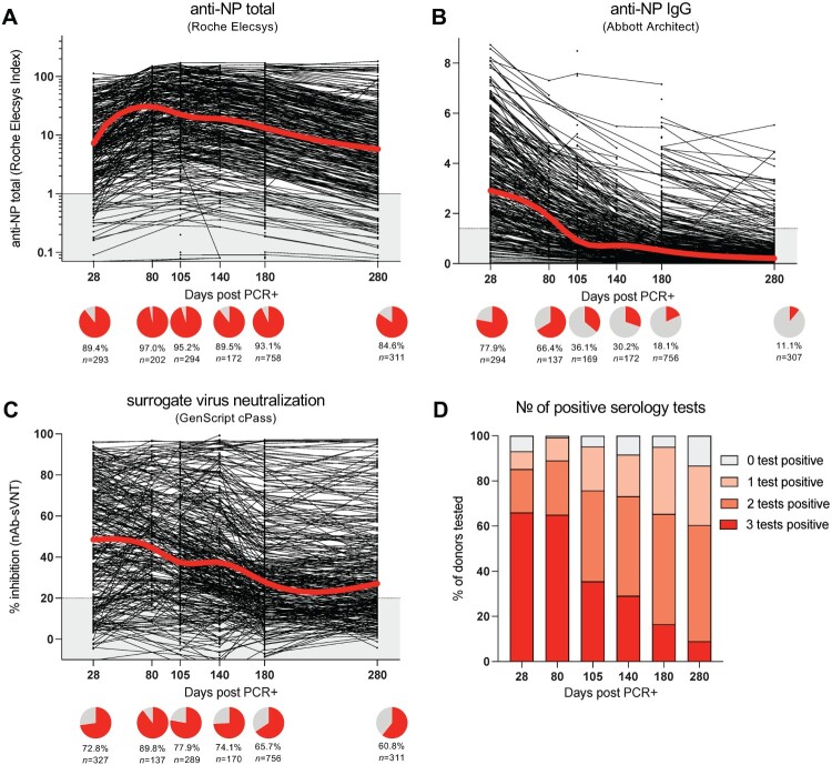
Results of serology testing are shown in Table 2 and Figure 1. The serology tests performed markedly differently (Table S1). The highest seropositive rates were seen with the total anti-NP test, which appeared to peak at 80 days post-infection and remained well above 90% positive even at day 180 (Figure 1(a)). The anti-NP IgG test had the lowest seropositive rates, with a significant decline of detectable antibodies after the initial day 28 timepoint (Figure 1(b)), whereas sVNT-NAbs also appeared to peak at 80 days post-infection, with a more gradual subsequent decline (Figure 1(c)). Similar antibody kinetics were also obtained in the subgroup of individuals that was tested at 5 consecutive timepoints (Figure S1). In total, >60% of study participants tested positive for all 3 antibody tests on days 28 and 80, and the proportion declined steadily through day 280 post PCR positivity (Figure 1(d)). Out of all participants that were tested at least twice, 30 (5.4%), 249 (45.0%), and 106 (19.1%) remained seronegative for total anti-NP, anti-NP IgG, and sVNT-NAbs, respectively. There were 43 participants who were seronegative on all tests throughout the period of testing.
Table 2.
Serology results.
| Tests | D28 positive resultsa (%) | D80 positive resultsa (%) | D105 positive resultsa (%) | D140 positive resultsa (%) | D180 positive resultsa (%) | D280 positive resultsa (%) |
|---|---|---|---|---|---|---|
| Total anti-NP (Elecsys® Anti-SARS-CoV-2, Roche) | 261 (89.7) | 133 (97.1) | 159 (94.1) | 151 (89.4) | 704 (93.1) | 263 (84.6) |
| Anti-NP IgG (Abbott Laboratories) | 227 (78.0) | 91 (66.4) | 61 (36.1) | 51 (30.2) | 137 (18.1) | 34 (10.9) |
| Surrogate virus neutralization test (cPass, GenScript) | 223 (76.6) | 123 (89.8) | 129 (76.3) | 125 (74.0) | 494 (65.2) | 189 (60.8) |
D; days; SARS-CoV-2; severe acute respiratory syndrome coronavirus 2, NP; nucleoprotein, IgG; Immunoglobulin G.
Excluding donors without all three serology tests at any timepoint (n = 53).
Figure 1.
Kinetics and decline of antibody responses to SARS-CoV-2 over nine months. Total anti-NP antibody titres were analysed using the Roche Elecsys test (a), anti-NP IgG antibody titres with the Abbott Architect (b), and virus neutralizing antibodies (sVNT) using the GenScript cPass test (c). All tested samples are shown as dots, the lines connect the samples of donors who were tested longitudinally. The red line indicates the median antibody titre index at the different timepoints post infection. The grey area shows the assay detection limit. The pie charts below the graph indicate the percentage of donors who tested positive for the antibody test at the different time points. (D) Summary of the percentage of donors who tested positive for none, 1, 2 or all 3 antibody tests performed at each timepoint post infection.
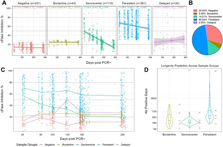
A wide heterogeneity of magnitude and decay of antibodies specific for RBD of spike protein was clearly visible. The heterogeneity was broadly categorized into five groups (Figure 2(a,b)): (1) Negative, whereby participants never developed detectable levels of NAbs throughout the entire follow-up period; (2) Borderline, whereby participants fluctuated between 20% and 30% NAbs but remained at a relatively flat trajectory throughout the entire follow-up period; 3. Seroreverters, whereby participants started at a range between 30% and 90% but dropped over time to be less than 20%; 4. Persistent, whereby participants started at the range between 30% and >90% and never dropped below 20% over the course of follow-up; and 5. Delayed, whereby participants showed increasing NAbs from 28 to 140 days post-PCR positive detection (Figure 2(c)), suggestive of a delayed immune response. The number and percentage of people in each group is as follows (Figure 2(b)): Negative = 231 (29.4%), Borderline = 43 (5.5%), Seroreverter = 110 (14.0%), Persistent = 381 (48.5%) and Delayed = 20 (2.5%). For the three groups with waning antibody kinetics (Borderline, Seroreverter, and Persistent groups), we estimated the longevity of NAbs (Figure 2(d)) and found that the median antibody-positive days ranged from 204 days in the Seroreverter group to 247 days in the Borderline group and 440 days in the Persistent group; yet there was a wide heterogeneity among the individuals in all three groups.
Figure 2.
Different SARS-CoV-2 neutralizing antibody profiles over nine months post infection. (a) Neutralizing antibody levels measured by percentage inhibition compared to negative control sample using cPass. Antibody levels were classified into five groups based on their kinetics and linear regression model for each group was applied. (b) The percentage of study participants in each group is as shown. (c) Group mean of the neutralizing antibody percentage is connected at days 28, 80, 105, 140, 180 and 280. Each point represents a single study participant. (d) Superimposed violin and box plots showing median, interquartile range, lowest and highest range of neutralizing antibody positive days in Borderline, Seroreverter and Persistent groups. p-value was calculated by Wilcoxon signed-rank test using the Persistent group as the reference group.
SARS-CoV-2-specific T cell analysis
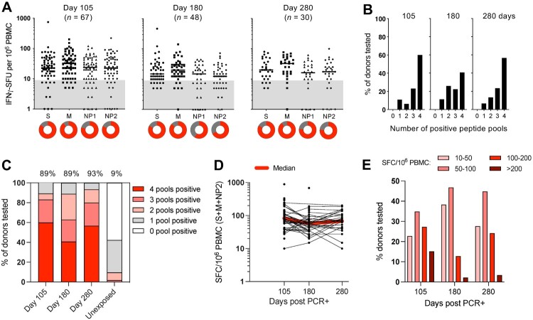
The frequency of T cells specific for spike (S), membrane (M), and nucleoprotein (NP; divided into two pools) were tested at days 105, 180, and 280 post-infection. Similar median frequencies of T cells reactive to the different peptide pools were detected, with the responses to membrane being the strongest (Figure 3(a)). This pattern of T cell responses to the different structural proteins was maintained over time. Figure 3(b) shows that all previously infected patients have T cells specific to at least 1 peptide pool, with the majority reacting to 2–4 peptide pools. In unexposed controls, cross-reactive T cells are found in about 40%, however, they react mostly only to a single peptide pool [18], thus unexposed donors can be distinguished from SARS-CoV-2 infected individuals, who instead mount a multi-specific T cell response directed towards different SARS-CoV-2 proteins (Figure 3(c)). Importantly, this multi-specific T cell response was maintained over time, and about 90% of all convalescents tested responded to at least 2 peptide pools at all three time points (Figure 3(c)). The overall median frequency of SARS-CoV-2 specific T cells was mostly stable over time (85.5 SFC/106 PBMC at day 105; 57.5 SFC/106 PBMC at day 180; 67.5 SFC/106 PBMC at day 280) (Figure 3(d)). Importantly, however, there was at least a log difference in the quantity of spots among different individuals (Figure 3(e); 22% patients 20–50 SFU/106 PBMC; 15% patients >200 SFU/106 PBMC).
Figure 3.
Kinetics of SARS-CoV-2-specific T cells over nine months post infection. (a) The frequencies of IFN-γ-spot forming cells (SFC) reactive to the peptide pools of Spike (S), Membrane (M) and Nucleoprotein (NP1 and NP2) are shown for donors tested at 105 (n = 67), 180 (n = 48) and 280 (n = 30) days post infection. Circles below represent the frequency of a positive (IFN-γ-SFC ≥10/106 PBMC) response (red) to the individual peptide pools. (b) Bar graphs show the percentage of donors reacting to the number of peptide pools tested. (c) Summary of the percentage of donors who tested positive for none, 1, 2, 3 or all 4 peptide pools tested at each timepoint post infection. Individuals who tested positive for at least 2 pools are were considered positive and the percentage of positive donors is indicated on the top of the graph. Cross-reactive T cell responses in unexposed donors (n = 51) are shown for comparison [18]. (d) The frequency of IFN-γ-SFC reactive to the peptide pools S, M and NP2 are shown. All tested samples are shown as dots, the lines connect the samples of donors who were tested longitudinally. The red line indicates the median T cell frequency at the different timepoints post infection. (e) The frequencies of donors with low (10–50 SFC/106 PBMC), medium (50–100 SFC/106 PBMC), high (100–200 SFC/106 PBMC), and very high (>200 SFC/106 PBMC) T cell responses are shown for the three timepoints post infection.
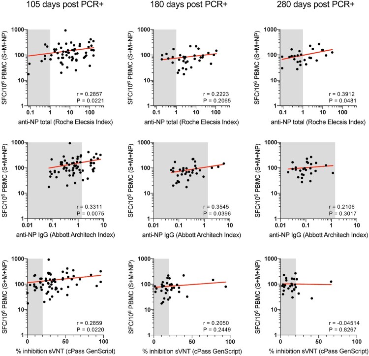
We then correlated the frequency of SARS-CoV-2 specific T cells with the different antibody titres (Figure 4, S2). Although there was a significant correlation between the T cell frequency and all antibodies tested at the early day 105 time point, this correlation was lost over time (>180 days). Furthermore, all individuals who became serological negative over time maintained a detectable SARS-CoV-2-specific T cell response (>15 SFU/106 PBMC) 9 months after infection.
Figure 4.
Correlation of SARS-CoV-2-specific T cells with levels of total anti-NP IgG, anti-NP IgG and sVNT-nAb. The frequency of SARS-CoV-2-specific T cells, as quantified by IFN-γ ELISpot, reactive to all (Spike, M, NP) proteins tested were correlated with levels of total anti-NP (upper panels), levels of anti-NP-IgG (middle panels) and levels of sVNT-nAb (lower panels) at 105, 180 and 280 days post infection. Spearman correlations.
Discussion
Coordinated activation of humoral and cellular virus-specific T cell responses is necessary for the efficient control of SARS-CoV-2 [23,24]. Our longitudinal analysis of virus-specific humoral and cellular immunity in individuals who controlled the infection with minimal symptoms supports this hypothesis. More than 95% of all the SARS-CoV-2 PCR+ individuals demonstrated positivity of at least one serological test performed 2 months after infection, and a similar level of SARS-CoV-2 cellular immunity (89%) was detected at 3 months post-infection.
Longitudinal analysis of humoral and cellular immunity in parallel revealed a different kinetic of decline among different antibodies and T cells. First, we observed a marked reduction in the ability of the Abbott CMIA assay to detect anti-NP IgG antibodies beyond 3 months after infection. Less than 40% of infected subjects were positive at day 105 and only 10% at 9 months. Total anti-NP antibody measurement with the Roche ECLIA assay and neutralizing antibody analysis with the sVNT cPass assay were more stable even though a progressive decrease of antibody titres was observed. T cells specific for different SARS-CoV-2 structural proteins were instead stable over time with the persistence of T cells able to recognize at least two different antigenic sites 9 months after infection in more than 90% of the tested individuals.
Interestingly, by selecting individuals preferentially with low/negative antibody titres, we could demonstrate that subjects with negative antibody tests conserved a detectable level of SARS-CoV-2-specific T cell response. These data confirm that the different components of the adaptive immune system during the memory phase are largely independently regulated [9]. Furthermore, the ability of SARS-CoV-2-specific T cells to persist longer than antibodies indicates that tests of cellular immune responses against different SARS-CoV-2 antigens might be a more robust predictor of previous exposure to SARS-CoV-2. A limitation of our T cell analysis was the inability, due to the use of a single ELISpot test, to differentiate between CD4 or CD8 T cell-mediated responses. Future analysis of T cell phenotype utilizing flow cytometry-based tests should be performed to understand whether individuals with low/negative antibody levels possess a preferential CD4 or CD8 T cell response.
One other interesting observation of this present study was the detection, in our studied population, of a marked heterogeneity of the magnitude of humoral and cellular immunity. This quantitative variation was observed despite the substantial similarity in age, genetic background, and symptoms after infection of the different individuals. Such levels of heterogeneity in immunity have been previously reported, but the population demographics was more heterogeneous in that cohort [16].
In this study, we observed a wide quantitative range of neutralization ability of the samples of the different individuals that also showed distinct kinetics of persistence or decay. Unlike previous observations that have linked differences in antibody titres with disease severity or sex [16,17,25], these variables cannot explain the heterogeneous patterns of neutralizing antibody response found in this study. Interestingly, we could not demonstrate any persistent correlations between the magnitude of humoral and cellular immunity, which was also detected over a wide range of T cell frequencies.
Due to the observational nature of this study, we did not further analyze the possible causes of the wide heterogeneity of humoral and cellular immunity in our cohort. Given that the migrant workers remained segregated from the rest of the community during the course of the study, and were routinely tested via PCR every two weeks, reinfection with SARS-CoV-2 is unlikely to account for the heterogeneity observed. The more plausible explanation is that the differences in virus-specific antibody and T cell magnitude detected in our homogeneous cohort are caused by the initial infectious viral load level possibly triggering a proportional activation of innate immune response. Unfortunately, we are unable to confirm this hypothesis as the confirmatory PCR tests in our cohort were performed in a number of different laboratories, and RT–PCR Ct values were not retained.
New data generated by detailed longitudinal analysis at single cell levels of immune reactions during mild SARS-CoV-2 infections have shown that the level of type I interferon (IFN) genes activation was directly proportional to the level of activated plasma cells and type II IFN-production [26]. One other possibility is that the different responses detected in different individuals might be caused by their preceding levels of cross-reactive immunity towards other coronaviruses. Immunity towards other coronavirus infections has been shown to modulate the immune response towards SARS-CoV-2 [27]. In Singapore, the incidence of individuals with pre-existing SARS-CoV-2 specific T cells that were reactive to spike and other SARS-CoV-2 proteins (NP, ORF-7) was also reported to be high (∼30% of unexposed donors) [13,18]. Recently, we have also shown that individuals who recovered from SARS-CoV-1 infection 17 years ago generated higher level of neutralizing antibodies with a much broader spectrum of neutralizing activities after receiving just one dose of vaccine against SARS-CoV-2 [28]. It can be speculated that such boosting can happen to any individual who had prior exposure to any of the sarbecoviruses which are antigenically related to SARS-CoV-2.
Irrespective of the underlying causes, the marked quantitative difference in virus-specific antibodies and T cells detected in our homogeneous cohort have implications for COVID-19 surveillance, vaccination strategies, and post-pandemic planning. The level of neutralizing antibodies and T cells is certainly an important immunological parameter of protection. Studies in animal models demonstrated that high neutralizing antibodies protect the animals from infection [29]. The demonstration that the decay of neutralizing antibodies titres is highly variable among different individuals suggests that longitudinal measurements should be performed over time to define the individuals who should receive vaccines to boost their neutralizing antibodies titres. On the other hand, the observation that T cells can persist over time despite the disappearance of antibodies is reassuring in terms of disease severity after reinfection. Even though T cells cannot protect from infection, their pivotal role in the protection from disease severity has been shown in natural infection of normal patients [30], oncological patients [31], and in vaccination [32]. Finally, the demonstration that serological analysis of SARS-CoV-2 infected individuals cannot be used to predict the level of virus-specific cellular immunity indicates that a proper profile of SARS-CoV-2 immunity necessitates a direct quantification of both arms of adaptive immunity.
Supplementary Material
Acknowledgements
We are very grateful for the support and patience of the migrant workers who participated in this study and acknowledge the uncertainty and difficulties they have experienced as a consequence of COVID-19. We would also like to thank the staff of Sengkang General Hospital as well as the staff at the dormitory for actively ensuring that the study was a success.
Funding Statement
This work was supported by grants from the National Medical Research Council COVID-19 Research Fund (MOH000-499-00 and MOH-000417). The study sponsor had no role in the design, implementation, analysis or write-up of the study.
Disclosure statement
No potential conflict of interest was reported by the author(s).
Potential conflicts of interest
N. Le Bert and A. Bertoletti reported a patent for a method to monitor SARS-CoV-2-specific T cells in biological samples pending. W.N. Chia and L-F. Wang are co-inventors of a patent on the surrogate virus neutralization test (sVNT), which has been commercialized under the trade name cPass.
References
- 1.Our World in Data . Cumulative confirmed COVID-19 deaths and cases. [cited 2021 Apr 6]. Available from: https://ourworldindata.org/grapher/cumulative-deaths-and-cases-covid-19?country=~OWID_WRL.
- 2.Buitrago-Garcia D, Egli-Gany D, Counotte MJ, et al. Occurrence and transmission potential of asymptomatic and presymptomatic SARS-CoV-2 infections: a living systematic review and meta-analysis. PLoS Med. 2020;17(9):e1003346. 10.1371/journal.pmed.1003346 [DOI] [PMC free article] [PubMed] [Google Scholar]
- 3.Gudbjartsson DF, Helgason A, Jonsson H, et al. Spread of SARS-CoV-2 in the Icelandic population. N Engl J Med. 2020;382:2302–2315. [cited 2021 Apr 6]. Available from: http://www.nejm.org/doi/ 10.1056/NEJMoa2006100. [DOI] [PMC free article] [PubMed] [Google Scholar]
- 4.Emery JC, Russell TW, Liu Y, et al. The contribution of asymptomatic SARS-CoV-2 infections to transmission on the Diamond Princess cruise ship. eLife. 2020;9:e58699. 10.7554/eLife.58699. [DOI] [PMC free article] [PubMed] [Google Scholar]
- 5.Ioannidis JPA. Infection fatality rate of COVID-19 inferred from seroprevalence data. Bull World Health Organ. 2021;99:19–33F. [cited 2021 Apr 6]. Available from: http://www.who.int/entity/bulletin/volumes/99/1/20-265892.pdf. [DOI] [PMC free article] [PubMed] [Google Scholar]
- 6.Hsu LY, Chia PY, Vasoo S.. A midpoint perspective on the COVID-19 pandemic. Singapore Med J. 2020;61:381–383. [cited 2021 Apr 6]. Available from: https://www.ncbi.nlm.nih.gov/pmc/articles/PMC7926611/. [DOI] [PMC free article] [PubMed] [Google Scholar]
- 7.Cromer D, Juno JA, Khoury D, et al. Prospects for durable immune control of SARS-CoV-2 and prevention of reinfection. Nat Rev Immunol. 2021;21:395–404. [cited 2021 Sept 10]. Available from: http://www.nature.com/articles/s41577-021-00550-x. [DOI] [PMC free article] [PubMed] [Google Scholar]
- 8.Breton G, Mendoza P, Hägglöf T, et al. Persistent cellular immunity to SARS-CoV-2 infection. J Exp Med. 2021;218(4):e20202515. Available from: https://www.ncbi.nlm.nih.gov/pmc/articles/PMC7845919/. [DOI] [PMC free article] [PubMed] [Google Scholar]
- 9.Dan JM, Mateus J, Kato Y, et al. Immunological memory to SARS-CoV-2 assessed for up to 8 months after infection. Science. 2021;371(6529):eabf4063. Available from: https://science.sciencemag.org/content/371/6529/eabf4063. [DOI] [PMC free article] [PubMed] [Google Scholar]
- 10.Bonifacius A, Tischer-Zimmermann S, Dragon AC, et al. COVID-19 immune signatures reveal stable antiviral T cell function despite declining humoral responses. Immunity. 2021;54:340–354.e6. [DOI] [PMC free article] [PubMed] [Google Scholar]
- 11.Sherina N, Piralla A, Du L, et al. Persistence of SARS-CoV-2-specific B and T cell responses in convalescent COVID-19 patients 6-8 months after the infection. Med (NY). 2021;2:281–295.e4. [DOI] [PMC free article] [PubMed] [Google Scholar]
- 12.Turner JS, Kim W, Kalaidina E, et al. SARS-CoV-2 infection induces long-lived bone marrow plasma cells in humans. Nature. 2021;595(7867):421–425. Available from: https://www.nature.com/articles/s41586-021-03647-4. [DOI] [PubMed] [Google Scholar]
- 13.Le Bert N, Tan AT, Kunasegaran K, et al. SARS-CoV-2-specific T cell immunity in cases of COVID-19 and SARS, and uninfected controls. Nature. 2020;584:457–462. [cited 2021 Apr 6]. Available from: https://www.nature.com/articles/s41586-020-2550-z. [DOI] [PubMed] [Google Scholar]
- 14.Vitale J, Mumoli N, Clerici P, et al. Assessment of SARS-CoV-2 reinfection 1 year after primary infection in a population in Lombardy, Italy. JAMA Intern Med. 2021;181(10):1407–1408. Available from: https://jamanetwork.com/journals/jamainternalmedicine/fullarticle/2780557. [DOI] [PMC free article] [PubMed] [Google Scholar]
- 15.Khoury DS, Cromer D, Reynaldi A, et al. Neutralizing antibody levels are highly predictive of immune protection from symptomatic SARS-CoV-2 infection. Nat Med. 2021;27(7):1205–1211. Available from: http://www.nature.com/articles/s41591-021-01377-8. [DOI] [PubMed] [Google Scholar]
- 16.Chia WN, Zhu F, Ong SWX, et al. Dynamics of SARS-CoV-2 neutralising antibody responses and duration of immunity: a longitudinal study. Lancet Microbe. 2021;2(6):e240-e249. Available from: https://linkinghub.elsevier.com/retrieve/pii/S2666524721000252. [DOI] [PMC free article] [PubMed] [Google Scholar]
- 17.Long Q-X, Tang X-J, Shi Q-L, et al. Clinical and immunological assessment of asymptomatic SARS-CoV-2 infections. Nat Med. 2020;26:1200–1204. [cited 2021 Apr 6]. Available from: https://www.nature.com/articles/s41591-020-0965-6. [DOI] [PubMed] [Google Scholar]
- 18.Le Bert N, Clapham HE, Tan AT, et al. Highly functional virus-specific cellular immune response in asymptomatic SARS-CoV-2 infection. J Exp Med. 2021;3;218(5):e20202617. Available from: 10.1084/jem.20202617. [DOI] [PMC free article] [PubMed] [Google Scholar]
- 19.Ministry of Health Singapore. COVID-19 situation report . (2020). [cited 2021 Oct 27]. Available from: https://covidsitrep.moh.gov.sg/.
- 20.Gazit S, Shlezinger R, Perez G, et al. Comparing SARS-CoV-2 natural immunity to vaccine-induced immunity: reinfections versus breakthrough infections. MedRXiV. 2021, Preprint. 10.1101/2021.08.24.21262415. [DOI] [PMC free article] [PubMed] [Google Scholar]
- 21.GenScript USA, Inc . cPass SARS-CoV-2 neutralization antibody detection kit. Version 4. New Jersey: Genscript USA, Inc.; 2020. Available from: https://www.fda.gov/media/143583/download.
- 22.Singapore Statutes Online, Government of Singapore . Infectious Diseases Act; 1977. [cited 2021 October 27]. Available from: https://sso.agc.gov.sg/Act/IDA1976.
- 23.Rydyznski Moderbacher C, Ramirez SI, Dan JM, et al. Antigen-Specific adaptive immunity to SARS-CoV-2 in acute COVID-19 and associations with age and disease severity. Cell. 2020;183:996–1012.e19. [DOI] [PMC free article] [PubMed] [Google Scholar]
- 24.Rodda LB, Netland J, Shehata L, et al. Functional SARS-CoV-2-specific immune memory persists after mild COVID-19. Cell. 2021;184:169–183.e17. [DOI] [PMC free article] [PubMed] [Google Scholar]
- 25.Takahashi T, Ellingson MK, Wong P, et al. Sex differences in immune responses that underlie COVID-19 disease outcomes. Nature. 2020;588:315–320. [cited 2021 June 5]. Available from: https://www.nature.com/articles/s41586-020-2700-3. [DOI] [PMC free article] [PubMed] [Google Scholar]
- 26.Talla A, Vasaikar SV, Lemos MP, et al. Longitudinal immune dynamics of mild COVID-19 define signatures of recovery and persistence. bioRxiv. 2021, Preprint. 10.1101/2021.05.26.442666. [DOI] [Google Scholar]
- 27.Ladner JT, Henson SN, Boyle AS, et al. Epitope-resolved profiling of the SARS-CoV-2 antibody response identifies cross-reactivity with endemic human coronaviruses. Cell Rep Med. 2021;2(1):100189. Available from: https://www.sciencedirect.com/science/article/pii/S2666379120302445. [DOI] [PMC free article] [PubMed] [Google Scholar]
- 28.Tan C-W, Chia W-N, Young BE, et al. Pan-Sarbecovirus neutralizing antibodies in BNT162b2-immunized SARS-CoV-1 survivors. N Engl J Med. 2021;385:1401–1406. Available from: https://www.nejm.org/doi/ 10.1056/NEJMoa2108453. [DOI] [PMC free article] [PubMed] [Google Scholar]
- 29.McMahan K, Yu J, Mercado NB, et al. Correlates of protection against SARS-CoV-2 in rhesus macaques. Nature. 2021;590:630–634. [DOI] [PMC free article] [PubMed] [Google Scholar]
- 30.Tan AT, Linster M, Tan CW, et al. Early induction of functional SARS-CoV-2-specific T cells associates with rapid viral clearance and mild disease in COVID-19 patients. Cell Rep. 2021;34(6):108728. [cited 2021 June 5]. Available from: https://linkinghub.elsevier.com/retrieve/pii/S2211124721000413. [DOI] [PMC free article] [PubMed] [Google Scholar]
- 31.Bange EM, Han NA, Wileyto P, et al. CD8+ t cells contribute to survival in patients with COVID-19 and hematologic cancer. Nat Med. 2021 Jul;27(7):1280–1289. Available from: http://www.nature.com/articles/s41591-021-01386-7. [DOI] [PMC free article] [PubMed] [Google Scholar]
- 32.Kalimuddin S, Tham CYL, Qui M, et al. Early T cell and binding antibody responses are associated with COVID-19 RNA vaccine efficacy onset. Med. 2021;2:682–688.e4. [cited 2021 June 5]. Available from: https://www.cell.com/med/abstract/S2666-6340(21)00152-5. [DOI] [PMC free article] [PubMed] [Google Scholar]
Associated Data
This section collects any data citations, data availability statements, or supplementary materials included in this article.


