Abstract
Accurate evaluation, diagnosis, and management of mandibular fractures is essential to effectively restore an individual's facial esthetics and function. Understanding of surgical anatomy, fracture fixation principles, and the nuances of specific fractures with respect to various patient populations can aid in adequately avoiding complications such as malocclusion, non-union, paresthesia, and revision procedures. This article reviews comprehensive mandibular fracture assessment, mandibular surgical anatomy, fracture fixation principles, management considerations, and commonly encountered complications. In addition, this article reviews emerging literature examining 3-dimensional printing and intraoperative imaging.
Keywords: mandible, mandibular fracture, maxillofacial injury, craniomaxillofacial trauma, facial trauma
Adequate treatment of mandible fractures not only restores an individual's ability to speak, chew, breathe, and sleep, but also reestablishes their occlusion and facial aesthetics. An analysis of the American College of Surgeons National Surgical Quality Improvement Program (ACS NSQIP) database showed that mandible fractures were the most common isolated facial fracture. 1 The causes of mandible fractures are varied and include motor vehicle accidents (MVAs), assault, domestic violence, falls, sports- and work-related accidents, ballistic injuries, and pathologic fractures. 1 2 3 4 One can further subgroup the etiology and severity of mandible fractures with respect to age, gender, socioeconomic status, substance use, and mechanism of injury. 3 5 For instance, men most often sustain mandible fractures as a result of assault, MVAs, and falls; whereas women sustain mandible fractures from MVAs, assault, and trauma. 2 Appreciation of the mechanism of injury and anatomy of the mandible will aid the plastic surgeon, oral and maxillofacial surgeon, or otolaryngologist in assessment and management of mandibular fractures. Mandibular fractures will vary in their severity according to number of sites involved, displacement, and comminution ( Fig. 1 ).
Fig. 1.
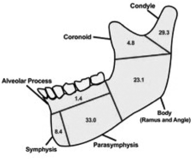
Frequency of mandibular fracture by location (Image courtesy: Avery et al. 80 ).
Evaluation
Initial Assessment - History
The diagnostic work-up of mandible fractures begins with a thorough primary survey as outlined by the Advanced Trauma Life Support (ATLS) protocols. 6 Mandible fractures are unique in that severe injuries, such as bilateral body fractures (“bucket handle”) can result in airway embarrassment ( Fig. 2 ). In these situations, stabilization of the airway may require tracheotomy. Life-threatening injuries, when present, need to be recognized and managed early before fracture assessment can begin. Before examination, the physician or physicians should be sure to don any necessary personal protective equipment (PPE) given the post-pandemic era we now live in, with the known increased risk of transmission related to manipulation of oronasal mucosal tissues. 7
Fig. 2.
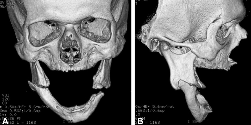
Bilateral mandibular fractures in an edentulous 55-year-old man. The anterior mandibular segment was pulled inferiorly by the suprahyoid muscles and resulted in airway obstruction.
A thorough history of present illness and past medical and surgical history will highlight any relevant medical conditions, previous trauma, bone disease, nutritional and metabolic disorders, and psychiatric conditions that may influence timing and management of the fracture. 8 In addition, the patient's premorbid dental history and occlusion needs should be accounted for. When available, photographs can aid in reduction of the patient's fracture to re-establish the premorbid occlusion.
Initial Assessment - Clinical Examination
Focused evaluation of the head and neck is a part of the secondary survey outlined by ATLS protocols. Examination should begin with inspection and palpation. The classical signs of inflammation, pain, swelling, and erythema will help guide the physician in thorough identification of potential injuries. After examining for any lacerations or sources bleeding that needs to be addressed urgently, the clinician should perform an in-depth fracture assessment. Extra- and intra-oral findings, in addition to a neurosensory examination, will help the physician in identification of fractures or fractures patterns that may be present.
Extra-oral Examination: An extra-oral assessment should begin by examining the face and mandible for any abnormal contours or step defects. Changes to the patient's facial profile and mandibular movements will cue the physician for types of fractures. For instance, a flattened facial profile may be due to a fractured mandibular body, angle, or ramus. A retruded chin may be caused by bilateral parasymphyseal fractures. An elongated face may be the result of bilateral subcondylar, angle, or body fractures. Any facial asymmetry should also signal the physician for the possibility of a mandible fracture ( Fig. 3 ).
Fig. 3.
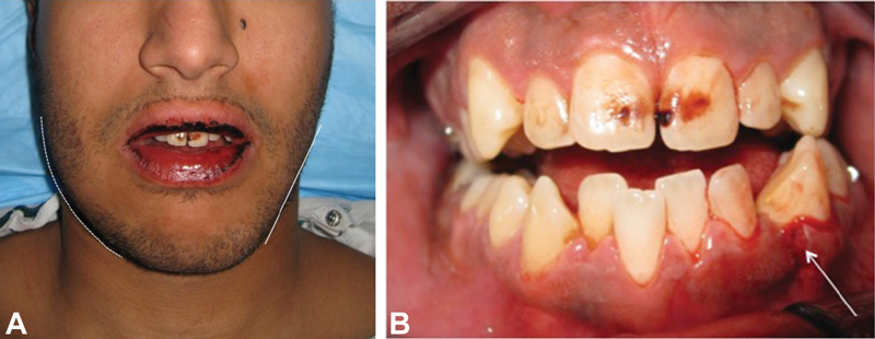
Frontal and intraoral views of a 19-year-old male patient with bilateral mandibular fractures involving the right mandibular angle and left mandibular parasymphysis. There is asymmetry of the lower facial soft tissues, with loss of the mandibular inferior border contour on the right (dashed line) relative to the left (solid line). Intraorally, there is a gingival laceration and step at the parasymphysis, between the left mandibular canine and lateral incisor (arrow).
Trismus ( Fig. 4 ), or limited mouth opening, and deviation on opening may be due to guarding of the muscles of mastication, non-functioning of muscles, or bony impingements. Deviation upon opening may signify a mandibular condylar fracture due to unopposed contraction of the contralateral lateral pterygoid muscle. Inability to fully open may be due to impingement of the coronoid process on the zygomatic arch when fractures of the ramus and coronoid process or depression of the zygomatic arch is present. On the other hand, inability to fully close may signify dentoalveolar process, angle, ramus, or symphysis fractures. Inability to fully bring one's teeth together may be due to an open bite that was present pre-injury; the presence of mammelons on the incisal edges of the anterior dentition may be a clue to determining a premorbid anterior open bite ( Fig. 5 ).
Fig. 4.
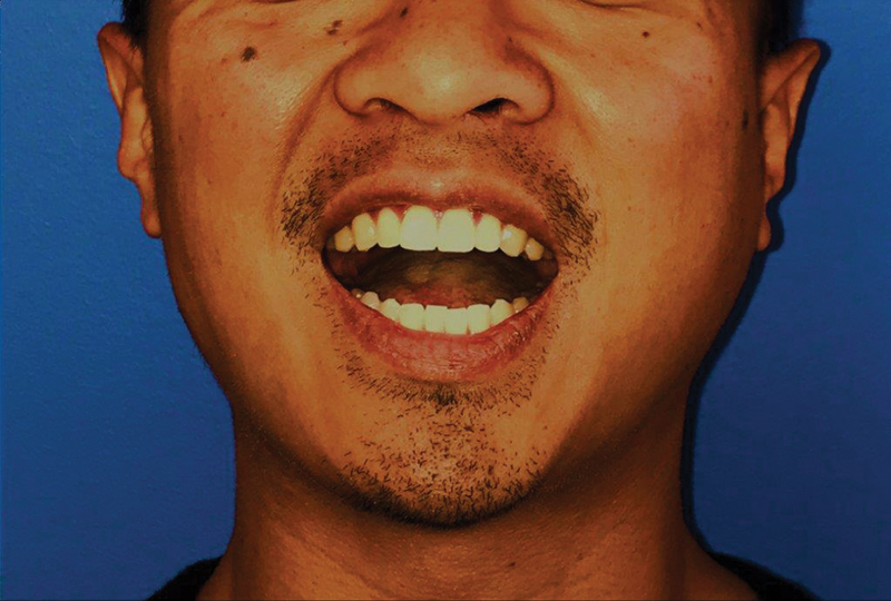
Trismus (limited mouth opening) may be a frequent exam finding in patients with mandibular fractures, particularly those involving the angle or ramus-condyle unit. This patient sustained a left-sided subcondylar fracture. Maximal incisal opening was 20 mm. The mandibular dental midline deviates toward the fracture with opening, due to the unopposed motion of the right lateral pterygoid muscle.
Fig. 5.
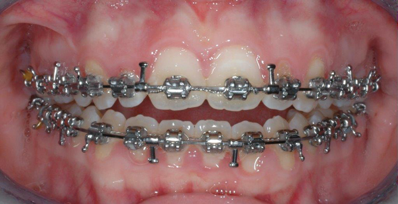
Assessment of premorbid occlusion is readily accomplished with careful history and, if available, dental or orthodontic records. Anterior open bites may be the result of bilateral fractures involving the condyles with posterior shortening of the rami. Anterior open bites that are premorbid may be identified by the presence of mammelons (rounded protuberances on the incisal edges) on the incisors, as seen in this orthognathic surgery patient.
Intra-oral Examination: The physician should be vigilant in intra-oral assessment. This includes assessment of the mandibular arch form and occlusion and identification of gingival lacerations, hematomas, or ecchymosis, and injuries to the teeth. The mandible is unique in that it is a continuous, U-shaped bone that crosses midline; deviations from this arch form may indicate a fracture. Any change in occlusion is highly suggestive of a mandible fracture. The mandible should be palpated bimanually to assess for fracture mobility.
The patient should be asked if their bite feels different. This can identify injuries to the teeth, dentoalveolar process, mandible, or temporomandibular joint (TMJ). Premature posterior contacts between the maxillary and mandibular dentition can result from bilateral mandible fractures of the angles or ramus-condyle units or signify the presence of a displaced maxillary fracture. Asking for premorbid photographs of the patient's premorbid occlusion can help to ensure accurate reduction of fractures based upon the occlusion.
Gingival lacerations, hematomas, or ecchymosis may indicate injury to the mandible ( Fig. 6 ). For instance, sublingual ecchymosis is a pathognomonic sign for symphysial, parasymphyseal, or body fractures. In addition, retromolar trigone ecchymosis can signify angle fractures. Segments of fractured teeth may indicate fractures to the dentoalveolar process or mandible itself. Fractured teeth, mobile teeth, and any grossly carious teeth in the line of fracture may require extraction for reduction and to prevent aspiration. Missing teeth should that have not been accounted for should be considered swallowed, aspirated, or displaced into soft tissue. Radiography and operative exploration may help identify lost teeth and may require removal to prevent infection or airway issues.
Fig. 6.
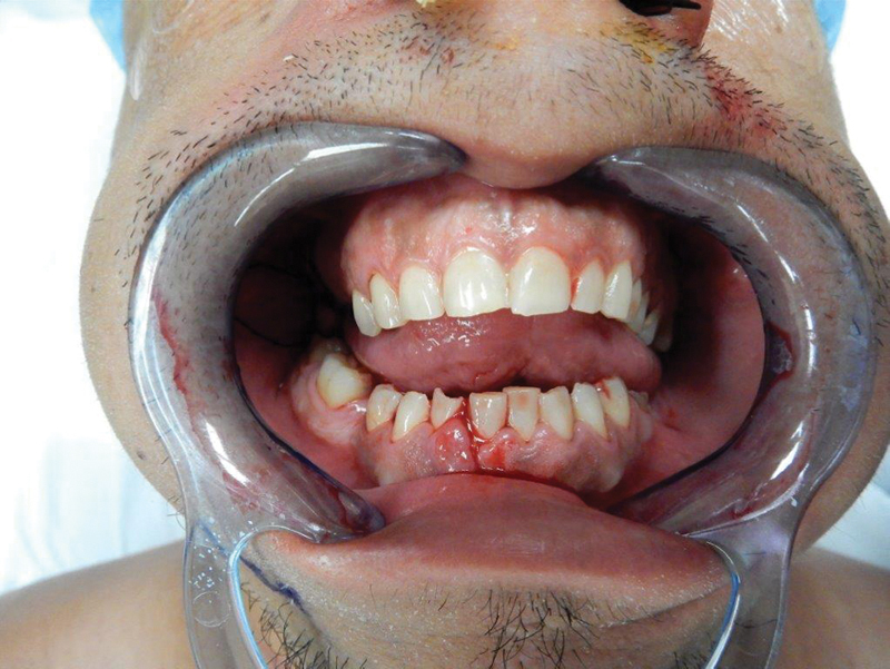
Intraoral assessment may reveal the site of the fracture. Gingival lacerations, vestibular or sublingual ecchymoses, and/or steps at the occlusal plane suggest a bony injury. Bimanual palpation across the suspected fracture site may demonstrate independent mobility of the mandibular segments. The presenting anterior open bite is likely related to the trauma, as there is evidence of wear on the incisal edges of the anterior dentition.
Any neurosensory deficits should be accurately documented. Paresthesia, dysesthesia, or anesthesia of the lower lip indicates injury to the inferior alveolar nerve distal to the mandibular foramen. Most injuries are neuropraxic in nature and related to transient ischemia, inflammation, and traction. Various classification systems, such as Sunderland grading and Medical Research Council Scale, can aid in diagnosis of nerve injuries and give not only the patient, but also the clinician the prognosis of return of sensation.
Initial Assessment - Radiographic Examination
Most patients with mandible fractures, especially in the setting of polytrauma, present to the emergency room and undergo radiographic assessment with computed tomographic (CT) imaging of the head and cervical spine (C-spine). Although CT imaging is now considered the gold standard, various other imaging studies can be helpful when CT is not available. These include plain films with Reverse Towne, Caldwell posteroanterior, lateral oblique, or occlusal views or panoramic radiograph (panorex). Panorex is advantageous in allowing visualization of the entire mandible including the subcondylar unit/TMJ unit ( Fig. 7 ); however, its availability may be limited in the acute setting. In addition, certain fracture patterns, particularly in the posterior mandible, may be missed on single-view panoramic radiography. When evaluating a patient with mandibular injury with only plain film imaging it is prudent to obtain atleast two views of the mandible.
Fig. 7.
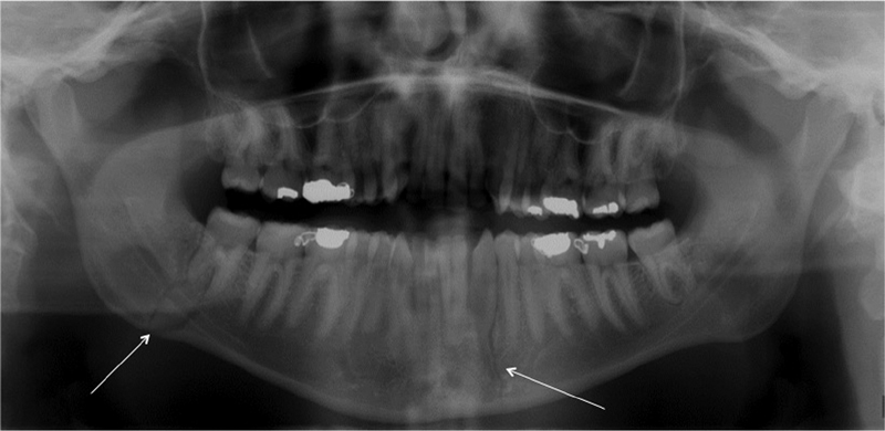
Panoramic radiographs may be useful for demonstrating fractures of the mandible, but frequently do not provide sufficient detail regarding displacement in multiple dimensions. This patient has right mandibular angle and left mandibular parasymphysis fractures, which are easily visualized on the panoramic view (arrows).
The advent of faster helical-CT imaging has 100% sensitivity in diagnosing mandible fractures compared with 86% sensitivity of panorex imaging. These CT images can be reformatted into three-dimensional reconstructions to further aid in operative planning of fracture management ( Fig. 8 ).
Fig. 8.
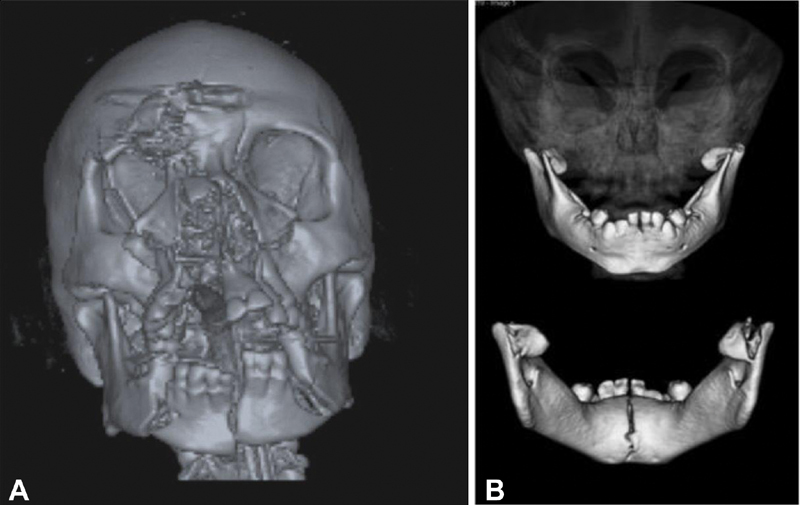
Multi-detector computed tomography scans have emerged as the gold standard for diagnosing mandibular injuries and are particularly useful in the setting of complex injuries such as high energy mechanisms ( A , facial gunshot wound). Three-dimensional imaging is also useful in pediatric patients, where plain film imaging may not be tolerated or may be non-diagnostic due to the overlap between developing teeth and fracture sites ( B , bilateral condylar head fractures and greenstick symphyseal fracture in an infant).
Surgical Anatomy
The osteology of the mandible, various muscle attachments and their influence, and presence of developing or permanent dentition, or lack of dentition, need to be understood for accurate treatment of mandible fractures. A full description of a mandibular fracture should include an assessment of its relationship to the external environment (ie, simple/closed, compound/open), type (ie, incomplete, greenstick, complete, comminuted), dentition (ie, primary, mixed, permanent, or lack of dentition), displacement, favorability, and location.
Bony Anatomy of the Mandible
The mandible is a U-shaped bone that crosses anatomical midline. The mandible has thirteen muscle attachments. Organized by function, these are jaw closers (temporalis, masseter, medial pterygoid), openers (digastric, lateral pterygoid), and glottic attachments (genioglossus and geniohyoid). The remaining muscles can influence displacement of fractures and may be involved in soft tissue closure (buccinator, platysma, mentalis, mylohyoid, depressor labii inferiorius, and depressor anguli oris).
The major vascular supply to the mandible during development is from the inferior alveolar artery, but transitions to the involved periosteum and muscle attachments as the body ages. During fixation of comminuted or atrophic mandible fractures, areas with poor blood supply, such as the body, careful soft tissue management is mandatory, as the blood supply to these regions is periosteal, rather than endosteal. Periosteal stripping in these areas should be minimized and done only to the extent necessary to apply fixation. Supra-periosteal placement of hardware has been studied in this context, but bears no discernable advantage for healing. The course of the facial artery and vein around the mandible in the antegonial notch should also be appreciated when treatment requires a transcervical approach.
The mandibular branch of the trigeminal nerve includes the auriculotemporal, buccal, inferior alveolar, and lingual nerves. Innervation of the mandible is supplied by the inferior alveolar nerve (IAN) and its branches, the mylohyoid, dental, incisive, and mental branches. The IAN enters the mandibular foramen on the medial aspect of the ramus behind the lingula, travels within a bony canal, and exits the mental foramen of the mandible near the apex of the first and second premolars. IAN injuries in the setting of mandible fractures have been reported to range from 5.7 to 58.5%. 9 10 11 12
Definition of Sites of Fracture
As defined by Dingman and Natvig, the mandible can be subdivided as follows ( Fig. 1E ): symphysis, bounded by vertical lines distal to canine; parasymphysis, distal aspect of the roots of lateral incisors to distal of roots of canine teeth; body, from distal roots of canine to line corresponding to anterior border of the masseter muscle (usually coincides with third molar); angle, bounded by anterior and posterior borders of the masseter muscle; ramus, bounded by superior aspect of the angle to sigmoid notch; condylar process, area superior to ramus; coronoid process, and alveolar process, the area that normally contains teeth. 13
Favorable versus Unfavorable Fractures
Mandibular angle and body fractures can be classified as vertically favorable or unfavorable or horizontally favorable or unfavorable. Favorability is determined by the direction of a fracture line and its relationship to muscle action on the fracture segments. Vertically favorable fractures resist the medial pull of the medial pterygoid muscle on the proximal segment in the vertical plane. Horizontally favorable fractures resist upward the vertical pull of the masseter, temporalis, and medial pterygoid muscles on the proximal segment in the horizontal plane. The more forward a fracture occurs in the body the more the upward displacement is counteracted by the downward pull of the mylohyoid muscles.
Fracture Fixation Principles
The mandible is the only moveable, load bearing bone of the skull. To properly treat mandible fractures, one must first understand basic fracture fixation principles. These can be grouped into tension versus compression and load-bearing versus load-sharing principles While a complex topic, the biomechanics and forces exerted on the mandible should be understood by the treating physician.
Tension versus Compression
At any time, there are counteracting forces of tension and compression on the mandible influenced by muscular attachments and loading. At rest, these forces are equal. While an oversimplification, forces of tension generally separate a fracture and forces of compression bring a fracture together. Under compression, fractures generally undergo rapid healing and a greater resistance to separation. However, without addressing tension forces, overcompression can compromise ideal bony healing leading to nonunion.
Studies have shown that in the region of the mandibular body tension exists along the alveolar border while compression exists along the inferior border of the mandible. Moving toward the symphysis and parasymphysis, these two opposing forces become mixed or even inverted due to the introduction of torsional, or rotational, forces.
Biomechanically, it is most advantageous to apply bicortical rigid fixation along the zone of tension. Bicortical rigid fixation along the alveolar border is not feasible due to the presence of tooth roots, thin cortical bone, and thin gingival tissue. The inferior border of the mandible is not constrained by these limitations, with the notable exception of pediatric patients in the primary or mixed dentition. Bicortical screw fixation in this region is extremely stable and then only requires placement of a tension band at the alveolar level (either a continuous arch bar at the dentition or a small plate with monocortical screws) to resist tensile forces.
Load Bearing versus Load Sharing
Fracture fixation can be divided into either load bearing or load sharing. Choosing which type depends on the bone quality, location of the fracture, comminution, or bone loss. With load bearing osteosynthesis, the plate bears 100% of all of the forces of function at the fracture site. Load bearing osteosynthesis is indicated in comminuted mandible fractures, segmental defects, complex fracture patterns, or fractures with compromised bone such as atrophic mandibles or patients with metabolic or endocrine disorders. Fixation is accomplished with 2.3mm-2.7mm diameter locking reconstruction plates ( Fig. 9 ).
Fig. 9.
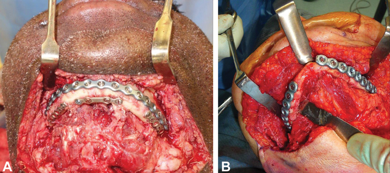
Various methods exist for obtaining rigid fixation of mandibular fractures. In high energy injuries, such as this comminuted mandibular fracture secondary to a gunshot wound ( A ), interfragmentary fixation was used to align the different segments of the mandibular and rigid fixation was achieved with the addition of a locking plate along the inferior border. In edentulous patients, reconstruction plates with bicortical screws should be placed as laterally and inferiorly as possible, to avoid interference with denture fabrication ( B ).
When using load sharing osteosynthesis, stability at the fracture site is shared between the plate and well-buttressed bone. Depending on the location, the functional load is either shared equally between bone and plate (e.g., angle fractures), or in more ideal situations the bone assumes a greater share of the functional load than the plate (e.g., body fractures in dentate mandibles). Here, fixation can be accomplished with 2.0mm diameter miniplate systems. Examples of load-sharing fixation include a single miniplate along the oblique ridge for angle fractures (ie, Champy technique), or a single miniplate and an arch bar (providing tension) for body or symphysial fractures, and lag screw fixation.
Lag Screw Fixation
The use of lag screws was popularized by Niederdellmann et al. in 1976. 14 Lag screws can be use in simple fractures where there is well-buttressed bone such as in symphysis or parasymphysis fractures. A lag screw has threads on only half the shaft so that the portion below the screw head is smooth and will not engage bone. Thus, the threads only engage the inner segment of bone and compress it against the outer segment. Typically, the two screws are placed, with minimal divergence between their long axes.
Rigid versus Non-rigid Fixation
Fixation can be grouped into rigid fixation, nonrigid fixation, or semirigid fixation. With rigid fixation, no bony callus if formed during healing and fracture segments are completely immobilized. In nonrigid fixation, micro-mobility of the fracture segments occurs and the fracture cap undergoes callus formation. Rigid fixation techniques include the use of plates and screws (miniplate and tension band with two screws on each side of the fracture), two lag screws, or reconstruction plates with three screws on each side of the fracture. A 2020 paper by Rughubar et al. compared the complication rates in patients with bilateral mandibular fractures randomized to either a combination of rigid fixation for an anterior fracture and nonrigid for the posterior fracture or nonrigid fixation for both fractures and found no significant difference; the risk of complications was significantly higher in patients with moderate to severe fracture displacement, regardless of treatment. 15
Fracture Management Techniques by Anatomic Site
Symphysis/Parasymphysis
Fractures of the anterior mandible can be addressed using either closed reduction, open reduction and internal fixation (ORIF), lag screws, or a combination of lag screws and miniplates. Closed reduction of fractures is most commonly achieved by applying Erich arch bars with circumdental stainless steel wires ( Fig. 10 ) or hybrid arch bars. The patient is then placed into maxillomandibular fixation using stainless steel wires loops or heavy elastics. While still acceptable for simple fractures, ORIF is the standard of care for management of most displaced mandibular injuries.
Fig. 10.
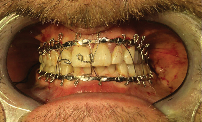
Intermaxillary fixation typically utilizes appliances that allow for coordinated alignment of the maxillary and mandibular dental arches. Intermaxillary fixation can be achieved with stainless steel wire loops, as shown here, or heavy elastics.
ORIF can be achieved using a variety of techniques. Semi-rigid fixation using application of transoral mini-plates can be applied in simple, non-displaced fractures. Rigid fixation with the use of a reconstruction plate and bicortical screws may be required in severely comminuted or displaced fractures. In severely comminuted fractures, a transcervical approach may be required for adequate reduction of fracture segments; simultaneous bone grafting may be considered for fractures with segmental bone loss.
In 1991, Ellis and Ghali presented a lag screw technique specific to the anterior mandible and favor this use over plate osteosynthesis in the absence of comminution or gap defects. 16 The lag screw technique is a form of rigid fixation that provides compression of fracture segments in the anterior mandible. Anatomic advantages such as the curvature of the mandible allowing for placement lag screws perpendicular to the fracture line, thick cortical bone, and lack of vital structures make this technique safe and reliable.
Body
As with anterior mandible fractures, body fractures can be treated with either a closed approach using MMF or ORIF. MMF can only be used in the presence of reliable, reproducible occlusion, using either the patient's own teeth or a mandibular prosthesis. It may be indicated in nondisplaced, favorable fractures, comminuted fractures, fractures in children with mixed dentition, or edentulous fractures (e.g., using dentures or gunning splints). This technique is generally contraindicated in displaced, unfavorable fractures, multiple fractures, or instances of malunion. In addition, certain systemic conditions such as psychiatric disease, neurologic problems like seizures disorders, pulmonary disease, or gastrointestinal disorders where aspiration of emesis is a concern preclude prolonged intermaxillary fixation.
In most clinical circumstances, management of displaced mandibular injuries necessitates. Fixation can be achieved solely via an intraoral approach with a single reconstruction plate (2.3 to 2.5mm, Fig. 11A ) at the inferior border of the mandible or two miniplates (superior and inferior border plate) along zones of tension and compression. A transbuccal puncture for screw fixation may be required to access more posterior body fractures. A review by Ellis of 682 patients treated with ORIF of body and/or symphysial fractures with two miniplates was associated with more postoperative complications (wound dehiscence, plate exposure, need for plate removal, and tooth root damage) compared with the use of a single, larger diameter plate. 17
Fig. 11.

Varied fixation strategies were used in this patient with a right mandibular body and left mandibular angle fracture. The body fracture was fixed with a single 2.3 mm thick locking plate with bicortical screws ( A ). The left mandibular angle fracture was managed using the Champy technique, a method of semi-rigid fixation that is frequently used for management of angle fractures that are favorably oriented. The technique utilizes a single miniplate with screws placed along the internal oblique ridge proximally and external oblique ridge distally ( B ). The proximal and distal screws are located at near 90 degree angles to each other ( C ).
In cases of severely displaced or comminuted fractures a transcervical or submandibular approach may be indicated. Risdon first described the submandibular approach in 1934, which consisted of a skin incision 2cm below the angle or inferior border of the mandible and 4–5cm in length. A skin crease should be identified to hide the scar. Making the incision roughly 2 cm below the inferior border of the mandible will generally avoid the marginal mandibular branch of the facial nerve when utilizing this approach ( Fig. 12 ). A cadaveric study by Dingman and Grabb identified this nerve branch is 1cm below the inferior border of the mandible 91% of the time when 18 posterior to the facial vessels. 19 Anterior to these vessels, the marginal mandibular branch is inferior to the mandibular border only 19% of the time. The Hayes-Martin technique of ligating and elevating the facial artery and vein generally protects this branch and allows direct access to the inferior border of the mandible.
Fig. 12.
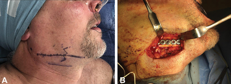
The transcervical, or Risdon, incision is typically placed 1.5–2 cm below the inferior border of the mandible ( A ), to prevent iatrogenic injury to the marginal mandibular nerve, which will run superior to this. When planning the extraoral approach in the setting of a displaced fracture, it is critical to place the incision at the anticipated location of the inferior border, as displacement will alter the position of the inferior border relative to its native location. Transcervical approaches to the mandible are frequently employed to allow adequate visualization of the buccal, inferior, and lingual aspects of the fracture ( B ).
Angle
Management of angle fractures is especially complex given the distracting forces of the muscles of mastication, lack of tooth-bearing segments, and possible presence of the third molar. If the fracture is unfavorable, treatment with only MMF would be contraindicated as the proximal segment may rotate. For years, rigid fixation with either a reconstruction plate or two miniplates was the treatment of choice. In 1976, Champy published an intraoral technique utilizing a single monocortical tension plate positioned proximally over the internal oblique ridge and distally over the external oblique ridge, with the screws in the segments oriented orthogonally relative to each other. 20 This form of semi-rigid fixation is ideal for nondisplaced or minimally displaced fractures with favorable orientation ( Fig. 11B-C ).
Ellis published a series of landmark prospective studies evaluating eight methods of treating mandible angle fractures over the course of several years and concluded that the use of either extraoral ORIF using a reconstruction plate or intraoral ORIF using a single miniplate yielded the fewest complications. 21 Later, he published a study comparing nonrigid fixation with 5–6 weeks of MMF, nonrigid fixation with a single miniplate (ie, Champy), or rigid fixation with 2 miniplates. The Champy technique yielded a complication rate of 2.5%; ORIF with a transcervical approach using a single reconstruction plate had a complication rate of 7.5%. The Champy technique also had the shortest operating time and was subjectively reported to be the easiest to perform. 22 The use of geometric miniplates was also found to decrease the risk of postoperative complications when compared with conventional miniplates. The more extensive dissection down to the inferior border of the mandibular angle, stripping the muscular attachments to bone and periosteum in this region and reducing the vascularity to the segments, may be one of the reasons why conventional plates lead to less favorable outcomes. 23
Ramus-Condyle Unit
Classification and management of condylar fractures is a controversial topic in craniomaxillofacial trauma. The two commonly used classification system are Lindahl and Spiessl. Lindahl's classification is based on three factors: level of fracture, amount of displacement, and relationship of the condylar head to the glenoid fossa. The three levels of fracture are condylar head (located within the joint capsule), condylar neck (inferior to the joint capsule and inferior attachment of the lateral pterygoid muscle), and subcondylar fracture (between the sigmoid notch and posterior aspect of the ramus). Spiessl's classification is based on the degree of displacement of fracture segments and dislocation of the condylar head from the fossa. 24 25
Management of condylar fractures is contentious and the subject of numerous studies. Treatment includes observation, closed reduction, ORIF by either transfacial or intraoral approaches. Endoscopic visualization may also be used. Singh et al. published a blinded, randomized controlled trial comparing ORIF or closed reduction and concluded that both groups yielded acceptable results. The surgical group achieved more accurate reduction, greater mouth opening/lateral excursion/protrusion, and less pain. No difference was found in occlusion between the two groups. 26
Absolute and relative indications for ORIF of condyle fractures were first described by Zide and Kent, but have been revised over the years. 27 Current absolute indications include bilateral fractures, severe dislocation, cases where closed reduction doesn't re-establish occlusion, concomitant fractures of other areas of the face that compromise occlusion, foreign bodies, or dislocation of the condyle into the middle cranial fossa. 28
Regardless of treatment choice early mobilization and physical therapy has been shown to decrease risk of ankylosis and trismus. 29 30 Decision-making for condylar injuries should prioritize early mobilization. In patients with intracapsular injuries and associated malocclusion, a short course of intermaxillary fixation (no longer than 7 days) can be considered, followed by aggressive mobilization to prevent ankylosis.
In patients with displaced condylar or subcondylar injuries, treatment with open or closed approaches is valid ( Fig. 13 ). When considering closed management, our approach is to re-establish occlusion with closed reduction and placement of intermaxillary fixation. This is followed by a period of tight intermaxillary fixation for 2 weeks, then 2 weeks of elastic (4–6 oz) fixation with jaw stretching exercises at meal times (3–5 minutes), and then 2 weeks of elastics use only at night, with jaw stretching exercises at meal times. The use of oral muscle relaxants during the subacute healing period may help patients with jaw stretching exercises.
Fig. 13.
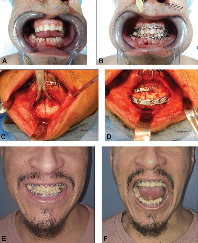
This patient sustained symphyseal and bilateral mandibular condylar fractures following an assault. The symphyseal fracture was managed with open reduction and placement of rigid internal fixation. The condylar fractures were managed with closed reduction and use of intermaxillary fixation with wires for 2 weeks, then heavy elastics for two weeks with initaition of jaw stretching exercises, and two weeks of night-time elastics with jaw stretching exercises 3 times daily. At 6 weeks post-operatively, the patient had a stable occlusion consistent with the preinjury state and preserved maximal incisal opening of 45 mm without deviation or pain.
Special Populations
Atrophic Mandible
It is estimated that roughly 8% of the United States population is edentulous. Atrophic, edentulous mandibular fractures represent 1% of all facial fractures. Although thought to be associated with advanced age, mandibular atrophy is more related to the duration of edentulism. The presence of tooth roots and forces of mastication send signals to inhibit bony resorption. 31 32 To be classified atrophic, the remaining bone height must be 15mm or less. Severe atrophy is classified as 10mm in height or less. 33 Decreased bony height correlates to increased incidence of nonunion, malunion, recurrent fractures, hardware failure, postoperative infection, and osteomyelitis. The incidence of these complications is between 10–20%. 33
ORIF has largely replaced other reduction techniques such as gunning splints, circum-mandibular wires around dentures, and external fixators. Load bearing osteosynthesis with a reconstruction plate for anatomic reduction provides a solid construct for atrophic mandible fractures ( Fig. 9B ). 34 35 In some cases, a large inferior border plate on the lateral border, coupled with a shallow vestibule may make wearing a denture less feasible. In these circumstances, placement of the reconstruction plate on the underside of the mandible, with screws oriented vertically or near vertically, may allow a patient to continue to wear a denture. This technique may also avoid potential intraoral hardware dehiscence. With any open approach, periosteal detachment should be limited to the buccal tissues to minimize devascularization. Autologous grafting can also be undertaken concurrently to aid in fracture healing and anticipation of dental rehabilitation.
Mandibular Injuries in Children
Mandible fractures in children follow different patterns than those observed in adults. In children, up to 50 to 80% of mandible fractures involve the condyle, subcondylar, or angle. The next most common are symphysis and parasymphysis fractures. 36 37 38 Body fractures are relatively rare. 39 40 41 Management must take into account eventual mandibular growth, presence of tooth buds, and eruption of permanent teeth. 42 The main goals of treatment of pediatric mandible fractures are to obtain bony union, restore occlusion, and prevent growth disturbances.
Treatment modalities for pediatric mandible fractures include physical therapy without MMF, a short period of MMF (7–14 days) followed by physical therapy, and ORIF. When considering intermaxillary fixation in children in the primary or mixed dentition, screw-retained devices should not be used, due to the risk of injury to the developing permanent dentition. Ivy loops, Risdon cables, Erich arch bars, sutures, and dental splints are all potential options for achieving intermaxillary fixation in children. Similarly, placement of internal fixation should account for the presence of the developing permanent teeth ( Fig. 14 ). In the dentate regions of the mandible, plates should be placed at or near the inferior border, with monocortical screws. If titanium fixation is used, it is prudent to consider removal at 2–3 months post-operatively in growing patients. Resorbable fixation in this population remains under investigation, but appears to result in stable healing when appropriately utilized.
Fig. 14.
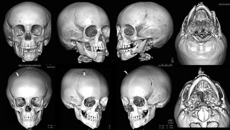
This two-year-old patient sustained displaced bilateral mandibular body fractures following a motor vehicle collision. The fractures were managed with open reduction and placement of internal fixation using miniplates with monocortical screws placed at the inferior border. Following appropriate healing, as evidenced clinically by lack of tenderness or mobility at the fracture sites and uniform mandibular movement, the plates were removed.
Similar to management in adults, treatment of pediatric condylar fractures remains controversial. Long-term favorable facial growth outcomes have been described with closed treatment of condylar fractures. Early mobilization is crucial to prevent hemarthrosis and ankylosis. TMJ ankylosis can be very difficult to treat and can children can result in profound facial deformities, particularly in growing children. 39 43 44
Management of pediatric mandibular injuries will be discussed more extensively later in this volume.
Complications
Complications associated with treatment of mandibular fractures may occur in up to 15% of cases. The most common complications include infection, hardware failure, osteomyelitis, malunion, nonunion, and wound dehiscence. 45 46
Infection
Postoperative infections are the most common complication associated with treatment of mandibular fractures. The most common organisms found in infected mandibular fractures are those species readily found in oral flora: Staphylococcus, Streptococcus, Klebsiella, and Actinomyces. In the acute period, infections are related to compromised soft tissues or teeth left in the fractures. Left untreated, infections may result in osteomyelitis. Contributing factors to complications include preoperative, perioperative, and postoperative oral hygiene, infected/fractured teeth in the line of fracture, alcoholism, metabolic disturbances, tobacco use, prolonged time between injury and definitive treatment, poor patient compliance with treatment, fracture severity, and inadequate reduction or fixation. 47 48 49
Numerous studies have shown that pre-operative antibiotics do not result in a decreased incidence of postoperative infections, hospital stay, or intensive care unit length of stay. 50 51 52 A study by Gaal et al. showed that limiting antibiotic exposure only to intraoperative antibiotic prophylaxis for patients undergoing transoral ORIF for treatment of open mandibular fractures was not associated with an increased risk of infection. 53
Malunion and Malocclusion
It is important for the provider to differentiate between malunion and nonunion as the treatment will differ. Malunion occurs when fracture segments are not properly reduced, not properly fixated, or in untreated fractures with delayed presentation allowing for bony union to take place in the absence of reduction. Malunion is most commonly evident clinically as malocclusion. Once recognized, malunion should be treated as early as possible. Malunion resulting in minor occlusal interferences can be treated with occlusal reduction and/or orthodontic therapy. In cases of severe malunion, fracture segments may need to osteotomized to obtain accurate, harmonic occlusion. To prevent malocclusion, obtaining premorbid photographs can assist the surgical team in obtaining the patient's premorbid occlusion, especially in cases where a premorbid malocclusion may have already existed, such as an anterior open bite.
Non-union
In contrast to malunion, non-union is a lack of osseous union between two or more fracture segments after an appropriate interval where osseous healing should be expected (e.g., 6–8 weeks in adults). Non-union is most common the result of fracture mobility, which results in a persistent inflammatory state that prevents normal bony repair of the defect. The overall incidence of nonunion is around 1–3%. 54 55
Clinically, if fracture segment mobility is observed early on within the dentate mandible, conservative management consisting of increasing the period of immobilization may resolve the nonunion. When this is not successful or gross mobility is noted, particularly in cases were fixation is inadequate in relation to the amount of bone, a second surgical intervention may be necessary. 56 57 58 Importantly, all infected material and soft tissue should be removed and the fracture sites should be debrided of immature callus to bleeding bone to ensure vascularity ( Fig. 15 ). 54 58 For segmental gaps that result from such debridement, autologous bone grafting or, in cases of large defects vascularized tissue transfer, may be indicated.
Fig. 15.

This edentulous patient developed an infected non-union following ORIF of a left mandibular body fracture. He was treated initially with resection of the involved devitalized bone and placement of a spanning reconstruction plate (left and center left). Following tailored treatment with intravenous antibiotics, the segmental defect was reconstructed using autologous iliac crest bone graft (center right and right).
Special Considerations
Teeth in Line of Fracture
Historically, the literature has shown repeatedly that healthy teeth in the line of the fracture does not increase the incidence of infection and may actually aid in the stabilization. A large prospective study of 253 patients with 422 mandibular fractures found a postoperative infection rate of 6.95% and found no association between the rate of infection and whether the tooth in the line of fracture was removed or not. 59 60 61 A systematic review by Fernandes et al. similarly failed to show any statistically significant difference in complication rates. 62 When teeth in the line of the fracture impair adequate reduction of the bony segments, are involved in an infected fracture, are mobile due to fracture or periodontal disease, have associated drainage, periapical radiolucency, or are themselves fractured, they should be removed. 61 62 63 64
For teeth otherwise involved in the lines of fractures, recent recommendations advise clinical and radiographic monitoring for at least 1 year and avoid unnecessary, costly endodontic procedures. 63 When a symptomatic tooth needs to be removed, it should ideally be removed once an adequate healing period has occurred, typically at a minimum of 3 months following reduction.
Three-dimensional Printing
Recent advances in computer-aided design and computer-aided manufacturing (CAD/CAM), in addition to the accessibility of three-dimensional (3D) printers, have found utility in management of complex CMF trauma. 65 The ability of 3D planning has the potential to not only reduce OR time and costs, but also aid in educating trainees in understanding the principles of fracture management. 66 67 68 69 70 King et al. showed that the use of 3D printers for fabrication of models to pre-bend plates drastically reduced operating time and costs ( Fig. 15 ). The mean intraoperative plate adaptation was reduced from roughly 23 minutes to 7 minutes resulting in a greater than 3-fold reduction in average operating room costs. 71
With the availability of in-house 3D printing capabilities at many institutions, the typical 1–2 week time required to send out imaging, plan, and obtain models, and pre-bend plates has been shortened to hours. Preliminary literature reports have demonstrated the utility of 3D-printed models for pre-bending fixation devices for simple or complex fractures, comminuted fractures, malunion, and even the fabrication of a dental splint to aid in circum-mandibular wiring for pediatric mandibular fracture fixation. 72 73
Intraoperative Imaging and Navigation
Surgical approaches to the mandible have anatomic limitations that may make visualization and accurate reduction challenging. Advances in mobile CBCT scanners, hybrid operating rooms with CT scanners, and intraoperative navigation systems allow for the provider to not only evaluate fracture reduction and fixation in real-time and make changes as needed. 65 74 75 76 However, one study examining cost-effectiveness and the use of intraoperative imaging found a cost break point at 17.7%. In other words, surgeons with complication rates of 17.7% or more would benefit from obtaining intraoperative imaging, while those with rates less than 17.7% would not. Interestingly, intraoperative CT was not cost-effective at any complication rate due to increased OR time. 77 In addition, the findings by Jain et al. found that the routine use of postoperative radiographs had no significant effect in management of maxillofacial fractures. 78 This idea has also been supported by the work of Courtemanche et al. 78 While these authors have demonstrated that post-operative imaging does not change management for patients with mandibular fractures following operative treatment, there remains a role for post-operative imaging to assess results and for educational purposes. In this context, surgeons should consider the balance between obtaining unnecessary imaging with the practical educational benefit for surgeon and trainee that comes from critical assessment of results using radiography.
Footnotes
Conflict of Interest None declared.
References
- 1.Kim K, Ibrahim A MS, Koolen P GL, Lee B T, Lin S J. Trends in facial fracture treatment using the American College of Surgeons National Surgical Quality Improvement Program database. Plast Reconstr Surg. 2014;133(03):627–638. doi: 10.1097/01.prs.0000438457.83345.e9. [DOI] [PubMed] [Google Scholar]
- 2.Afrooz P N, Bykowski M R, James I B, Daniali L N, Clavijo-Alvarez J A. The Epidemiology of Mandibular Fractures in the United States, Part 1: A Review of 13,142 Cases from the US National Trauma Data Bank. J Oral Maxillofac Surg. 2015;73(12):2361–2366. doi: 10.1016/j.joms.2015.04.032. [DOI] [PubMed] [Google Scholar]
- 3.Iida S, Kogo M, Sugiura T, Mima T, Matsuya T. Retrospective analysis of 1502 patients with facial fractures. Int J Oral Maxillofac Surg. 2001;30(04):286–290. doi: 10.1054/ijom.2001.0056. [DOI] [PubMed] [Google Scholar]
- 4.Kaura S, Kaur P, Bahl R, Bansal S, Sangha P. Retrospective Study of Facial Fractures. Ann Maxillofac Surg. 2018;8(01):78–82. doi: 10.4103/ams.ams_73_17. [DOI] [PMC free article] [PubMed] [Google Scholar]
- 5.Kim B J, Lee S I, Chung C M. A Retrospective Analysis of 303 Cases of Facial Bone Fracture: Socioeconomic Status and Injury Characteristics. Arch Craniofac Surg. 2015;16(03):136–142. doi: 10.7181/acfs.2015.16.3.136. [DOI] [PMC free article] [PubMed] [Google Scholar]
- 6.Das D, Salazar L. Maxillofacial Trauma: Managing Potentially Dangerous And Disfiguring Complex Injuries. Emerg Med Pract. 2017;19(04):1–24. [PubMed] [Google Scholar]
- 7.Panesar K, Dodson T, Lynch J, Bryson-Cahn C, Chew L, Dillon J. Evolution of COVID-19 Guidelines for University of Washington Oral and Maxillofacial Surgery Patient Care. J Oral Maxillofac Surg. 2020;78(07):1136–1146. doi: 10.1016/j.joms.2020.04.034. [DOI] [PMC free article] [PubMed] [Google Scholar]
- 8.Miloro M, Basi D, Halpern L, Kang D.Patient Assessment J Oral Maxillofac Surg 201775(8S):e12–e33. [DOI] [PubMed] [Google Scholar]
- 9.Boffano P, Roccia F, Gallesio C, Karagozoglu K, Forouzanfar T. Inferior alveolar nerve injuries associated with mandibular fractures at risk: a two-center retrospective study. Craniomaxillofac Trauma Reconstr. 2014;7(04):280–283. doi: 10.1055/s-0034-1375169. [DOI] [PMC free article] [PubMed] [Google Scholar]
- 10.Halpern L R, Kaban L B, Dodson T B. Perioperative neurosensory changes associated with treatment of mandibular fractures. J Oral Maxillofac Surg. 2004;62(05):576–581. doi: 10.1016/j.joms.2003.12.006. [DOI] [PubMed] [Google Scholar]
- 11.Schultze-Mosgau S, Erbe M, Rudolph D, Ott R, Neukam F W. Prospective study on post-traumatic and postoperative sensory disturbances of the inferior alveolar nerve and infraorbital nerve in mandibular and midfacial fractures. J Craniomaxillofac Surg. 1999;27(02):86–93. doi: 10.1016/s1010-5182(99)80019-5. [DOI] [PubMed] [Google Scholar]
- 12.Marchena J M, Padwa B L, Kaban L B.Sensory abnormalities associated with mandibular fractures: incidence and natural history J Oral Maxillofac Surg 19985607822–825., discussion 825–826 [DOI] [PubMed] [Google Scholar]
- 13.Dingman R ONP. Surgery of facial fractures. Br J Surg. 1964;51(07):560. [Google Scholar]
- 14.Niederdellmann H, Schilli W, Düker J, Akuamoa-Boateng E. Osteosynthesis of mandibular fractures using lag screws. Int J Oral Surg. 1976;5(03):117–121. doi: 10.1016/s0300-9785(76)80059-2. [DOI] [PubMed] [Google Scholar]
- 15.Rughubar V, Vares Y, Singh P. Combination of Rigid and Nonrigid Fixation Versus Nonrigid Fixation for Bilateral Mandibular Fractures: A Multicenter Randomized Controlled Trial. J Oral Maxillofac Surg. 2020;78(10):1781–1794. doi: 10.1016/j.joms.2020.05.012. [DOI] [PubMed] [Google Scholar]
- 16.Ellis E, III, Ghali G E.Lag screw fixation of anterior mandibular fractures J Oral Maxillofac Surg 1991490113–21., discussion 21–22 [DOI] [PubMed] [Google Scholar]
- 17.Ellis E., III A study of 2 bone plating methods for fractures of the mandibular symphysis/body. J Oral Maxillofac Surg. 2011;69(07):1978–1987. doi: 10.1016/j.joms.2011.01.032. [DOI] [PubMed] [Google Scholar]
- 18.Ellis E., III Open reduction and internal fixation of combined angle and body/symphysis fractures of the mandible: how much fixation is enough? J Oral Maxillofac Surg. 2013;71(04):726–733. doi: 10.1016/j.joms.2012.09.017. [DOI] [PubMed] [Google Scholar]
- 19.Dingman R O, Grabb W C. Surgical anatomy of the mandibular ramus of the facial nerve based on the dissection of 100 facial halves. Plast Reconstr Surg Transplant Bull. 1962;29:266–272. doi: 10.1097/00006534-196203000-00005. [DOI] [PubMed] [Google Scholar]
- 20.Champy M, Lodde J P. [Mandibular synthesis. Placement of the synthesis as a function of mandibular stress] Rev Stomatol Chir Maxillofac. 1976;77(08):971–976. [PubMed] [Google Scholar]
- 21.Ellis E., III Treatment methods for fractures of the mandibular angle. Int J Oral Maxillofac Surg. 1999;28(04):243–252. [PubMed] [Google Scholar]
- 22.Ellis E., III A prospective study of 3 treatment methods for isolated fractures of the mandibular angle. J Oral Maxillofac Surg. 2010;68(11):2743–2754. doi: 10.1016/j.joms.2010.05.080. [DOI] [PubMed] [Google Scholar]
- 23.Al-Moraissi E A, Ellis E., III What method for management of unilateral mandibular angle fractures has the lowest rate of postoperative complications? A systematic review and meta-analysis. J Oral Maxillofac Surg. 2014;72(11):2197–2211. doi: 10.1016/j.joms.2014.05.023. [DOI] [PubMed] [Google Scholar]
- 24.McLeod N M, Keenan M. Towards a consensus for classification of mandibular condyle fractures. J Craniomaxillofac Surg. 2021;49(04):251–255. doi: 10.1016/j.jcms.2021.01.017. [DOI] [PubMed] [Google Scholar]
- 25.Dahlström L, Kahnberg K E, Lindahl L. 15 years follow-up on condylar fractures. Int J Oral Maxillofac Surg. 1989;18(01):18–23. doi: 10.1016/s0901-5027(89)80009-8. [DOI] [PubMed] [Google Scholar]
- 26.Singh V, Bhagol A, Goel M, Kumar I, Verma A. Outcomes of open versus closed treatment of mandibular subcondylar fractures: a prospective randomized study. J Oral Maxillofac Surg. 2010;68(06):1304–1309. doi: 10.1016/j.joms.2010.01.001. [DOI] [PubMed] [Google Scholar]
- 27.Zide M F, Kent J N. Indications for open reduction of mandibular condyle fractures. J Oral Maxillofac Surg. 1983;41(02):89–98. doi: 10.1016/0278-2391(83)90214-8. [DOI] [PubMed] [Google Scholar]
- 28.Strohl A M, Kellman R M. Current Management of Subcondylar Fractures of the Mandible, Including Endoscopic Repair. Facial Plast Surg Clin North Am. 2017;25(04):577–580. doi: 10.1016/j.fsc.2017.06.008. [DOI] [PubMed] [Google Scholar]
- 29.Shakya S, Zhang X, Liu L. Key points in surgical management of mandibular condylar fractures. Chin J Traumatol. 2020;23(02):63–70. doi: 10.1016/j.cjtee.2019.08.006. [DOI] [PMC free article] [PubMed] [Google Scholar]
- 30.He D, Ellis E, III, Zhang Y. Etiology of temporomandibular joint ankylosis secondary to condylar fractures: the role of concomitant mandibular fractures. J Oral Maxillofac Surg. 2008;66(01):77–84. doi: 10.1016/j.joms.2007.08.013. [DOI] [PubMed] [Google Scholar]
- 31.Brucoli M, Boffano P, Romeo I. The epidemiology of edentulous atrophic mandibular fractures in Europe. J Craniomaxillofac Surg. 2019;47(12):1929–1934. doi: 10.1016/j.jcms.2019.11.021. [DOI] [PubMed] [Google Scholar]
- 32.Goldschmidt M J, Castiglione C L, Assael L A, Litt M D. Craniomaxillofacial trauma in the elderly. J Oral Maxillofac Surg. 1995;53(10):1145–1149. doi: 10.1016/0278-2391(95)90620-7. [DOI] [PubMed] [Google Scholar]
- 33.Ellis E, III, Price C. Treatment protocol for fractures of the atrophic mandible. J Oral Maxillofac Surg. 2008;66(03):421–435. doi: 10.1016/j.joms.2007.08.042. [DOI] [PubMed] [Google Scholar]
- 34.Florentino V GB, Abreu D F, Ribeiro N RB. Surgical Treatment of Bilateral Atrophic Mandible Fracture. J Craniofac Surg. 2020;31(08):e753–e755. doi: 10.1097/SCS.0000000000006630. [DOI] [PubMed] [Google Scholar]
- 35.Brucoli M, Boffano P, Romeo I. Surgical management of unilateral body fractures of the edentulous atrophic mandible. Oral Maxillofac Surg. 2020;24(01):65–71. doi: 10.1007/s10006-019-00824-8. [DOI] [PubMed] [Google Scholar]
- 36.Cleveland C N, Kelly A, DeGiovanni J, Ong A A, Carr M M. Maxillofacial trauma in children: Association between age and mandibular fracture site. Am J Otolaryngol. 2021;42(02):102874. doi: 10.1016/j.amjoto.2020.102874. [DOI] [PubMed] [Google Scholar]
- 37.Posnick J C, Wells M, Pron G E.Pediatric facial fractures: evolving patterns of treatment J Oral Maxillofac Surg 19935108836–844., discussion 844–845 [DOI] [PubMed] [Google Scholar]
- 38.Thorén H, Iizuka T, Hallikainen D, Lindqvist C. Different patterns of mandibular fractures in children. An analysis of 220 fractures in 157 patients. J Craniomaxillofac Surg. 1992;20(07):292–296. doi: 10.1016/s1010-5182(05)80398-1. [DOI] [PubMed] [Google Scholar]
- 39.Zhou W, An J, He Y, Zhang Y. Analysis of pediatric maxillofacial trauma in North China: Epidemiology, pattern, and management. Injury. 2020;51(07):1561–1567. doi: 10.1016/j.injury.2020.04.053. [DOI] [PubMed] [Google Scholar]
- 40.Ghosh R, Gopalkrishnan K, Anand J. Pediatric Facial Fractures: A 10-year Study. J Maxillofac Oral Surg. 2018;17(02):158–163. doi: 10.1007/s12663-016-0965-8. [DOI] [PMC free article] [PubMed] [Google Scholar]
- 41.Kambalimath H V, Agarwal S M, Kambalimath D H, Singh M, Jain N, Michael P. Maxillofacial Injuries in Children: A 10 year Retrospective Study. J Maxillofac Oral Surg. 2013;12(02):140–144. doi: 10.1007/s12663-012-0402-6. [DOI] [PMC free article] [PubMed] [Google Scholar]
- 42.Berlin R S, Dalena M M, Oleck N C.Facial Fractures and Mixed Dentition - What Are the Implications of Dentition Status in Pediatric Facial Fracture Management?J Craniofac Surg 2021 Publish Ahead of Print [DOI] [PubMed]
- 43.Yesantharao P S, Lopez J, Reategui A. Open Reduction, Internal Fixation of Isolated Mandible Angle Fractures in Growing Children. J Craniofac Surg. 2020;31(07):1946–1950. doi: 10.1097/SCS.0000000000006892. [DOI] [PubMed] [Google Scholar]
- 44.Bilgen F, Ural A, Bekerecioğlu M. Our Treatment Approach in Pediatric Maxillofacial Traumas. J Craniofac Surg. 2019;30(08):2368–2371. doi: 10.1097/SCS.0000000000005896. [DOI] [PubMed] [Google Scholar]
- 45.Lamphier J, Ziccardi V, Ruvo A, Janel M.Complications of mandibular fractures in an urban teaching center J Oral Maxillofac Surg 20036107745–749., discussion 749–750 [DOI] [PubMed] [Google Scholar]
- 46.Bicsák Á, Abel D, Tack L, Smponias V, Hassfeld S, Bonitz L. Complications after osteosynthesis of craniofacial fractures-an analysis from the years 2015-2017. Oral Maxillofac Surg. 2021;25(02):199–206. doi: 10.1007/s10006-020-00903-1. [DOI] [PubMed] [Google Scholar]
- 47.Shridharani S M, Berli J, Manson P N, Tufaro A P, Rodriguez E D. The Role of Postoperative Antibiotics in Mandible Fractures: A Systematic Review of the Literature. Ann Plast Surg. 2015;75(03):353–357. doi: 10.1097/SAP.0000000000000135. [DOI] [PubMed] [Google Scholar]
- 48.Kyzas P A. Use of antibiotics in the treatment of mandible fractures: a systematic review. J Oral Maxillofac Surg. 2011;69(04):1129–1145. doi: 10.1016/j.joms.2010.02.059. [DOI] [PubMed] [Google Scholar]
- 49.Odom E B, Snyder-Warwick A K. Mandible Fracture Complications and Infection: The Influence of Demographics and Modifiable Factors. Plast Reconstr Surg. 2016;138(02):282e–289e. doi: 10.1097/PRS.0000000000002385. [DOI] [PubMed] [Google Scholar]
- 50.Zosa B M, Ladhani H A, Sajankila N, Elliott C W, Claridge J A. Pre-Operative Antibiotic Agents for Facial Fractures: Is More than One Day Necessary? Surg Infect (Larchmt) 2021;22(05):516–522. doi: 10.1089/sur.2020.036. [DOI] [PubMed] [Google Scholar]
- 51.Wick E H, Deutsch B, Kallogjeri D, Chi J J, Branham G H. Effectiveness of Prophylactic Preoperative Antibiotics in Mandible Fracture Repair: A National Database Study. Otolaryngol Head Neck Surg. 2021;•••:1.94599821100427E15. doi: 10.1177/01945998211004270. [DOI] [PMC free article] [PubMed] [Google Scholar]
- 52.Zein Eddine S B, Cooper-Johnson K, Ericksen F. Antibiotic Duration and Outcome Complications for Surgical Site Infection Prevention in Traumatic Mandible Fracture. J Surg Res. 2020;247:524–529. doi: 10.1016/j.jss.2019.09.050. [DOI] [PubMed] [Google Scholar]
- 53.Gaal A, Bailey B, Patel Y. Limiting Antibiotics When Managing Mandible Fractures May Not Increase Infection Risk. J Oral Maxillofac Surg. 2016;74(10):2008–2018. doi: 10.1016/j.joms.2016.05.019. [DOI] [PubMed] [Google Scholar]
- 54.Bochlogyros P N. Non-union of fractures of the mandible. J Maxillofac Surg. 1985;13(04):189–193. doi: 10.1016/s0301-0503(85)80046-1. [DOI] [PubMed] [Google Scholar]
- 55.Melmed E P, Koonin A J. Fractures of the mandible. A review of 909 cases. Plast Reconstr Surg. 1975;56(03):323–327. doi: 10.1097/00006534-197509000-00011. [DOI] [PubMed] [Google Scholar]
- 56.Hsieh T Y, Funamura J L, Dedhia R, Durbin-Johnson B, Dunbar C, Tollefson T T. Risk Factors Associated With Complications After Treatment of Mandible Fractures. JAMA Facial Plast Surg. 2019;21(03):213–220. doi: 10.1001/jamafacial.2018.1836. [DOI] [PMC free article] [PubMed] [Google Scholar]
- 57.Furr A M, Schweinfurth J M, May W L. Factors associated with long-term complications after repair of mandibular fractures. Laryngoscope. 2006;116(03):427–430. doi: 10.1097/01.MLG.0000194844.87268.ED. [DOI] [PubMed] [Google Scholar]
- 58.Ellis E, III, Muniz O, Anand K. Treatment considerations for comminuted mandibular fractures. J Oral Maxillofac Surg. 2003;61(08):861–870. doi: 10.1016/s0278-2391(03)00249-0. [DOI] [PubMed] [Google Scholar]
- 59.Alichniewicz A, Jabloński M. [Management of a tooth in the line of fracture in the body of the mandible] Czas Stomatol. 1967;20(08):831–836. [PubMed] [Google Scholar]
- 60.Wilkie C A, Diecidue A A, Simses R J. Management of teeth in the line of mandibular fracture. J Oral Surg (Chic) 1953;11(03):227–230. [PubMed] [Google Scholar]
- 61.James R B, Fredrickson C, Kent J N. Prospective study of mandibular fractures. J Oral Surg. 1981;39(04):275–281. [PubMed] [Google Scholar]
- 62.Fernandes I A, Souza G M, Silva de Rezende V. Effect of third molars in the line of mandibular angle fractures on postoperative complications: systematic review and meta-analysis. Int J Oral Maxillofac Surg. 2020;49(04):471–482. doi: 10.1016/j.ijom.2019.09.017. [DOI] [PubMed] [Google Scholar]
- 63.Hosgor H, Coskunses F M, Akin D. Evaluation of the Prognosis of the Teeth in the Mandibular Fracture Line. Craniomaxillofac Trauma Reconstr. 2021;14(02):144–149. doi: 10.1177/1943387520952673. [DOI] [PMC free article] [PubMed] [Google Scholar]
- 64.Khavanin N, Jazayeri H, Xu T. Management of Teeth in the Line of Mandibular Angle Fractures Treated with Open Reduction and Internal Fixation: A Systematic Review and Meta-Analysis. Plast Reconstr Surg. 2019;144(06):1393–1402. doi: 10.1097/PRS.0000000000006255. [DOI] [PubMed] [Google Scholar]
- 65.Cuddy K, Khatib B, Bell R B. Use of Intraoperative Computed Tomography in Craniomaxillofacial Trauma Surgery. J Oral Maxillofac Surg. 2018;76(05):1016–1025. doi: 10.1016/j.joms.2017.12.004. [DOI] [PubMed] [Google Scholar]
- 66.Sinha P, Skolnick G, Patel K B, Branham G H, Chi J JA. A 3-Dimensional-Printed Short-Segment Template Prototype for Mandibular Fracture Repair. JAMA Facial Plast Surg. 2018;20(05):373–380. doi: 10.1001/jamafacial.2018.0238. [DOI] [PMC free article] [PubMed] [Google Scholar]
- 67.Kraeima J, Glas H H, Witjes M JH, Schepman K P. Patient-specific pre-contouring of osteosynthesis plates for mandibular reconstruction: Using a three-dimensional key printed solution. J Craniomaxillofac Surg. 2018;46(06):1037–1040. doi: 10.1016/j.jcms.2018.03.022. [DOI] [PubMed] [Google Scholar]
- 68.Elegbede A, Diaconu S C, McNichols C HL. Office-Based Three-Dimensional Printing Workflow for Craniomaxillofacial Fracture Repair. J Craniofac Surg. 2018;29(05):e440–e444. doi: 10.1097/SCS.0000000000004460. [DOI] [PubMed] [Google Scholar]
- 69.van de Velde W L, Schepers R H, van Minnen B. [The 3D-printed dental splint: a valuable tool in the surgical treatment of malocclusion after polytrauma] Ned Tijdschr Tandheelkd. 2016;123(01):19–23. doi: 10.5177/ntvt.2016.01.15132. [DOI] [PubMed] [Google Scholar]
- 70.el-Gengehi M, Seif S A. Evaluation of the Accuracy of Computer-Guided Mandibular Fracture Reduction. J Craniofac Surg. 2015;26(05):1587–1591. doi: 10.1097/SCS.0000000000001773. [DOI] [PubMed] [Google Scholar]
- 71.King B J, Park E P, Christensen B J, Danrad R. On-Site 3-Dimensional Printing and Preoperative Adaptation Decrease Operative Time for Mandibular Fracture Repair. J Oral Maxillofac Surg. 2018;76(09):19500–1.95E11. doi: 10.1016/j.joms.2018.05.009. [DOI] [PubMed] [Google Scholar]
- 72.Ding Q, Fu Z Z, Yue J, Xu Y X, Xue L F, Xiao W L.Virtual Surgical Planning and Three-Dimensional Printing to Aid the Anatomical Reduction of an Old Malunited Fracture of the MandibleJ Craniofac Surg2021 [DOI] [PubMed]
- 73.Ma J, Ma L, Wang Z, Zhu X, Wang W. The use of 3D-printed titanium mesh tray in treating complex comminuted mandibular fractures: A case report. Medicine (Baltimore) 2017;96(27):e7250. doi: 10.1097/MD.0000000000007250. [DOI] [PMC free article] [PubMed] [Google Scholar]
- 74.Goguet Q, Lee S H, Longis J, Corre P, Bertin H. Intraoperative imaging and navigation with mobile cone-beam CT in maxillofacial surgery. Oral Maxillofac Surg. 2019;23(04):487–491. doi: 10.1007/s10006-019-00765-2. [DOI] [PubMed] [Google Scholar]
- 75.Gebhard F, Riepl C, Richter P. [The hybrid operating room. Home of high-end intraoperative imaging] Unfallchirurg. 2012;115(02):107–120. doi: 10.1007/s00113-011-2118-3. [DOI] [PubMed] [Google Scholar]
- 76.Rabie A, Ibrahim A M, Lee B T, Lin S J. Use of intraoperative computed tomography in complex facial fracture reduction and fixation. J Craniofac Surg. 2011;22(04):1466–1467. doi: 10.1097/SCS.0b013e31821d1982. [DOI] [PubMed] [Google Scholar]
- 77.Barrett D M, Halbert T W, Fiorillo C E, Park S S, Christophel J J. Cost-based decision analysis of postreduction imaging in the management of mandibular fractures. JAMA Facial Plast Surg. 2015;17(01):28–32. doi: 10.1001/jamafacial.2014.782. [DOI] [PubMed] [Google Scholar]
- 78.Jain M K, Alexander M. The need of postoperative radiographs in maxillofacial fractures–a prospective multicentric study. Br J Oral Maxillofac Surg. 2009;47(07):525–529. doi: 10.1016/j.bjoms.2008.11.010. [DOI] [PubMed] [Google Scholar]
- 79.Courtemanche D J, Barton R, Li D, McNeill G, Heran M KS. Routine Postoperative Imaging Is Not Indicated in the Management of Mandibular Fractures. J Oral Maxillofac Surg. 2017;75(04):770–774. doi: 10.1016/j.joms.2016.12.024. [DOI] [PubMed] [Google Scholar]
- 80.Avery L L, Susarla S M, Novelline R A. Multidetector and threedimensional CT evaluation of the patient with maxillofacial injury. Radiol Clin North Am. 2011;49(01):183–203. doi: 10.1016/j.rcl.2010.07.014. [DOI] [PubMed] [Google Scholar]


