

Abstract
Despite the availability of a vaccine, pertussis is still a worldwide health problem. Outer membrane vesicles (OMVs) in gram-negative bacteria can stimulate the immune system due to several outer membrane proteins and are very good candidates in vaccine development. OMVs obtained from Bordetella pertussis contain several antigens, which are considered immunogenic, and could make them a potential candidate for vaccine production. The current study aimed to compare the current OMV extraction method (with ultracentrifuge) and a modified extraction method (without ultracentrifuge) and to evaluate the physicochemical properties as well as the expression of their main virulence factors. Vaccinal strain BP134 grown on Bordet Gengo agar were inoculated in Modified Stainer-Scholte medium for mass cultivation. OMVs were prepared using two different methods. They were then stained and examined with a transmission electron microscope. Protein contents were measured by the Bradford method, and then the protein profile was evaluated by SDS-PAGE. The presence of immunogenic antigens was detected by Western blotting. The size and shape of the OMVs obtained from the modified method without the use of ultracentrifuge were similar to the current method and had a size between 40 and 200 nm. The total protein yields of the OMV isolated using the current and modified methods were 800 and 600 µg/ml, respectively. Evaluating the protein profile of extracted OMVs showed the presence of different proteins. Finally, the presence of PTX, PRN, and FHA was observed in OMVs extracted from both methods. Comparison of the two OMV extraction methods showed that the obtained vesicles have a suitable and similar shape and size as well as the expression of three important pathogenic factors as immunogens. Despite the relatively low reduction in protein yield as the modified method does not require ultracentrifuge, this extraction method can be used as a suitable alternative for extracting the outer membrane vesicles from B. pertussis, especially in developing countries. It should be noted that further experiments including immunogenicity determination of OMVs obtained as vaccine candidates in animal models are required.
Keywords: B. pertussis, outer membrane vesicle, virulence factors
1. Introduction
Pertussis, or whooping cough, is a highly contagious bacterial respiratory tract infection caused by Bordetella pertussis ( 1 ). Some of the major virulence factors which play important roles in its pathogenesis are pertussis toxin (PTX), pertactin (PRN), and filamentous hemagglutinin (FHA). PTX induces protective immunity and plays an important role in the disease development ( 2 ). PRN is a surface protein involved in mediating adherence to the epithelium of the respiratory tract ( 3 ). FHA is also a major adhesion factor on the surface of B. pertussis ( 4 ).
This respiratory disease was a major cause of infant mortality worldwide before the vaccine was introduced in the 1940s. Widespread vaccination with the first generation of vaccines, whole cell pertussis (wP) vaccines that consisted of detoxified killed whole bacteria, significantly reduced morbidity attributed to the disease. However, in the 1970s, in some countries, concerns about the reactogenicity of wP vaccines led to rising rates of vaccine refusal and, consequently, to increased pertussis incidence ( 5 , 6 ).
Since the 1980s, many countries have replaced wP vaccines with less reactogenic acellular pertussis (aP) vaccines. Acellular vaccines are composed of pertussis toxin (PTX) as a major protective antigen and other surface proteins such as bivalent, trivalent, and pentavalent pertussis vaccines. In general, five-component vaccines (pentavalent) (including pertussis toxin, filamentous hemagglutinin, pertactin, and fimbriae 2 and 3) are considered more effective than bivalent or trivalent vaccines ( 7 - 9 ).
Unexpectedly, despite the high vaccination rate, recent years have seen a large number of pertussis outbreaks that had not been seen since pre-vaccine days. Indeed, pertussis is now recognized as a reemergent disease and is still among the main causes of death in children worldwide ( 10 - 14 ).
This reemergence is due to short-term immunity induced by aP vaccines. While wP vaccines induce Th1/Th17 responses that lead to lung clearance and long-lasting immunity, aP vaccines mainly induce a Th2 response. Because B. pertussis is an intracellular bacterium, a Th1 response must be induced for lung clearance. To overcome these problems, a new generation of vaccines needs to be developed, as the outer membrane vesicles (OMVs) of B. pertussis contain phospholipids, lipooligosaccharides, nucleic acid, and several immunogenic antigens. Moreover, they have shown a basal level of protection against B. pertussis that also induces a Th1 response, potentially making them an attractive alternative over the currently available vaccines ( 15 , 16 ).
The current study aimed to extract outer membrane vesicles (OMVs) from B. pertussis using a modified procedure without ultracentrifuging at a very high speed in comparison with the current method which uses ultracentrifuge and evaluate the physicochemical properties and the expression of the main virulence factors, PTX, PRN, and FHA of the extracted OMVs as potential vaccine candidates.
2. Material and Methods
2.1. Bacterial Strains and Cultures
B. pertussis vaccinal strain BP134 was obtained from Razi Vaccine and Serum Research Institute (RVSRI). This strain was grown on Bordet-Gengou agar (BGA; Difco) supplemented with 10% defibrinated sheep blood at 37 °C for 72 h. Then colonies were confirmed with biochemical tests and slide agglutination using B. pertussis antiserum (Difco) for the final approval. Bacterial colonies were subcultured on the same medium for 48 h and then inoculated in liquid Modified Stainer-Scholte medium (MSS) with methyl-β-cyclodextrin at 200 rpm in a Beckman-Coulter shaker (BeckmanCoulter, Brea, CA) until decelerating phases (optical densities, OD600 between 0.7 and 1.0) were reached for the large-scale production of cultures ( 17 , 18 ).
2.2. Isolation of OMVs
With the current method used in previous studies, sequential ultra-centrifugation at speeds above 100,000 g has been used to isolate OMVs as described before ( 15 , 16 , 18 ). In this modified method, however, simple steps with centrifuging at a lower speed was used as described in the following.
First, 600 ml of MSS broth was inoculated with 20 ml of a decelerating phase culture of B. pertussis for large-scale production. After about 30 h, the decelerating phase was reached at 36 °C with aeration at 200 rpm (Beckman-Coulter, Brea, CA). The cultures were pelleted by centrifugation at 8000 g for 30 min at 4 °C, and then the pellets were washed twice in phosphate-buffered saline (PBS) to eliminate cell debris. The pellet (1 g wet weight) was subsequently resuspended in 7.5 ml of TE buffer (Tris-HCl, EDTA, pH 8.5) and homogenized completely to make a uniform suspension. Next, the suspension was incubated at room temperature for 30 min, sonicated for 10 min (MSE sonicator, 4-5 pm amplitude, 20 kHz on ice with intervals of 30 s), and then centrifuged at 10,000 g for 20 min at 4 °C. The pellets were washed with TE, centrifuged, and the supernatants pelleted at 60,000 g for 2 h at 4 °C. Subsequently, the resulting pellets were resuspended in 0.1 M Tris, 10 mM EDTA, DOC (5 g/L) buffer, and mixed again several times by pipetting to make a homogenized suspension which was incubated for 10 min then centrifuged for 2 h at 60,000 g at 4 °C. Afterward, the supernatant was separated carefully in a new tube and treated with TE buffer and centrifuged again for 1 h at 60,000 g at 4 °C. The pellets were dissolved in 5 ml of 3% sucrose and passed through 0.22 µm pore size filters (PVDF, syringe filters, Germany). The filtered sample containing the OMVs was inactivated by heating in a water-bath at 56 °C for 30 min. The suspensions were then spread on blood agar and Bordet-Gengou agar plates and incubated at 37 °C for 48 h to confirm bacterial inactivation. Each extraction method was repeated four times, and in all repeats, the physiochemical properties of the OMVs extracted by the two methods, such as shape, size, SDS-PAGE, and Western profiles, were determined ( 15 , 16 , 18 ).
2.3. Protein Assay
The proteins in the outer membrane vesicles were quantified by Bradford assay with bovine serum albumin (BSA-Sigma) as standard using Nanodrop (Thermo Scientific, Wilmington, DE, USA) ( 15 , 16 , 18 ).
2.4. Transmission Electron Microscopy (TEM)
The OMV preparations were suspended in 0.1 M ammonium acetate (pH 7.0), and a drop was placed on a grid coated with a carbon-reinforced formvar film. After 30 s evaporation, the excess fluid was removed by absorption with filter paper and the grids stained with 2% (w/v) phosphotungstic acid. The grids were examined on a Zeiss EM10C TEM (Germany) operating at an accelerating voltage of 100 kV ( 15 , 18 ).
2.5. SDS-PAGE and Western Blotting
Protein profiles of the extracted OMVs were studied by SDS-PAGE followed by 12% sodium dodecyl sulfate-polyacrylamide gel electrophoresis (SDS-PAGE) used for the separation of protein molecules based on their molecular weight. After electrophoresis, proteins were stained by 0.1% (w/v) Coomassie blue (DNAbiotech, IRAN) with gentle shaking for 1 h. The gels were then washed three times with methanol 40% (v/v) and acetic acid 10% (v/v) in double-distilled water (ddw) for 20 min. Washed solution was discharged and gels were fixed with a solution containing 2% phosphoric acid (v/v), 18% ethanol (v/v), and 15% ammonium sulfate (w/v) in ddW for 30 min with minor modifications ( 15 ). Moreover, western immunoblotting analyses using specific antibodies against PTX, FHA, and PRN were employed to characterize and describe OMV properties.
The proteins from the polyacrylamide gel were transferred to the polyvinylidene difluoride (PVDF) membrane which was blocked overnight with 5% skimmed milk in PBS. Membranes were then washed three times with PBS in 0.05% Tween 20 (PBST) and next, exposed to mouse monoclonal immune sera directed against the PTX, PRN, and FHA (NIBSC No. 97/572, 97/558, and 97/564, respectively) for 1 h at 37 °C. Membranes were washed three times with PBST, followed by incubation with rabbit anti-sheep horseradish peroxidase-conjugated (HRP) antibody (PADZA Company, Iran) at a 1:1000 dilution for 1 h at room temperature. Finally, after washing three times with PBST, the color reaction was ultimately generated in the presence of metal-enhanced 3,3`-diaminobenzidines (DAB) substrate.
3. Results
OMVs were isolated from B. pertussis vaccinal strain BP134 using the current method and a modified method. Both obtained samples were negatively stained and examined with an electron microscope with a mean size of 70 nm and ranging from 40 to 200 nm (Figure 1A and 1B).
Figure 1.

Negatively stained Bordetella pertussis OMVs examined with an electron microscope. (A) shows the OMV obtained from B. pertussis with the current method. (B) shows the OMV obtained from B. pertussis with the modified method. Vesicle sizes range from 40 to 200 nm.
At least four independent extractions of OMVs using the two methods and characterization were carried out. Similar morphologies were observed in all cases; the OMVs’ size range was consistent from batch to batch and similar to previously described OMV preparations ( 15 ).
The amounts of the membrane vesicle's protein were 800 and 600 µg/ml, respectively.
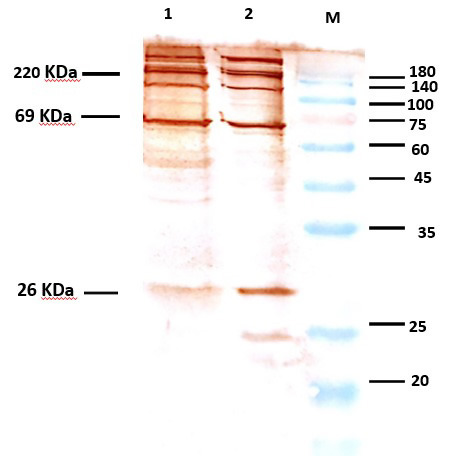
To further characterize the OMV properties, including the virulence factors, the electrophoretic pattern was assessed by 12% SDS-PAGE. Bands similar to each other and to other studies, including 32, 69, and 140-180 kDa, were observed (Figure 2).
Figure 2.

SDS-PAGE Pattern of OMV containing PTXS1, Prn, FHA. Lane 1 shows the OMV obtained from B. pertussis using the modified method, and lane 2 shows the OMV obtained with the current protocol. M: Marker. Molecular weights are indicated at the right.
Western blotting was performed using specific monoclonal antibodies against PTX, PRN, and FHA to confirm the presence of known pertussis immunogens in OMVs which finally confirmed and verified the presence of these important antigens in extracted OMVs by two methods as also described by previous authors (18) (Figure 3).
Figure 3.
Western blot of OMVs using anti-PTX S1, anti-PRN, and anti-FHA. Lane 1 shows the OMV obtained from B. pertussis with the modified method, and lane 2 shows the OMV obtained from the current protocol. Molecular weights are indicated at the right.
4. Discussion
The resurgence of pertussis has been reported in several countries that have shifted from wP to aP vaccines. The side effects associated with whole-cell vaccines as well as their inadequacy to induce protective immunity led to the development of aP vaccines, but they, too, have some disadvantages, such as low efficacy, waning antibodies after immunization, providing immunity against a limited number of antigens, and inducing a humoral response. Because of these drawbacks, much research has focused on other parts of the bacterium ( 14 , 19 ).
From the vaccine point of view, OMVs derived from B. pertussis, which contain main bacterial surface antigens, have been shown to successfully exhibit a basal level of protection and induce Th1, Th2, Th17, similar to wP vaccines. As OMVs are spherical nanoparticles, they are expected to exert an improved uptake of the antigen by antigen-presenting cells than the bacterial whole cell ( 16 ). Another attractive feature of OMVs is its advantage over the currently used aP vaccines, as they are capable of conferring both long-lasting immunity as well as protection against different strain genotypes that result in better protection. Therefore, OMVs could be considered as good vaccine candidates against B. pertussis ( 20 ).
Accordingly, several studies have revealed that because meningococcal OMVs express immunogenic antigens, they could be naturally taken advantage of for the important features required for a good vaccine. There are currently two licensed vaccines for serogroup B meningococcal disease based on OMVs. The efficacy and safety of these OMV vaccines have been proven ( 21 - 24 ). As OMVs are stable even at room temperature and they don't require cold chain or buffer solution, the possibility of employing OMVs as a good vaccine candidate for the prevention of diseases should be considered ( 25 ).
Roberts and Moreno ( 15 ) recently demonstrated that OMVs derived from B. pertussis can protect against an intranasal pertussis challenge when administered by either the intraperitoneal or intranasal route in a mouse model of infection. Moreover, the isolation of OMVs has important advantages over purified proteins, as OMV extraction based on simple steps eliminates the need for costly prior purification of each antigen, which must be done for the current acellular pertussis vaccines ( 26 ).
This research is a comparison study of the current OMV extraction method with the modified method. At least five independent OMV extractions were carried out using each method, and similar morphologies and size ranges (from 40 to 200 nm) were observed in all.
Analysis of the OMVs isolated in this study demonstrated that they were numerous nanoparticle vesicles, which were perfectly fitted with B. pertussis OMVs used in other studies and also with each other ( 15 , 18 , 26 , 27 ).
The main protective bacterial antigens in the development of effective pertussis vaccines are virulence factors such as filamentous hemagglutinin, fimbriae, and pertactin, which allow B. pertussis to bind to the ciliated epithelial cells in the upper respiratory tract ( 1 , 28 ). However, the presence of PTX, PRN, and FHA in OMVs is very important ( 29 , 30 ).
Our findings show the expression of PTX, PRN, and FHA in OMVs isolated from the vaccinal strain with the current method and the modified method.
As OMVs reported herein contained several protective immunogens, they might be considered as a possible basic material for the development of a B. pertussis vaccine.
The current results have revealed no noticeable differences in their size or shape, nor in their protein profile; however, the modified product yield showed a slight difference. It is worth noting that this method was modified from previous methods to avoid the need for ultracentrifuging at high speed, which is an expensive and advanced technology not generally available in laboratories or research centers. Therefore, this modified method can be a valuable alternative method, especially in developing countries.
It is obvious that further studies and challenges are necessary to determine the potential immunogenic effect of OMVs in formulation with different adjuvants and the probability of producing immune responses and the efficiency of the OMV as a vaccine candidate to improve the current B. pertussis vaccines.
Authors' Contribution
Study concept and design: F. Sh. and M. N.
Acquisition of data: M. S. S.
Analysis and interpretation of data: M. S. S.
Drafting of the manuscript: M. S. S. and M. N.
Critical revision of the manuscript for important intellectual content: M. S. S., M. N., F. Sh. and F. E.
Statistical analysis: M. S. S., M. N., F. Sh. and F. E.
Administrative, technical, and material support: M. N., F. Sh. and S. R. B.
Ethics
All procedures performed in studies involving animals were in accordance with the ethical standards of the Razi Vaccine and Serum Research Institute.
Grant Support
This article was extracted from Ph.D's thesis in Microbiology entitled "Preparation and immunological evaluation of OMVs from predominant strain of Bordetella pertussis as vaccine candidate in laboratory animal ". This research was supported by Razi Vaccine and Serum Research Institute, and Pasteur institute.
Acknowledgement
The authors would like to appreciate the staff of the Aerobic Bacterial Research and Vaccine Production department and department of Immunology & Microbiology of Razi Vaccine and Serum Research Institute and Microbiology department of Pasteur institute for their cooperation.
Conflict of Interest: The authors declare that they have no conflict of interest.
References
- 1.Howard C. Bordetella pertussis: Epidemiology, Virulence Factors, Pathogenesis, Treatments, and Vaccines. 2016 [Google Scholar]
- 2.Mooi FR. Bordetella pertussis and vaccination: the persistence of a genetically monomorphic pathogen. Infect Genet Evol. 2010;10(1):36–49. doi: 10.1016/j.meegid.2009.10.007. [DOI] [PubMed] [Google Scholar]
- 3.Breakwell L, Kelso P, Finley C, Schoenfeld S, Goode B, Misegades LK, et al. Pertussis Vaccine Effectiveness in the Setting of Pertactin-Deficient Pertussis. Pediatrics. 2016:e20153973. doi: 10.1542/peds.2015-3973. [DOI] [PubMed] [Google Scholar]
- 4.Zaretzky FR, Gray MC, Hewlett EL. Mechanism of association of adenylate cyclase toxin with the surface of Bordetella pertussis: a role for toxin–filamentous haemagglutinin interaction. Mol Microbiol. 2002;45(6):1589–98. doi: 10.1046/j.1365-2958.2002.03107.x. [DOI] [PubMed] [Google Scholar]
- 5.Bolotin S, Harvill ET, Crowcroft NS. What to do about pertussis vaccines? Linking what we know about pertussis vaccine effectiveness, immunology and disease transmission to create a better vaccine. Pathog Dis. 2015;73(8) doi: 10.1093/femspd/ftv057. [DOI] [PMC free article] [PubMed] [Google Scholar]
- 6.Edwards K, editor. Pertussis vaccines. 42nd Annual Meeting; 2004: Idsa [Google Scholar]
- 7.Gustafsson L, Hallander HO, Olin P, Reizenstein E, Storsaeter J. A controlled trial of a two-component acellular, a five-component acellular, and a whole-cell pertussis vaccine. N Engl J Med. 1996;334(6):349–56. doi: 10.1056/NEJM199602083340602. [DOI] [PubMed] [Google Scholar]
- 8.Olin P, Rasmussen F, Gustafsson L, Hallander HO, Heijbel H. Randomised controlled trial of two-component, three-component, and five-component acellular pertussis vaccines compared with whole-cell pertussis vaccine. Lancet. 1997;350(9091):1569–77. doi: 10.1016/s0140-6736(97)06508-2. [DOI] [PubMed] [Google Scholar]
- 9.Poolman JT, Hallander HO. Acellular pertussis vaccines and the role of pertactin and fimbriae. Expert Rev Vaccines. 2007;6(1):47–56. doi: 10.1586/14760584.6.1.47. [DOI] [PubMed] [Google Scholar]
- 10.Wendelboe AM, Van Rie A, Salmaso S, Englund JA. Duration of immunity against pertussis after natural infection or vaccination. Pediatr Infect Dis J. 2005;24(5):S58–S61. doi: 10.1097/01.inf.0000160914.59160.41. [DOI] [PubMed] [Google Scholar]
- 11.Mooi FR, Van Loo I, Gent Mv, He Q, Bart MJ, Heuvelman KJ, et al. Bordetella pertussis strains with increased toxin production associated with pertussis resurgence. Emerg Infect Dis. 2009;15(8):1206–13. doi: 10.3201/eid1508.081511. [DOI] [PMC free article] [PubMed] [Google Scholar]
- 12.Bart MJ, van Gent M, van der Heide HG, Boekhorst J, Hermans P, Parkhill J, et al. Comparative genomics of prevaccination and modern Bordetella pertussis strains. BMC Genomics. 2010;11(1) doi: 10.1186/1471-2164-11-627. [DOI] [PMC free article] [PubMed] [Google Scholar]
- 13.Witt MA, Arias L, Katz PH, Truong ET, Witt DJ. Reduced risk of pertussis among persons ever vaccinated with whole cell pertussis vaccine compared to recipients of acellular pertussis vaccines in a large US cohort. Clin Infect Dis. 2013;56(9):1248–54. doi: 10.1093/cid/cit046. [DOI] [PubMed] [Google Scholar]
- 14.Mooi F, Van Der Maas N, De Melker H. Pertussis resurgence: waning immunity and pathogen adaptation–two sides of the same coin. Epidemiol Infect. 2014;142(04):685–94. doi: 10.1017/S0950268813000071. [DOI] [PMC free article] [PubMed] [Google Scholar]
- 15.Roberts R, Moreno G, Bottero D, Gaillard ME, Fingermann M, Graieb A, et al. Outer membrane vesicles as acellular vaccine against pertussis. Vaccine. 2008;26(36):4639–46. doi: 10.1016/j.vaccine.2008.07.004. [DOI] [PubMed] [Google Scholar]
- 16.Gaillard ME, Bottero D, Errea A, Ormazábal M, Zurita ME, Moreno G, et al. Acellular pertussis vaccine based on outer membrane vesicles capable of conferring both long-lasting immunity and protection against different strain genotypes. Vaccine. 2014;32(8):931–7. doi: 10.1016/j.vaccine.2013.12.048. [DOI] [PubMed] [Google Scholar]
- 17.Nikbin VS, Shahcheraghi F, Lotfi MN, Zahraei SM, Parzadeh M. Comparison of culture and real-time PCR for detection of Bordetella pertussis isolated from patients in Iran. Iran J Microbiol. 2013;5(3):209–214. [PMC free article] [PubMed] [Google Scholar]
- 18.Hozbor D, Rodriguez M, Fernandez J, Lagares A, Guiso N, Yantorno OJCm. Release of outer membrane vesicles from Bordetella pertussis. Curr Microbiol. 1999;38(5):273–8. doi: 10.1007/pl00006801. [DOI] [PubMed] [Google Scholar]
- 19.Sealey KL, Belcher T, Preston A. Bordetella pertussis epidemiology and evolution in the light of pertussis resurgence. Infect Genet Evol. 2016;40:136–43. doi: 10.1016/j.meegid.2016.02.032. [DOI] [PubMed] [Google Scholar]
- 20.Bottero D, Gaillard M, Zurita E, Moreno G, Martinez DS, Bartel E, et al. Characterization of the immune response induced by pertussis OMVs-based vaccine. Vaccine. 2016;34(28):3303–9. doi: 10.1016/j.vaccine.2016.04.079. [DOI] [PubMed] [Google Scholar]
- 21.Sandbu S, Feiring B, Oster P, Helland OS, Bakke HS, Næss LM, et al. Immunogenicity and safety of a combination of two serogroup B meningococcal outer membrane vesicle vaccines. Clin Vaccine Immunol. 2007;14(9):1062–9. doi: 10.1128/CVI.00094-07. [DOI] [PMC free article] [PubMed] [Google Scholar]
- 22.de Kleijn ED, de Groot R, Labadie J, Lafeber AB, van den Dobbelsteen G, van Alphen L, et al. Immunogenicity and safety of a hexavalent meningococcal outer-membrane-vesicle vaccine in children of 2–3 and 7–8 years of age. Vaccine. 2000;18(15):1456–66. doi: 10.1016/s0264-410x(99)00423-5. [DOI] [PubMed] [Google Scholar]
- 23.Nøkleby H, Aavitsland P, O’hallahan J, Feiring B, Tilman S, Oster P. Safety review: two outer membrane vesicle (OMV) vaccines against systemic Neisseria meningitidis serogroup B disease. Vaccine. 2007;25(16):3080–4. doi: 10.1016/j.vaccine.2007.01.022. [DOI] [PubMed] [Google Scholar]
- 24.Van de Waterbeemd B, Streefland M, Van der Ley P, Zomer B, Van Dijken H, Martens D, et al. Improved OMV vaccine against Neisseria meningitidis using genetically engineered strains and a detergent-free purification process. Vaccine. 2010;28(30):4810–6. doi: 10.1016/j.vaccine.2010.04.082. [DOI] [PubMed] [Google Scholar]
- 25.Thornton V, Lennon D, Rasanathan K, O’hallahan J, Oster P, Stewart J, et al. Safety and immunogenicity of New Zealand strain meningococcal serogroup B OMV vaccine in healthy adults: beginning of epidemic control. Vaccine. 2006;24(9):1395–400. doi: 10.1016/j.vaccine.2005.09.043. [DOI] [PubMed] [Google Scholar]
- 26.Bottero D, Gaillard ME, Errea A, Moreno G, Zurita E, Pianciola L, et al. Outer membrane vesicles derived from Bordetella parapertussis as an acellular vaccine against Bordetella parapertussis and Bordetella pertussis infection. Vaccine. 2013;31(45):5262–8. doi: 10.1016/j.vaccine.2013.08.059. [DOI] [PubMed] [Google Scholar]
- 27.Hozbor DF. Outer membrane vesicles: an attractive candidate for pertussis vaccines. Expert Rev Vaccines. 2017;16(3):193–6. doi: 10.1080/14760584.2017.1276832. [DOI] [PubMed] [Google Scholar]
- 28.Shrivastava R, Miller JF. Virulence factor secretion and translocation by Bordetella species. Curr Opin Microbiol. 2009;12(1):88–93. doi: 10.1016/j.mib.2009.01.001. [DOI] [PMC free article] [PubMed] [Google Scholar]
- 29.Pawloski L, Queenan A, Cassiday P, Lynch A, Harrison M, Shang W, et al. Prevalence and molecular characterization of pertactin-deficient Bordetella pertussis in the United States. Clin Vaccine Immunol. 2014;21(2):119–25. doi: 10.1128/CVI.00717-13. [DOI] [PMC free article] [PubMed] [Google Scholar]
- 30.Smith AM, Guzmán CA, Walker MJ. The virulence factors of Bordetella pertussis: a matter of control. FEMS Microbiol Rev. 2001;25(3):309–33. doi: 10.1111/j.1574-6976.2001.tb00580.x. [DOI] [PubMed] [Google Scholar]