Abstract
This erratum corrects the Monte Carlo simulation results reported in our recently published manuscript [Biomed. Opt. Express 12, 1205 (2021) 10.1364/BOE.415054], which affects the results in Section 3.1. We also correct a summary statement regarding Table 4. These corrections do not alter the main findings of our publication.
We recently published the results of an empirical methodology to assess laser-related tissue damage applied to liver tissue [1]. In Section 3.1 we report results of Monte Carlo simulations, which were performed with the incorrect optical properties. Specifically, the absorption and scattering values for human skin were reported incorrectly in Table 1, and the units for the absorption and scattering values were input as mm−1 rather than cm−1. We provide an updated Table 1 here to reflect the correct values and their respective units. In addition, we clarify here that the optical properties for human skin are from the dermis, and the scattering coefficient was calculated from the reported reduced scattering at 750 nm in Ref. [2] based on the following equation:
| (1) |
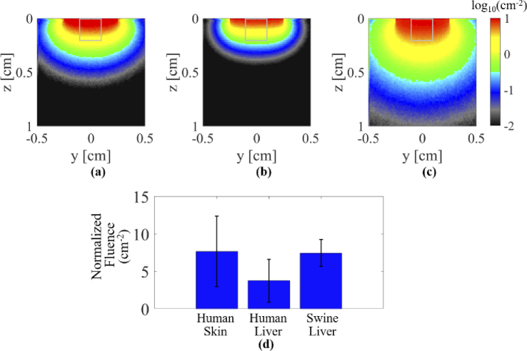
where and are the scattering and reduced scattering coefficients, respectively, and is the anisotropy, which was assumed to be 0.8 following Ref. [2]. The output of these simulations was an image that displayed the normalized fluence in units of . The mean and standard deviation of this normalized fluence was quantified within a tissue depth up to 0.2 cm within a 2D slice parallel to the light propagation direction to assess relative fluence differences across tissue types. With the correct simulation parameters, Fig. 2 in [1] should be updated as shown in Fig. 1.
Table 1. Optical properties modeled with Monte Carlo simulations.
Fig. 1.
(Replacement for Fig. 2 in original manuscript [1]) Simulated fluence profiles for (a) human skin tissue, (b) human liver tissue, (c) swine liver tissue in log compressed units of cm−2. (d) Mean ± one standard deviation of normalized fluence measured within the regions of interest outlined on each simulated profile result.
While reviewing these changes, we noticed that the number of swine reported in Table 4 with minimal to mild inflammation was miscounted in the text. Therefore, Section 3.4 should state, “Swine 2 and 3 experienced... minimal to mild inflammation on 4 [not 6] out of 12 laser application sites as indicated in Table 4."
These corrections do not alter the details reported in the abstract, discussion, and conclusion sections of [1], and they do not affect the main findings of our manuscript.
Funding
Alfred P. Sloan Foundation10.13039/100000879; National Science Foundation10.13039/100000001 (ECCS-175152); National Institutes of Health10.13039/100000002 (T32GM007057-44).
References
- 1.Huang J., Wiacek A., Kempski K. M., Palmer T., Izzi J., Beck S., Bell M. A. L., “Empirical assessment of laser safety for photoacoustic-guided liver surgeries,” Biomed. Opt. Express 12(3), 1205–1216 (2021). 10.1364/BOE.415054 [DOI] [PMC free article] [PubMed] [Google Scholar]
- 2.Salomatina E. V., Jiang B., Novak J., Yaroslavsky A. N., “Optical properties of normal and cancerous human skin in the visible and near-infrared spectral range,” J. Biomed. Opt. 11(6), 064026 (2006). 10.1117/1.2398928 [DOI] [PubMed] [Google Scholar]
- 3.Carneiro I., Carvalho S., Henrique R., Oliveira L., Tuchin V. V., “Measuring optical properties of human liver between 400 and 1000 nm,” Quantum Electron. 49(1), 13–19 (2019). 10.1070/QEL16903 [DOI] [Google Scholar]
- 4.Ritz J.-P., Roggan A., Isbert C., Müller G., Buhr H. J., Germer C.-T., “Optical properties of native and coagulated porcine liver tissue between 400 and 2400 nm,” Lasers Surg. Med. 29(3), 205–212 (2001). 10.1002/lsm.1134 [DOI] [PubMed] [Google Scholar]


