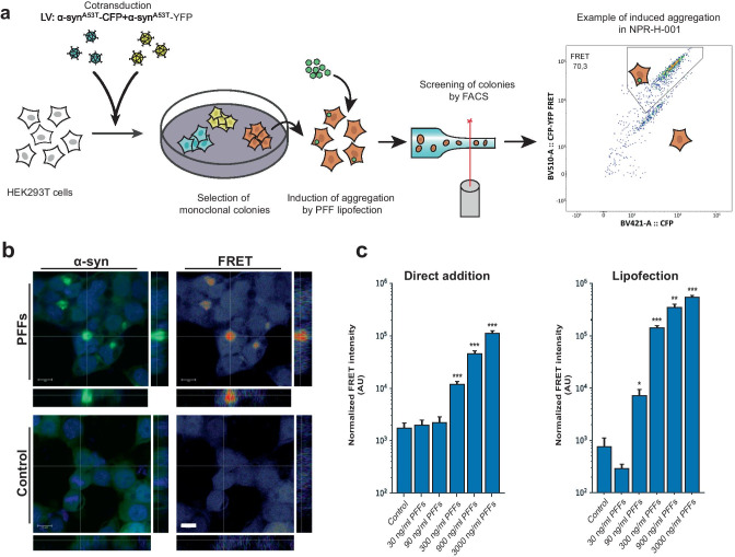
Fig. 1.
Monoclonal α-syn aggregate FRET reporter cells allow inclusion-specific detection of aggregates. a HEK293T cells were simultaneously transduced with lentivirus encoding α-synA53T-CFP and -YFP. Monoclonal cell lines were established by FACS single-cell sorting. Clonal lines were compared for brightness and signal-to-noise ratio of FRET by induction of aggregation by lipofection of α-syn PFFs to the biosensor lines. b Confocal microscopy confirmed a strong induction of FRET signal overlapping with the YFP signal, and no induced FRET signatures when treated with monomeric α-syn. cSensitivity assessment by PFF titration and FRET detection by flow cytometry of both direct addition and lipofection with PFFs (direct addition n = 6, lipofection n = 4). Bar chart values show mean ± SD, *p < 0.05, **p < 0.005, ***p < 0.001. Statistical testing was performed using Brown–Forsythe and Welch one-way ANOVA for multiple comparisons to a control group

