PLOS ONE logoLink to PLOS ONE
. 2021 Nov 23;16(11):e0249979. doi: 10.1371/journal.pone.0249979

Detection of cross-reactive immunoglobulin A against the severe acute respiratory syndrome-coronavirus-2 spike 1 subunit in saliva

Keiichi Tsukinoki 1,*, Tatsuo Yamamoto 2, Keisuke Handa 3, Mariko Iwamiya 4, Juri Saruta 1, Satoshi Ino 5, Takashi Sakurai 6
Editor: Etsuro Ito7
PMCID: PMC8610234  PMID: 34813596

Abstract

Abundant secretory immunoglobulin A (SIgA) in the mucus, breast milk, and saliva provides immunity against infection of mucosal surfaces. Pre-pandemic breast milk samples containing SIgA have been reported to cross-react with SARS-CoV-2; however, it remains unknown whether SIgA showing the cross-reaction with SARS-CoV-2 exists in saliva. We aimed to clarify whether SIgA in saliva cross-reacts with SARS-CoV-2 spike 1 subunit in individuals who have not been infected with this virus. The study involved 137 (men, n = 101; women, n = 36; mean age, 38.7; age range, 24–65 years) dentists and doctors from Kanagawa Dental University Hospital. Saliva and blood samples were analyzed by polymerase chain reaction (PCR) and immunochromatography for IgG and IgM, respectively. We then identified patients with saliva samples that were confirmed to be PCR-negative and IgM-negative for SARS-CoV-2. The cross-reactivity of IgA-positive saliva samples with SARS-CoV-2 was determined by enzyme-linked immunosorbent assay using a biotin-labeled spike recombinant protein (S1-mFc) covering the receptor-binding domain of SARS-CoV-2. The proportion of SARS-CoV-2 cross-reactive IgA-positive individuals was 46.7%, which correlated negatively with age (r = –0.218, p = 0.01). The proportion of IgA-positive individuals aged ≥50 years was significantly lower than that of patients aged ≤49 years (p = 0.008). SIgA was purified from the saliva of patients, which could partially suppress the binding of SARS-CoV-2 spike protein to the angiotensin converting enzyme-2 receptor. This study demonstrates the presence of SARS-CoV-2 cross-reactive SIgA in the saliva of individuals who had never been infected with the virus, suggesting that SIgA may help prevent SARS-CoV-2 infection.

Introduction

Secretory immunoglobulin A (SIgA) prevents infections through mucosal immunity—an aspect of the immune system. SIgA, comprising dimeric IgA, a J chain, and a secretory component, is secreted from glandular tissues such as the salivary glands and mammary glands onto mucosal surfaces, where it plays a central role in preventing the entry of antigens from the mucosa [1]. Severe acute respiratory syndrome coronavirus 2 (SARS-CoV-2) infects humans via the oral and nasal cavities, and the lungs [2]. The squamous cells of the tongue and periodontal tissues express angiotensin-converting enzyme-2 (ACE-2), a SARS-CoV-2 receptor, transmembrane protease serine 2 (TMPRSS2), and furin, which are proteases that promote infection [3], and saliva can harbor SARS-CoV-2 [4]. Saliva also contains several substances that suppress infection (such as lactoferrin, lysozyme, and SIgA, which is the most abundant) to potentially prevent the virus from entering the oral cavity [5].

Cross-reactive SIgA (CRsA) against SARS-CoV-2 was identified in breast milk before the COVID-19 pandemic [6, 7]. Furthermore, SARS-CoV-2-reactive CD4+ T cells were detected in approximately 40%–60% of unexposed individuals before the pandemic, suggesting that T cells have cross-reactivity to common cold coronaviruses and SARS-CoV-2 [8]. Later findings [9, 10] suggested that prior infection with coronavirus creates an immunological memory that is associated with IgG cross-reactivity.

Infection with SARS-CoV-2 causes COVID-19, which manifests as a unique spectrum of symptoms, ranging from asymptomatic to fatal acute respiratory failure [11]. The severity and prevalence of SARS-CoV-2 infection noticeably differs among age groups and countries [12]. Immune mechanisms might explain this wide disparity, but they are not yet fully understood. Immunoglobulin G (IgG) can eliminate SARS-CoV-2; thus, there is an urgent need for vaccine development against this virus [13]. However, mucosal immunity conferred by SIgA has not been investigated from the perspective of recovery from SARS-CoV-2 infection and its prevention. New findings in this area might facilitate a deeper understanding of COVID-19 characteristics.

Therefore, in this study, we aimed to develop an enzyme-linked immunosorbent assay (ELISA) for detecting SIgA that has cross-reactivity with SARS-CoV-2, and it could be used to reveal whether non-infected individuals harbor salivary SIgA that cross-reacts with the SARS-CoV-2 spike 1 subunit.

Methods

Participant selection

We tested saliva and blood samples using polymerase chain reaction (PCR) and immunochromatography, respectively. Individuals with saliva samples that were confirmed to be negative for COVID-19 by PCR and IgM testing were included in the study. The participants were 5 doctors and 132 dentists from Kanagawa Dental University Hospital. Individuals with IgA nephropathy, selective IgA deficiency, and autoimmune diseases, or who had cold-like symptoms within the past 2 weeks were excluded. The 137 participants, comprising 101 men and 36 women (mean age, 38.7; range, 24–65 years), provided fully informed consent. This study was approved by the Kanagawa Dental University Research Ethics Review Board (approval number: #690). This study was registered in the Japanese clinical trial UMIN-CTR (approval number: #R000046461) registry, which meets the ICMJE standards.

Saliva collection for ELISA

We collected samples using Salivettes® (Sarstedt AG & Co., KG, Nümbrecht, Germany) in a fixed room of the hospital between 9 a.m. and 12 p.m. in August 2020 under infection control protocols. The participants were instructed to refrain from eating, drinking, and brushing their teeth for at least 1 h before sample collection. The saliva samples were immediately centrifuged at 2,000 × g for 15 min, and then stored at –80°C.

Design of ELISA for CRsA against SARS-CoV-2 spike protein

We modified an ELISA system that could detect IgA cross-reactivity to influenza viruses using the human IgA ELISA quantitation set (#E88-102; Bethyl Laboratories, Montgomery, TX, USA) reported by Yamamoto et al. [14]. The saliva samples were diluted 500-fold in carbonate-bicarbonate buffer and incubated for 1 h at 25°C. ELISA plate wells were washed five times with wash solution. The antigen was spike 1-mFc recombinant protein (#40591-V05H1; Sino Biological, Beijing, China) comprising the SARS-CoV-2 spike 1 subunit with the spike protein receptor-binding domain (RBD). The antigen was labeled with biotin using a kit as described by the manufacturer (#BK01; Dojindo Laboratories, Kumamoto, Japan). Biotin-labeled spike 1 was added to the ELISA plate with 1 μg/mL saliva sample per well and incubated for 1 h at 25°C. The wells were washed five times with wash solution. Streptavidin-horseradish peroxidase conjugate (SA202; Millipore, USA; dilution, 1:1000) was then added to the wells and reacted for 1 h at 25°C. TMB substrate solution was added to wells, allowed to react for 15 min at 25°C, and the reaction was quenched with stop solution. Spike 1 protein-bound IgA was detected at 450 nm using a microplate absorbance reader (Bio-Rad Laboratories, Hercules, CA, USA). Background absorbance from the negative control containing phosphate-buffered saline was subtracted from the absorbance of all saliva samples.

IgA purification and western blotting

We purified SIgA using an IgA purification kit (#20395; Thermo Fisher Scientific, Waltham, MA, USA) as described by the manufacturer. Purified saliva samples were added to the mixture of sample buffer (#NP0008; Thermo Fisher Scientific) and sample reducing agent (#NP0009; Thermo Fisher Scientific). The samples were heated for 5 min at 96°C and run on a sodium dodecyl sulfate-polyacrylamide gel electrophoresis (SDS-PAGE) gel using a standard protocol. The gel was stained with Coomassie brilliant blue (#178–00551; FUJIFILM Wako Chemicals, Osaka, Japan). Antibodies specific for the heavy chain of IgA were used to determine whether the purified substance was IgA. We further confirmed that the detected IgA was the secretory type (SIgA) using an antibody specific to the secretory component. Western blotting was employed using the following primary antibodies: anti-human IgA rabbit monoclonal antibody (ab184863; Abcam Plc, Cambridge, UK; 1:500 dilution) and anti-human IgA SC mouse monoclonal antibody (ab3924; Abcam, 1:500 dilution). Horseradish peroxidase-conjugated anti-rabbit polyclonal antibody (#P0448; Dako, Glostrup, Denmark; 1:1000 dilution) or anti-mouse monoclonal antibody (#P0447; Dako; 1:1000 dilution) was used as the secondary antibody.

Ability of SIgA antibody to inhibit ACE-2-spike protein binding

To investigate whether ACE-2 binding to spike protein is inhibited by purified SIgA, we selected and pooled the top 20 antibody-positive and the bottom 20 antibody-negative samples based on the quantitative ELISA results. The final concentrations of antibodies in the pooled positive and negative saliva samples were 93.6 and 63.3 μg/mL, respectively.

The ability of SIgA to inhibit the binding of ACE-2 to the SARS-CoV-2 spike protein was then assessed using a SARS-CoV-2 spike-ACE-2 binding assay kit (#COV-SACE2-1; RayBiotech, Peachtree Corners, GA, USA), according to the manufacturer’s instructions. A SARS-CoV-2 spike-neutralizing rabbit IgG monoclonal antibody (#40592-R001; Sino Biological) was prepared as a positive control, and the neutralizing antibody was added at concentrations of 0, 0.0125, 0.025, 0.05, 0.1, and 0.2 μg/mL.

The percent binding inhibition (BI%) was determined as follows: [1 –(OD of test regent well/OD of no inhibitor in the positive control well)] × 100, according to the manufacturer’s instruction.

Questionnaire

The participants completed a self-administered questionnaire before saliva collection to determine whether they had previously been inoculated with Bacillus Calmette–Guérin (BCG), hepatitis B, and influenza vaccines within the past year. In addition, we requested the participants to fill out the questionnaire with their age and gender.

Statistical analysis

Negative values were set to 0 to determine relative ELISA values of CRsA, and the association with age was analyzed using Spearman rank correlations. Associations between the relative positive (>0) and negative (0) values of CRsA in the ELISA and binary data, age group, sex, BCG vaccination status, hepatitis B vaccination status, and influenza vaccination status were examined. Associations with vaccination history were examined using chi-square or Fisher exact tests. We summarized the variables of hepatitis B and influenza vaccines as neither, either, or both to examine associations between the number of vaccinations and IgA positivity or negativity. The variables of BCG, hepatitis B vaccine, and influenza vaccine were summarized as zero, one, two, or three to examine associations between the number of vaccinations and IgA positivity or negativity. The significance level was set to 5%. All data were statistically analyzed using SPSS version 26 (IBM Corp., Armonk, NY, USA).

Results

CRsA against the SARS-CoV-2 spike protein

The relative value of CRsA determined using the ELISA was set to 0 as the negative value. The CRsA was positive in 64 (46.7%) and was negative (absorbance of 0 or below) in 73 (53.3%) of the 137 samples.

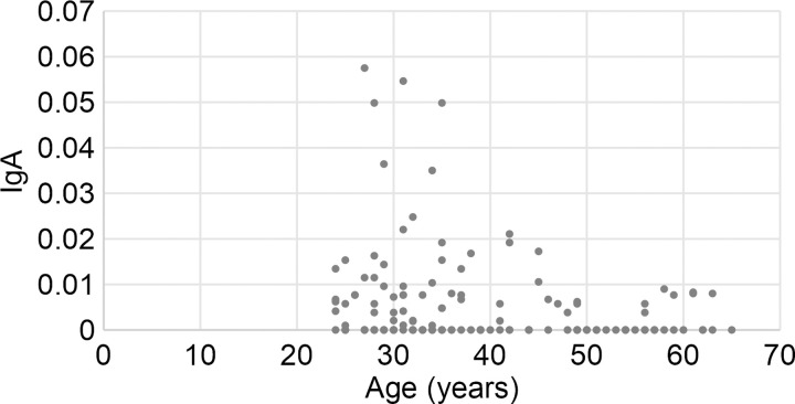
Fig 1 shows the plots showing age and cross-reactive IgA. Age significantly negatively correlated with relative CRsA (r = –0.218, p = 0.01). The positive rate of CRsA was significantly lower in participants aged ≥50 years than in those aged ≤49 years (p = 0.008), as shown in Table 1. The association between vaccines and CRsA positivity or negativity was not significant.

Fig 1. Associations between participant age and positive or negative rate of CRsA.

Fig 1

Vertical axis: Relative amount of IgA determined as the absorbance of saliva samples from CRsA-positive participants. Negative samples are shown as 0. The absorbance was considerably high among many participants aged ≤49 years.

Table 1. Associations of CrSA positivity with age, sex, and vaccination status.

Variable Total IgA-positive p*
n n %
Age (years) 20–29 32 19 59.4 0.081
30–39 54 27 50.0
40–49 23 11 47.8
50–59 20 4 20.0
≥60 8 3 37.5
20–49 109 57 52.3 0.008
≥50 28 7 25.0
Sex Male 101 48 47.5 0.452
Female 36 16 44.4
Vaccination
 BCG Yes 102 49 48.0 0.781
No 8 4 50.0
Unknown 27 11 40.7
 Hepatitis B Yes 124 58 46.8 0.992
No 11 5 45.5
Unknown 2 1 50.0
 Influenza Yes 127 60 47.2 0.458
No 10 4 40.0
 Hepatitis B and Influenza Neither 1 1 100.0 0.114
Either 19 7 36.8
Both 115 55 47.8
 BCG, Hepatitis B, and Influenza One 3 1 33.3 0.809
Two 21 11 52.4
Three 84 40 47.6

*Chi-square or Fisher exact test.

Characterization of purified saliva

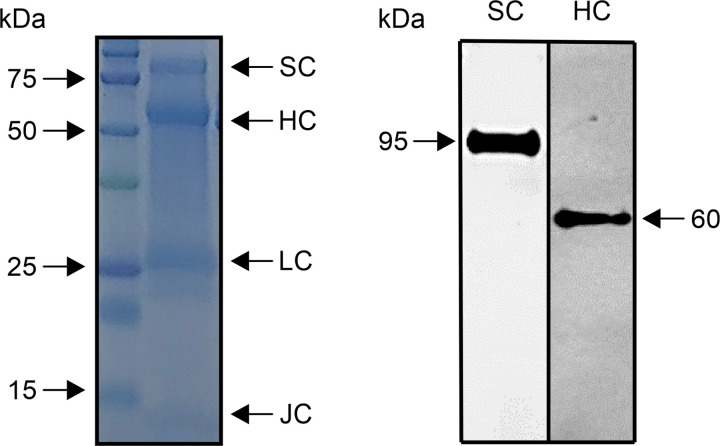
Some IgA bands were detected in the purified saliva samples by SDS-PAGE (Fig 2, left panel). Signals of the secretory component (approximately 95 kDa), IgA heavy chain (approximately 60 kDa), IgA light chain (approximately 26 kDa), and J chain (approximately 13 kDa) were identified. Western blotting revealed a single band of approximately 60 kDa, confirming the presence of IgA heavy chain in the purified saliva (Fig 2, right panel). In addition, a single band of approximately 95 kDa was identified as the secretory component of SIgA (Fig 2, right panel). These confirmed that purified saliva contained SIgA components.

Fig 2. Characterization of purified saliva.

Fig 2

The left panel shows protein bands of various molecular weights separated by SDS-PAGE. These bands were confirmed to correspond to the molecular weight of the secretory component (SC), IgA heavy chain (HC), IgA light chain, and J chain. The right panel shows the specific signals of SC and HC determined by western blotting.

CRsA inhibition tests

In the positive control, the range of absorbance values after the addition of spike-neutralizing antibody at different concentrations (0–0.2 μg/mL) was 2.155–0.493, with the concentration-dependent decrease confirming absorbance values. The neutralizing antibody against the spike protein showed an absorbance of 2.155 at an antibody concentration of 0 μg/mL. This absorbance indicates the absorbance when ACE-2 and the spike protein are strongly bound by ELISA. As the CRsA-negative saliva sample showed an absorbance of 2.402, which was higher than 2.155, it indicated that the binding between ACE-2 and the spike protein was not inhibited. The CRsA-positive saliva sample had an absorbance of 1.678, demonstrating the inhibition of binding between ACE-2 and the spike protein. The BI% was 21.7% for CRsA-positive saliva, but was –11% for CRsA-negative saliva. These results indicate that saliva SIgA1 antibody partially inhibited the spike protein.

Discussion

In this study, we found that 46.7% of saliva samples from 137 participants who had not been infected with SARS-CoV-2 contained CRsA against SARS-CoV-2 spike 1. The spike 1 region that we examined included the RBD that binds to the SARS-CoV-2 receptor ACE-2. This region is important for preventing infections and could thus be a target for vaccine development [14]. A previous ELISA for detecting SARS-CoV-2 CRsA in breast milk showed positivity rates of 100% and 80% using total spike protein and RBD as the antigen, respectively [6]. However, the values of the saliva samples in the present study were lower than both values obtained in the previous study. This might be because more SIgA is produced in breast milk than in saliva [15]. The abundance of CRsA in breast milk is a proven post-vaccination response [16], which could further explain why the CRsA positivity rate was lower in saliva than breast milk.

The participants who tested negative for COVID-19 determined by PCR and antibody tests did not subsequently develop COVID-19 at the time of submission of this article. Thus, we believe that they had not been exposed to SARS-CoV-2. As such, we revealed SIgA antibodies against the spike 1 protein of SARS-CoV-2 in individuals with no history of SARS-CoV-2 infection. Only one other study found salivary IgA antibodies that cross-react with the spike protein of SARS-CoV-2 before the COVID-19 pandemic [17]. The nucleocapsid proteins of coronaviruses are highly homologous, but their spike proteins share little commonality [18]. Patients with a history of infection with HCoV-OC43 and other coronaviruses have antibodies against SARS-CoV-1 and antibodies that cross-react to the SARS-CoV-1 nucleocapsid protein, respectively [19]. Furthermore, while HCoV-NL63 uses ACE-2 as a receptor, its spike protein shares little homology with that of SARS-CoV-2 [20]. However, the cross-reactivity of receptor-binding motif 3 of NL63 and COV2-SPIKE421–434 of SARS-CoV-2 has recently been reported [20]. As a past common coronavirus infection can induce antibodies that are cross-reactive to the spike and nucleocapsid proteins, it is reasonable that saliva would contain antibodies such as SIgA with cross-reactivity to SARS-CoV-2. However, the epitope responsible for salivary CRsA cross-reactivity remains to be clarified.

In the present study, we found that the CRsA level decreased with age. This might be explained by the fact that the IgA level decreases with age [21]. In addition, cross-reactive IgG antibodies were identified in 62%, 43.75%, and 5.72% of serum samples from individuals aged 1–16, 17–25, and ≥26 years, respectively, before the SARS-CoV-2 pandemic [9]. Because children are infected with common coronaviruses more frequently, this seems to indicate that more exposure leads to more individuals with cross-reactive antibodies. Moreover, cross-reactive T cells for SARS-CoV-2 are rare in elderly individuals [22]. These results can explain why CRsA is prevalent in younger individuals but uncommon in elderly individuals. This could offer a partial explanation to the mechanism underlying the fact that COVID-19 is less frequently severe and often asymptomatic in children [23] and adolescents [12].

IgA purified from saliva contains SIgA1, as determined by SDS-PAGE and western blotting. The purification kit used in this study is simple and versatile, but it has the limitation of not being able to measure IgA2 activity. In this study, we found that the binding between ACE-2 and spike protein was not inhibited in individuals without CRsA. In contrast, cross-reactive SIgA1 inhibited the binding of ACE-2 to the spike protein, demonstrating that SIgA1 functions as a neutralizing antibody against SARS-CoV-2. As blood IgA may prevent SARS-CoV-2 infection through a neutralization reaction [24], SIgA2 may have a similar function. Although this result demonstrates only partial suppression, this is the first report to indicate that SIgA may suppress SARS-CoV-2 in oral infection. Furthermore, if the neutralizing activity of SIgA containing SIgA1 and SIgA2 is investigated, the inhibition rate may increase.

Although saliva contains SARS-CoV-2, it also contains infection inhibitory factors [3]. Saliva lactoferrin is an infection suppressor that binds to SARS-CoV-2 [25]. Because SIgA has an antigen-processing function that is synergistic with lactoferrin, lysozyme, and peroxidase, salivary anti-bacterial or anti-viral factors might enhance the action of CRsA [26]. On one hand, early SARS-CoV-2-specific humoral responses are dominated by IgA antibodies, indicating that they play an important role in immunity after SARS-CoV-2 infection [27]. On the other hand, neutralizing IgA antibodies against SARS-CoV-2 persist in saliva for 49–73 days after the occurrence of symptoms [27]. Spike 1-CRsA in saliva is also associated with the severity of pneumonia in patients with COVID-19 [17]. Saliva can be collected easily and noninvasively, and SIgA is a practical test specimen because it is resistant to degradation and does not have strict transport conditions. Developing a method to easily and non-invasively measure SIgA in saliva might be important for the diagnosis or risk prediction of SARS-CoV-2 infection and responses to vaccines in the future.

A limitation of the present study was the low number of participants. Although vaccines stimulate the production of cross-reactive antibodies [7], we found no significant association between vaccines and cross-reactive antibodies. Future investigations should compare individuals who are not involved in medical care (without experience of vaccination).

Conclusions

In this study, we identified the SARS-CoV-2 cross-reactive IgA spike protein in the saliva from individuals who did not have COVID-19. Elderly participants showed lower levels of SARS-CoV-2 spike protein-CRsA than younger participants. Salivary IgA might block the binding of ACE-2 to the spike protein. We revealed the importance of IgA as an inhibitor of SARS-CoV-2 infection in the oral cavity. Our findings are novel, as only a few studies have examined anti-SARS-CoV-2 antibodies in this compartment, despite the fact that the oral cavity is a recognized viral replication site [28].

Supporting information

S1 Fig. Original gel.

The slide shows the original gel image in Fig 2. Western blotting of (A) SDS-PAGE-separated components, (B) secretory protein, and (C) H chain; + indicates saliva purified with jacarin and–indicates unpurified saliva. Only saliva purified with jacarin is shown in Fig 2, which was cropped. No image processing was performed. M is a molecular weight marker.

(TIF)

S2 Fig. Percentage of biding inhibition.

CRsA-positive saliva showed the neutralization activity for 21.72% (right bar). No apparent neutralization activity was observed in the CRsA-negative saliva (left bar).

(TIF)

Acknowledgments

We are grateful to Ms. Makiko Yamada at the Research Support Center of Graduate School of Dentistry, Kanagawa Dental University for providing valuable technical assistance.

Data Availability

All relevant data are within the manuscript and its Supporting Information files.

Funding Statement

The authors received from Kanagawa Dental University fund for this work. The funders had no role in study design, data collection and analysis, decision to publish, or preparation of the manuscript.

References

  • 1.Yamamoto Y, To M, Hayashi T, Shimizu T, Kamata Y, Saruta J, et al. Intake of indigestible carbohydrates influence SIgA response and polymeric Ig receptor expression in the rat submandibular gland. Brit J Nutr. 2015;113: 1859–1902. [DOI] [PubMed] [Google Scholar]
  • 2.Shi Y, Wang G, Cai X, Deng JW, Zheng L, Zhu HH, et al. An overview of COVID-19. J Zhejiang Univ Sci B. 2020;21: 343–360. doi: 10.1631/jzus.B2000083 [DOI] [PMC free article] [PubMed] [Google Scholar]
  • 3.Sakaguchi W, Kubota N, Shimizu T, Saruta J, Fuchida S, Kawata A, et al. Existence of SARS-CoV-2 entry molecules in the oral cavity. Int J Mol Sci. 2020;21(17): 6000. doi: 10.3390/ijms21176000 [DOI] [PMC free article] [PubMed] [Google Scholar]
  • 4.Xu R, Cui B, Duan X, Zhang P, Zhou X, Yuan Q. Saliva: potential diagnostic value and transmission of 2019-nCoV. Int J Oral Sci. 2020;12: 11. doi: 10.1038/s41368-020-0080-z [DOI] [PMC free article] [PubMed] [Google Scholar]
  • 5.Hayashi T, To M, Saruta J, Sato C, Yamamoto Y, Kondo Y, et al. Salivary lactoferrin is transferred into the brain via the sublingual route. Biosci Biotechnol Biochem. 2017;81: 1300–1304. doi: 10.1080/09168451.2017.1308241 [DOI] [PubMed] [Google Scholar]
  • 6.Fox A, Marino J, Amanat F, Krammer F, Hahn-Holbrook J, Zolla-Pazner S, et al. Robust and specific secretory IgA against SARS-CoV-2 detected in human milk. iScience. 2020;23(11): 101735. doi: 10.1016/j.isci.2020.101735 [DOI] [PMC free article] [PubMed] [Google Scholar]
  • 7.Veronique DM, Dung MD, Gabrielle BM, David AS, Antti S, Kirsi MJ, et al. Difference in levels of SARS-CoV-2 S1 and S2 subunits- and nucleocapsid protein-reactive SIgM/IgM, IgG and SIgA/IgA antibodies in human milk. J Perinatol. 2020;1: 1–10. doi: 10.1038/s41372-020-00805-w [DOI] [PMC free article] [PubMed] [Google Scholar]
  • 8.Grifoni A, Weiskopf D, Ramirez SI, Mateus J Dan JM, Moderbacher CR, et al. Target of T cell responses SARS-CoV-2 coronavirus in humans with COVID-19 disease and unexposed individuals. Cell. 2020;181: 1489–1501. doi: 10.1016/j.cell.2020.05.015 [DOI] [PMC free article] [PubMed] [Google Scholar]
  • 9.Ng KW, Faulkner N, Cornish GH, Rosa A, Harvey R, Hussain S, et al. Preexisting and de novo humoral immunity to SARS-CoV-2 in humans. Science. 2020;370: 1339–1343. doi: 10.1126/science.abe1107 [DOI] [PMC free article] [PubMed] [Google Scholar]
  • 10.Zhang J, Wu Q, Liu Z, Wang Q, Wu J, Hu Y, et al. Spike-specific circulating T follicular helper cell and cross-neutralizing antibody responses in COVID-19-convalescent individuals. Nat Microbiol. 2021;6(1): 51–58. doi: 10.1038/s41564-020-00824-5 [DOI] [PubMed] [Google Scholar]
  • 11.Tyrpsteen W, Cleemput JV, van Snippenberg W, Gerio S, Vandekehove L. On the whereabouts of SARS-CoV-2 in the human body: A systematic review. PLoS Pathog. 2020;16(10): e1009037. doi: 10.1371/journal.ppat.1009037 [DOI] [PMC free article] [PubMed] [Google Scholar]
  • 12.Watanabe S, Naito Y, Yamamoto T. Host factors that aggravate COVID-19 pneumonia. Int J Family Med Primary Care. 2020;1(3): 1–4. [Google Scholar]
  • 13.Wang Y, Wang L, Cao H, Liu C. SARS-CoV-2 S1 is superior to the RBD as a COVID-19 subunit vaccine antigen. J Med Virol. 2021; 93(2):892–898. doi: 10.1002/jmv.26320 [DOI] [PMC free article] [PubMed] [Google Scholar]
  • 14.Ejemel M, Li Q, Hou S, Schiller ZA, Tree JA, Wallace A, et al. A cross-reactive human IgA monoclonal antibody blocks SARS-CoV-2 spike-ACE2 interaction. Nat Commun. 2020;11: 4198. doi: 10.1038/s41467-020-18058-8 [DOI] [PMC free article] [PubMed] [Google Scholar]
  • 15.Brandtzaeg P. The secretory immune system of lactating human mammary glands compared with other exocrine organs. Ann N Y Acad Sci. 1983;30(409): 353–382. doi: 10.1111/j.1749-6632.1983.tb26883.x [DOI] [PubMed] [Google Scholar]
  • 16.Jertborn M, Svennerholm AM, Holmgren J. Saliva, breast milk, and serum antibody responses as indirect measures of intestinal immunity after oral cholera vaccination of natural disease. J Clin Microbiol. 1986;24(2): 203–209. doi: 10.1128/jcm.24.2.203-209.1986 [DOI] [PMC free article] [PubMed] [Google Scholar]
  • 17.Varadhachary A, Chatterjee D, Garza J, Garr RP, Foley C, Letkeman A, et al. Salivary anti-SARS-CoV-2 IgA as an accessible biomarker of mucosal immunity against COVID-19. medRxiv. 2020;2020.08.07.20170258. doi: 10.1101/2020.08.07.20170258 [DOI] [PMC free article] [PubMed] [Google Scholar]
  • 18.Kang S, Yang M, Hong Z, Zhang L, Huang Z, Chen X, et al. Crystal structure of SARS-CoV-2 nucleocapsid protein RNA binding domain reveals potential unique drug targeting sites. BioRxiv. 10.1101/2020.03.06.977876 [DOI] [PMC free article] [PubMed] [Google Scholar]
  • 19.Patrick D, Petric M, Skowronski DM, Guasparini R, Booth TF, Krajden M, et al. An outbreak of human coronavirus OC43 infection and serological cross-reactivity with SARS coronavirus. Can J Infect Dis Med Microbiol. 2006;17(6): 330–336. doi: 10.1155/2006/152612 [DOI] [PMC free article] [PubMed] [Google Scholar]
  • 20.Simula ER, Manca MA, Jasemi S, Uzzau S, Rubino S, Manchia P, et al. HCoV-NL63 and SARS-CoV-2 share recognized epitopes by the humoral response in sera of people collected pre- and during CoV-2 pandemic. Microorganisms. 2020;8(12): 1993. doi: 10.3390/microorganisms8121993 [DOI] [PMC free article] [PubMed] [Google Scholar]
  • 21.Tanida T, Ueta E, Tobiume A, Hamada T, Rao F, Osaki T. Influence of aging on candida growth and adhesion regulatory agents in saliva. J Oral Pathol Med. 2001;30: 328–335. doi: 10.1034/j.1600-0714.2001.300602.x [DOI] [PubMed] [Google Scholar]
  • 22.Saletti G, Gerlach T, Jansen JM, Molle A, Elbahesh H, Ludlow M, et al. Older adults lack SARS CoV-2 cross-reactive T lymphocyte directed to human OC43 and NL63. Sci Rep. 2020;10: 21447. doi: 10.1038/s41598-020-78506-9 [DOI] [PMC free article] [PubMed] [Google Scholar]
  • 23.Tosif S, Neeland MR, Sutton P, Licciardi P, Sarkar S, Selva KJ, et al. Immune responses to SARS-CoV-2 in three children of parents with symptomatic COVID-19. Nat Commun. 2020;11: 5703. doi: 10.1038/s41467-020-19545-8 [DOI] [PMC free article] [PubMed] [Google Scholar]
  • 24.Wang C, Li W, Dubravka D, Okabe NMA, van Haperen R, Osterhaus ADME, et al. A human monoclonal antibody blocking SARS-CoV-2 infection. Nat Commun. 2020;11: 2251. doi: 10.1038/s41467-020-16256-y [DOI] [PMC free article] [PubMed] [Google Scholar]
  • 25.Hu Y, Meng X, Zhang F, Xiang Y, Wang J. The in vitro antiviral activity of lactoferrin against common human coronaviruses and SARS-CoV-2 is mediated by targeting the heparan sulfate co-receptor. Emerg Microbes Infect. 2021;10(1): 317–330. doi: 10.1080/22221751.2021.1888660 [DOI] [PMC free article] [PubMed] [Google Scholar]
  • 26.Rudney JD, Smith QT. Relationship between levels of lysozyme, lactoferrin, salivary peroxidase and secretory immunoglobulin A in stimulated parotid saliva. Infect Immun. 1985;49: 469–475. doi: 10.1128/iai.49.3.469-475.1985 [DOI] [PMC free article] [PubMed] [Google Scholar]
  • 27.Sterlin D, Mathian A, Miyara M, Mohr A, Anna F, Clear L, et al. IgA dominates the early neutralizing antibody response to SARS-CoV-2. Sci Transl Med. 2020;13(577): eabd2223. doi: 10.1126/scitranslmed.abd2223 [DOI] [PMC free article] [PubMed] [Google Scholar]
  • 28.Isho B, Abe KT, Zuo M, Jamal A, Rathod B, Wang JH, et al. Persistence of serum and saliva antibody responses to SARS-CoV-2 spike antigens in COVID-19 patients. Sci Immunol. 2020;5(52): eabe5511. doi: 10.1126/sciimmunol.abe5511 [DOI] [PMC free article] [PubMed] [Google Scholar]

Decision Letter 0

Etsuro Ito

26 Apr 2021

PONE-D-21-10603

Detection of cross-reactive IgA in saliva against SARS-CoV-2 spike1 subunit

PLOS ONE

Dear Dr. Tsukinoki,

Thank you for submitting your manuscript to PLOS ONE. After careful consideration, we feel that it has merit but does not fully meet PLOS ONE’s publication criteria as it currently stands. Therefore, we invite you to submit a revised version of the manuscrPlease describe the characterization of the antibodies in detail.

Please describe the characterization of the antibodies in detail.

Please submit your revised manuscript by Jun 10 2021 11:59PM. If you will need more time than this to complete your revisions, please reply to this message or contact the journal office at plosone@plos.org. When you're ready to submit your revision, log on to https://www.editorialmanager.com/pone/ and select the 'Submissions Needing Revision' folder to locate your manuscript file.

Please include the following items when submitting your revised manuscript:

  • A rebuttal letter that responds to each point raised by the academic editor and reviewer(s). You should upload this letter as a separate file labeled 'Response to Reviewers'.

  • A marked-up copy of your manuscript that highlights changes made to the original version. You should upload this as a separate file labeled 'Revised Manuscript with Track Changes'.

  • An unmarked version of your revised paper without tracked changes. You should upload this as a separate file labeled 'Manuscript'.

If you would like to make changes to your financial disclosure, please include your updated statement in your cover letter. Guidelines for resubmitting your figure files are available below the reviewer comments at the end of this letter.

If applicable, we recommend that you deposit your laboratory protocols in protocols.io to enhance the reproducibility of your results. Protocols.io assigns your protocol its own identifier (DOI) so that it can be cited independently in the future. For instructions see: http://journals.plos.org/plosone/s/submission-guidelines#loc-laboratory-protocols. Additionally, PLOS ONE offers an option for publishing peer-reviewed Lab Protocol articles, which describe protocols hosted on protocols.io. Read more information on sharing protocols at https://plos.org/protocols?utm_medium=editorial-email&utm_source=authorletters&utm_campaign=protocols.

We look forward to receiving your revised manuscript.

Kind regards,

Etsuro Ito

Academic Editor

PLOS ONE

Journal Requirements:

When submitting your revision, we need you to address these additional requirements.

  1. Please ensure that your manuscript meets PLOS ONE's style requirements, including those for file naming. The PLOS ONE style templates can be found at

https://journals.plos.org/plosone/s/file?id=wjVg/PLOSOne_formatting_sample_main_body.pdf and

https://journals.plos.org/plosone/s/file?id=ba62/PLOSOne_formatting_sample_title_authors_affiliations.pdf

  1. PLOS requires an ORCID iD for the corresponding author in Editorial Manager on papers submitted after December 6th, 2016. Please ensure that you have an ORCID iD and that it is validated in Editorial Manager. To do this, go to ‘Update my Information’ (in the upper left-hand corner of the main menu), and click on the Fetch/Validate link next to the ORCID field. This will take you to the ORCID site and allow you to create a new iD or authenticate a pre-existing iD in Editorial Manager. Please see the following video for instructions on linking an ORCID iD to your Editorial Manager account: https://www.youtube.com/watch?v=_xcclfuvtxQ

3. Please provide additional details regarding participant consent. In the ethics statement in the Methods and online submission information, please ensure that you have specified what type you obtained (for instance, written or verbal, and if verbal, how it was documented and witnessed). If your study included minors, state whether you obtained consent from parents or guardians. If the need for consent was waived by the ethics committee, please include this information.

Once you have amended this/these statement(s) in the Methods section of the manuscript, please add the same text to the “Ethics Statement” field of the submission form (via “Edit Submission”).

For additional information about PLOS ONE ethical requirements for human subjects research, please refer to http://journals.plos.org/plosone/s/submission-guidelines#loc-human-subjects-research.

4. Please amend the manuscript submission data (via Edit Submission) to include author Juri Saruta.

5. We note you have included a table to which you do not refer in the text of your manuscript. Please ensure that you refer to Table 1 in your text; if accepted, production will need this reference to link the reader to the Table.

6. Please ensure that you refer to Figure 1 in your text as, if accepted, production will need this reference to link the reader to the figure.

7. We note that you have included the phrase “data not shown” in your manuscript. Unfortunately, this does not meet our data sharing requirements. PLOS does not permit references to inaccessible data. We require that authors provide all relevant data within the paper, Supporting Information files, or in an acceptable, public repository. Please add a citation to support this phrase or upload the data that corresponds with these findings to a stable repository (such as Figshare or Dryad) and provide and URLs, DOIs, or accession numbers that may be used to access these data. Or, if the data are not a core part of the research being presented in your study, we ask that you remove the phrase that refers to these data.

Reviewers' comments:

Reviewer's Responses to Questions

Comments to the Author

1. Is the manuscript technically sound, and do the data support the conclusions?

The manuscript must describe a technically sound piece of scientific research with data that supports the conclusions. Experiments must have been conducted rigorously, with appropriate controls, replication, and sample sizes. The conclusions must be drawn appropriately based on the data presented.

Reviewer #1: Partly

Reviewer #2: Yes

**********

2. Has the statistical analysis been performed appropriately and rigorously?

Reviewer #1: Yes

Reviewer #2: Yes

**********

3. Have the authors made all data underlying the findings in their manuscript fully available?

The PLOS Data policy requires authors to make all data underlying the findings described in their manuscript fully available without restriction, with rare exception (please refer to the Data Availability Statement in the manuscript PDF file). The data should be provided as part of the manuscript or its supporting information, or deposited to a public repository. For example, in addition to summary statistics, the data points behind means, medians and variance measures should be available. If there are restrictions on publicly sharing data—e.g. participant privacy or use of data from a third party—those must be specified.

Reviewer #1: Yes

Reviewer #2: Yes

**********

4. Is the manuscript presented in an intelligible fashion and written in standard English?

PLOS ONE does not copyedit accepted manuscripts, so the language in submitted articles must be clear, correct, and unambiguous. Any typographical or grammatical errors should be corrected at revision, so please note any specific errors here.

Reviewer #1: Yes

Reviewer #2: Yes

**********

5. Review Comments to the Author

Please use the space provided to explain your answers to the questions above. You may also include additional comments for the author, including concerns about dual publication, research ethics, or publication ethics. (Please upload your review as an attachment if it exceeds 20,000 characters)

Reviewer #1: This manuscript simply assessed the cross-binding activity of salivary IgA antibodies to spike-1 protein of the SARS-CoV-2 virus. The findings are interesting; however, the authors should address several points in order to confirm the conclusion and to improve the quality of the manuscript.

Major Points:

1. It is essential to assess a more precise characterization of salivary IgA antibodies. For example, since the authors collected whole saliva samples from COVID-free participants, the distribution of monomeric versus polymeric forms of IgA antibody should be determined. Further, among the polymeric IgA antibodies, the distribution of dimeric and others should be analyzed.

2. Along the same line above, as the author described in the Discussion section, it is essential to assess the presence of other coronavirus-specific IgA antibody responses in saliva.

3. Since the IgA purification kit used in this study fails to collect IgA2 antibodies, the role of this subclass remains unknown. Since it has been shown that approximately up to 40 % of salivary IgA antibodies consist of the IgA2 subclass, it is essential to assess this subclass for the inhibition of spike-1 mediated binding to ACE-2.

4. The results of the inhibition of spike-1 mediated binding to ACE-2 by IgA antibodies should be presented in the figures or tables.

5. The authors described “purified IgA as a single band of ~60 kDa,” in the text (page 12, line 181). Since the molecular weight of secretory IgA (dimeric) is approximate ~390 kDa, even that of monomeric IgA is ~160 kDa, it is most likely that the authors employed unknown protein for the inhibition assay.

Minor Point:

1. Secretory IgA should be abbreviate as SIgA.

Reviewer #2: The manuscript by Lee et al. entitled “Detection of cross-reactive IgA in saliva against SARS-CoV-2 spike1 subunit” demonstrated the possibility that sIgA could bind to spike protein and prevent SARS-CoV-2 infection. It is of interest that participants of age 50 and over age 50 significantly showed a lower positive rate of CrsA than those of age 49 and under age 49. Moreover, the CrsA-positive sample can partially inhibit binding between ACE-2 and spike protein. The manuscript was clearly presented and well-written. The present information should be valuable for the clinician.

**********

6. PLOS authors have the option to publish the peer review history of their article (what does this mean?). If published, this will include your full peer review and any attached files.

If you choose “no”, your identity will remain anonymous but your review may still be made public.

Do you want your identity to be public for this peer review? For information about this choice, including consent withdrawal, please see our Privacy Policy.

Reviewer #1: No

Reviewer #2: No

While revising your submission, please upload your figure files to the Preflight Analysis and Conversion Engine (PACE) digital diagnostic tool, https://pacev2.apexcovantage.com/. PACE helps ensure that figures meet PLOS requirements. To use PACE, you must first register as a user. Registration is free. Then, login and navigate to the UPLOAD tab, where you will find detailed instructions on how to use the tool. If you encounter any issues or have any questions when using PACE, please email PLOS at figures@plos.org. Please note that Supporting Information files do not need this step.

PLoS One. 2021 Nov 23;16(11):e0249979. doi: 10.1371/journal.pone.0249979.r002

Author response to Decision Letter 0


1 Jul 2021

Response to the Editor:

IgA in human saliva exists as a secretory antibody and a serotype. Secretory IgA (SIgA) is secreted over the mucosal surface and protects the host against infection. Protease-resistant secretory component (SC) is required for IgA to function at the mucosal surface. Therefore, SIgA covers most of the mucosal surface, and IgA in saliva basically signifies SIgA. On the contrary, IgA without SC is the main component of serum. The ELISA used in this study was not specific to SIgA, but was specific to IgA and recognized SIgA as well. Although saliva also contains IgG, our ELISA specifically detected IgA in saliva and was unaffected by the presence of other antibodies. Nonetheless, SDS-PAGE and western blotting were performed to determine the characteristics of the detected antibody, and additional experiments revealed that IgA purified from saliva contained SIgA. 

Figure 1 was removed from the revised version since this was repeated data in the text. In the supporting information, original gels data was added.

1: This paper meets the style requirements of PLOS ONE.

2: I have obtained an ORCID.

3: The study participant's consent has been documented in the consent form; hence, I have clarified this aspect in the text and ethics statement field. Furthermore, no minors were included in the study.

4: I have added Saruta Juri to the list of authors.

5: "Table 1" and "Fig. 1" have been added to the text.

6: All data have been provided. Moreover, the original gel data have been provided as supporting data.

Response to the Reviewers:

Major Points

Response to Comment 1

Human saliva contains both SIgA secreted by the salivary glands and serum IgA. However, most of the IgA in saliva is considered to be SIgA. The concerned sample was that of a healthy individual who was negative for COVID-19, and I believe that the conventional concept of IgA applies here. Therefore, the distribution of SIgA and serum IgA in saliva was clear.

However, IgA in saliva purified using jacarin contained SIgA fragments, as CBB staining confirmed SC, light chain, heavy chain, and J chain signals. Furthermore, since the presence of SC is characteristic to the presence of a secretory antibody, western blotting of SC expression was performed to validate the signals noted through CBB staining. These results indicated that saliva purification using jacarin led to the detection of SIgA. The above-mentioned method (Line 122-136) and results (Line 191-204) have been added in the revised manuscript.

In addition, SIgA in saliva mainly exists as dimers. It has been reported that pentamer IgA is formed after intranasal vaccination; however, since these persons were not vaccinated, the distribution of antibody multimers was not evaluated.

Response to Comment 2:

As the reviewers point out, it makes sense to look at reactions towards other coronaviruses. On other hand, since it has been pointed out that the appearance of a cross-reactive antibody is related to the previous experience of common coronavirus disease, it is not surprising that cross-reactivity against SARS-CoV-2 occurs in saliva.

It has been previously reported (Sci Immunol, ref 28) that the relationship between SARS-CoV-2 and the oral cavity (host) remains unclear. Thus, our study aimed to show whether cross-reactive antibodies against SARS-CoV-2 were present in host saliva.

Since there is little knowledge about COVID-19, our findings, especially those pertaining to the age factor, are very important for determining the emergence of cross-reactive antibodies in COVID-19.

Response to Comment 3:

A jacarin purification kit was used to easily purify IgA from saliva. As the reviewers pointed out, IgA2 is not detected with this reagent kit. Here, the weaker neutralizing activity in the healthy subject may be due to the exclusion of IgA2 detection. However, it became clear, at least in this study, that IgA1 has neutralizing activity. In other words, our study demonstrated that IgA2 might also have neutralizing activity, and we believe that this is an important implication for IgA research. Therefore, the third paragraph of the discussion section has been significantly changed (Line 260-270). In the line 40, ‘partially’ was added, since data of IgA2 no included.

Response to Comment 4:

The binding inhibition rate (BI%) was determined according to the reagent kit manufacturer’s instructions using the formula: BI% = [1 – (OD of test reagent well/OD of positive control)] � 100. This information has been added to the method (Line 151-152), results (Line 216-218), and S2 (Line 412-414) in the revised manuscript.

Response to Comment 5:

The anti-IgA antibody used in the western blot analysis was prepared to target the constant region (250–350) of the heavy chain of human IgA as the antigen. Therefore, the signal generated in the western blot analysis represented the heavy chain of IgA, and the molecular weight was 55–60 kDa, which was not concerning. Furthermore, since SDS is used for sample processing, the S-S bonds were also separated.

In conclusion, since a singular fragment of the heavy chain was detected after SDS treatment, it can be considered that the western blot analysis recognized the IgA heavy chain.

Minor Points:

Response to Comment 1:

I have changed “sIgA” to “SIgA” at the relevant instances.

Attachment

Submitted filename: Response_to_Reviewers.docx

Decision Letter 1

Etsuro Ito

13 Jul 2021

PONE-D-21-10603R1

Detection of cross-reactive Immunoglobin A against the Severe Acute Respiratory Syndrome-Coronavirus-2 spike 1 subunit in saliva

PLOS ONE

Dear Dr. Tsukinoki,

Thank you for submitting your manuscript to PLOS ONE. After careful consideration, we feel that it has merit but does not fully meet PLOS ONE’s publication criteria as it currently stands. Therefore, we invite you to submit a revised version of the manuscript that addresses the points raised during the review process.

Please provide your reply to the reviewer. I will decide my decision after carefully reading your reply.

Please submit your revised manuscript by Aug 27 2021 11:59PM. If you will need more time than this to complete your revisions, please reply to this message or contact the journal office at plosone@plos.org. When you're ready to submit your revision, log on to https://www.editorialmanager.com/pone/ and select the 'Submissions Needing Revision' folder to locate your manuscript file.

Please include the following items when submitting your revised manuscript:

  • A rebuttal letter that responds to each point raised by the academic editor and reviewer(s). You should upload this letter as a separate file labeled 'Response to Reviewers'.

  • A marked-up copy of your manuscript that highlights changes made to the original version. You should upload this as a separate file labeled 'Revised Manuscript with Track Changes'.

  • An unmarked version of your revised paper without tracked changes. You should upload this as a separate file labeled 'Manuscript'.

If you would like to make changes to your financial disclosure, please include your updated statement in your cover letter. Guidelines for resubmitting your figure files are available below the reviewer comments at the end of this letter.

If applicable, we recommend that you deposit your laboratory protocols in protocols.io to enhance the reproducibility of your results. Protocols.io assigns your protocol its own identifier (DOI) so that it can be cited independently in the future. For instructions see: http://journals.plos.org/plosone/s/submission-guidelines#loc-laboratory-protocols. Additionally, PLOS ONE offers an option for publishing peer-reviewed Lab Protocol articles, which describe protocols hosted on protocols.io. Read more information on sharing protocols at https://plos.org/protocols?utm_medium=editorial-email&utm_source=authorletters&utm_campaign=protocols.

We look forward to receiving your revised manuscript.

Kind regards,

Etsuro Ito

Academic Editor

PLOS ONE

Journal Requirements:

Please review your reference list to ensure that it is complete and correct. If you have cited papers that have been retracted, please include the rationale for doing so in the manuscript text, or remove these references and replace them with relevant current references. Any changes to the reference list should be mentioned in the rebuttal letter that accompanies your revised manuscript. If you need to cite a retracted article, indicate the article’s retracted status in the References list and also include a citation and full reference for the retraction notice.

Reviewers' comments:

Reviewer's Responses to Questions

Comments to the Author

1. If the authors have adequately addressed your comments raised in a previous round of review and you feel that this manuscript is now acceptable for publication, you may indicate that here to bypass the “Comments to the Author” section, enter your conflict of interest statement in the “Confidential to Editor” section, and submit your "Accept" recommendation.

Reviewer #1: (No Response)

**********

2. Is the manuscript technically sound, and do the data support the conclusions?

The manuscript must describe a technically sound piece of scientific research with data that supports the conclusions. Experiments must have been conducted rigorously, with appropriate controls, replication, and sample sizes. The conclusions must be drawn appropriately based on the data presented.

Reviewer #1: Partly

**********

3. Has the statistical analysis been performed appropriately and rigorously?

Reviewer #1: No

**********

4. Have the authors made all data underlying the findings in their manuscript fully available?

The PLOS Data policy requires authors to make all data underlying the findings described in their manuscript fully available without restriction, with rare exception (please refer to the Data Availability Statement in the manuscript PDF file). The data should be provided as part of the manuscript or its supporting information, or deposited to a public repository. For example, in addition to summary statistics, the data points behind means, medians and variance measures should be available. If there are restrictions on publicly sharing data—e.g. participant privacy or use of data from a third party—those must be specified.

Reviewer #1: Yes

**********

5. Is the manuscript presented in an intelligible fashion and written in standard English?

PLOS ONE does not copyedit accepted manuscripts, so the language in submitted articles must be clear, correct, and unambiguous. Any typographical or grammatical errors should be corrected at revision, so please note any specific errors here.

Reviewer #1: No

**********

6. Review Comments to the Author

Please use the space provided to explain your answers to the questions above. You may also include additional comments for the author, including concerns about dual publication, research ethics, or publication ethics. (Please upload your review as an attachment if it exceeds 20,000 characters)

Reviewer #1: Since the natural (antigen-non-specific) SIgA antibodies cross-reacted with Spike 1 subunit, it is significantly important to delineate the molecular characteristics of SIgA antibodies based on their polymeric styles. However, the authors failed to provide these essential results. The authors stated that salivary SIgA is mainly dimeric form without any direct evidence.

It is essential to assess the presence of common coronavirus-specific IgA antibody responses in saliva in order to interpret the outcomes of the current findings.

The authors failed to provide the role of IgA2 subclass antibodies for the inhibition of spike-1 mediated binding to ACE-2.

Binding inhibition assay was poorly performed. Since 20 of each positive and negative sample were assessed, individual results should be plotted with the mean values. Further, statistical analysis should be performed. The results should be moved into the main body.

**********

7. PLOS authors have the option to publish the peer review history of their article (what does this mean?). If published, this will include your full peer review and any attached files.

If you choose “no”, your identity will remain anonymous but your review may still be made public.

Do you want your identity to be public for this peer review? For information about this choice, including consent withdrawal, please see our Privacy Policy.

Reviewer #1: No

While revising your submission, please upload your figure files to the Preflight Analysis and Conversion Engine (PACE) digital diagnostic tool, https://pacev2.apexcovantage.com/. PACE helps ensure that figures meet PLOS requirements. To use PACE, you must first register as a user. Registration is free. Then, login and navigate to the UPLOAD tab, where you will find detailed instructions on how to use the tool. If you encounter any issues or have any questions when using PACE, please email PLOS at figures@plos.org. Please note that Supporting Information files do not need this step.

PLoS One. 2021 Nov 23;16(11):e0249979. doi: 10.1371/journal.pone.0249979.r004

Author response to Decision Letter 1


18 Sep 2021

Saliva is a valid biological fluid for testing the presence of SARS-CoV-2 mRNA. However, even though the oral cavity is the site of infection and viral replication of SARS-CoV-2, little is known about the local antigen-antibody response. Japan is presently in a state of emergency because of the delta strain. Isho et al. (Science Immunol, 2020;5(52): eabe551.) investigated the persistence of salivary antibody responses to SARS-CoV-2 spike antigens in COVID-19 patients but there have been no reports of studies in healthy (non-infected) individuals. In particular, as vaccination progresses, findings in uninfected individuals will not be available due to the effects of the vaccine.

Our study examined the presence of crossed IgA antibodies against SARS-CoV-2 in uninfected individuals. To date, there have been few reports on this topic. Of particular interest is the association between age and crossed IgA antibodies: there was a significant difference in the presence of crossed IgA antibodies between individuals over 50 years old and those under 49 years old, with more crossed IgA antibodies in the younger individuals. This finding is consistent with the distribution of COVID-19 patients, suggesting that the measurement of crossed IgA antibodies may help predict susceptibility to infectious diseases in the future. Furthermore, the development of drugs that increase crossed IgA antibody levels could lead to the development of preventive and therapeutic drugs. This paper contains findings that are potentially important for the control of SARS-CoV-2; therefore, we request that it be accepted as soon as possible.

We have responded to the reviewers' questions as best as possible and have considered additional testing. However, owing to the spread of the delta strain, there are obstacles to various research activities, and additional testing is not possible. Even with the current research results, the paper is extremely important, and we ask for your favorable consideration.

Response to reviewers

We are very grateful to the reviewers for their careful peer review.

1. Salivary IgA is actively taken up by pIgRs in glandular atrial cells after dimeric IgA is produced by plasmacytoid cells, binds secretory components, and is discharged into conduits as sIgA. In the blood, IgA is predominantly monomeric, but on the mucosal surface, sIgA is mostly present (90% in parotid saliva). In addition, the secretory components (SC) results of western blotting showed the presence of sIgA, corresponding to the molecular characteristic mentioned by the reviewer.

2. A large amount of saliva is required to search for four or more common coronaviruses. However, due to the declaration of a state of emergency, it is very difficult to collect saliva. Past coronavirus infections may explain the presence of crossed IgA antibodies and this point has been well proven in previous studies as shown in the discussion.

3. As pointed out by the reviewer, the role of IgA2 has not been shown in the study, so this fact has now been included in the discussion as a limitation.

4.  In the binding inhibition assay, the test was performed using pooled saliva samples. The mean of the negative results is zero; we believe this does not allow for statistical analysis for comparison with positive results.

Attachment

Submitted filename: Response to Reviewers.docx

Decision Letter 2

Etsuro Ito

7 Oct 2021

Detection of cross-reactive Immunoglobin A against the Severe Acute Respiratory Syndrome-Coronavirus-2 spike 1 subunit in saliva

PONE-D-21-10603R2

Dear Dr. Tsukinoki,

We’re pleased to inform you that your manuscript has been judged scientifically suitable for publication and will be formally accepted for publication once it meets all outstanding technical requirements.

Within one week, you’ll receive an e-mail detailing the required amendments. When these have been addressed, you’ll receive a formal acceptance letter and your manuscript will be scheduled for publication.

An invoice for payment will follow shortly after the formal acceptance. To ensure an efficient process, please log into Editorial Manager at http://www.editorialmanager.com/pone/, click the 'Update My Information' link at the top of the page, and double check that your user information is up-to-date. If you have any billing related questions, please contact our Author Billing department directly at authorbilling@plos.org.

If your institution or institutions have a press office, please notify them about your upcoming paper to help maximize its impact. If they’ll be preparing press materials, please inform our press team as soon as possible -- no later than 48 hours after receiving the formal acceptance. Your manuscript will remain under strict press embargo until 2 pm Eastern Time on the date of publication. For more information, please contact onepress@plos.org.

Kind regards,

Etsuro Ito

Academic Editor

PLOS ONE

Acceptance letter

Etsuro Ito

9 Nov 2021

PONE-D-21-10603R2

Detection of cross-reactive Immunoglobulin A against the Severe Acute Respiratory Syndrome-Coronavirus-2 spike 1 subunit in saliva

Dear Dr. Tsukinoki:

I'm pleased to inform you that your manuscript has been deemed suitable for publication in PLOS ONE. Congratulations! Your manuscript is now with our production department.

If your institution or institutions have a press office, please let them know about your upcoming paper now to help maximize its impact. If they'll be preparing press materials, please inform our press team within the next 48 hours. Your manuscript will remain under strict press embargo until 2 pm Eastern Time on the date of publication. For more information please contact onepress@plos.org.

If we can help with anything else, please email us at plosone@plos.org.

Thank you for submitting your work to PLOS ONE and supporting open access.

Kind regards,

PLOS ONE Editorial Office Staff

on behalf of

Prof. Etsuro Ito

Academic Editor

PLOS ONE

Associated Data

    This section collects any data citations, data availability statements, or supplementary materials included in this article.

    Supplementary Materials

    S1 Fig. Original gel.

    The slide shows the original gel image in Fig 2. Western blotting of (A) SDS-PAGE-separated components, (B) secretory protein, and (C) H chain; + indicates saliva purified with jacarin and–indicates unpurified saliva. Only saliva purified with jacarin is shown in Fig 2, which was cropped. No image processing was performed. M is a molecular weight marker.

    (TIF)

    S2 Fig. Percentage of biding inhibition.

    CRsA-positive saliva showed the neutralization activity for 21.72% (right bar). No apparent neutralization activity was observed in the CRsA-negative saliva (left bar).

    (TIF)

    Attachment

    Submitted filename: Response_to_Reviewers.docx

    Attachment

    Submitted filename: Response to Reviewers.docx

    Data Availability Statement

    All relevant data are within the manuscript and its Supporting Information files.


    Articles from PLoS ONE are provided here courtesy of PLOS

    RESOURCES