Abstract
Objective:
To investigate whether root-filled teeth are similar to vital pulp teeth in terms of apical root resorption (ARR) after orthodontic treatment.
Materials and Methods:
An original sample of cone beam computed tomography (CBCT) images of 1256 roots from 30 orthodontic patients were analyzed. The inclusion criteria demanded root-filled teeth and their contralateral vital teeth, while teeth with history of trauma had to be excluded to comply with exclusion criteria. CBCT images of root-filled teeth were compared before and after orthodontic treatment in a split-mouth design study. Tooth measurements were made with multiplanar reconstruction using axial-guided navigation. The statistical difference between the treatment effects was compared using the paired t-test.
Results:
Twenty posterior root-filled teeth and their contralaterals with vital pulp were selected before orthodontic treatment from six adolescents (two boys and four girls; mean [SD] age 12.8 [1.8] years). No differences were detected between filled and vital root lengths before treatment (P = .4364). The mean differences in root length between preorthodontic and postorthodontic treatment in filled- and vital roots were −0.30 mm and −0.16 mm, respectively, without any statistical difference (P = .4197) between them.
Conclusion:
There appears to be no increase in ARR after orthodontic treatment in root-filled teeth with no earlier ARR.
Keywords: Root resorption, Orthodontics, Endodontics, Cone beam computed tomography
INTRODUCTION
Concern about apical root resorption (ARR) as a result of orthodontic treatment is justified by its high incidence levels.1,2 ARR, an irreversible orthodontic side effect, is typically identified by radiographic methods as the shortening of the root from the apex, brought about by clast cell activity.3 Different degrees of severity, varying from mild to severe, can occur after orthodontic treatment. The most preoccupying is severe ARR, diagnosed as the loss of more than one-third of the original root length and which affects less than 5% of anterior teeth.1
Although ARR is multifactorial and not yet fully understood, many studies have tried to identify the risk factors which involve ARR during orthodontic treatment. In general, such factors can be classified as either mechanical or biological. Mechanical factors are related to the magnitude, direction, and duration of orthodontic force,4 while biological factors include a history of traumatic injury,5 follicle with ectopic tooth eruption,6 presence of periapical lesions,7 root morphologies, previous root resorption,8 individual susceptibility,9 and genetic predisposition.10 It has been asked if root-filled teeth could lead to a higher or lesser occurrence of ARR.
Animal studies are controversial in that they show similar11–13 or lesser14 ARR levels in root-filled teeth than in vital teeth. In addition, earlier clinical studies comparing ARR levels in humans following fixed orthodontic treatment in root-filled and contralateral vital teeth have not proven consensual. Spurrier et al.15 and Mirabella and Årtun16 found a resorption protector effect in teeth with root canal fillings which compared with that of vital teeth, whereas Esteves et al.17and Llamas-Carreras et al.18,19 found no statistically significant difference. In contrast, Wickwire et al.20 reported a greater frequency of ARR in the endodontically treated teeth in a noncontrolled clinical study.
Recent systematic reviews21,22 on this topic agree that the available literature is scarce and that root-filled teeth do not increase the risk of ARR. On the other hand, evidence for less resorption in endodontically treated teeth following orthodontic treatment is not fully conclusive. Furthermore, these critiques are based on primary studies using conventional radiographs, which may underestimate the amount of apical structure loss.23 To date, no study has compared root-filled and vital teeth using three-dimensional imaging methods, such as cone beam computed tomography (CBCT).
Against this background, the question of whether the isolated effect of endodontic treatment can influence the course of ARR during orthodontic treatment is real and relevant. Considering the lack of more reliable tools to detect ARR in previous studies, the aim of this study was to test the hypothesis that root-filled teeth are not individual predisposing factors for ARR after orthodontic treatment.
MATERIALS AND METHODS
Ethics Statement
This retrospective and intraindividual study was designed and approved by the Research Ethics Committee of the Federal University of Goiás for human studies (Brazil, 235/2010). In addition, written informed consent was obtained from the parents prior to the moment of orthodontic treatment.
Sample Characteristics
The initial sample was composed of CBCT images of 30 healthy adolescents who had been recruited for orthodontic treatment and had been evaluated in an earlier study.2 All patients presented permanent dentition and Class I malocclusion with moderate dental crowding. They were treated without extractions for an average of 22 months (from April 2009 to February 2011). Both interproximal stripping and dental arch expansion were done using a straight-wire technique (Roth prescription, 0.022 × 0.0028-inch slot, Abzil, São José do Rio Preto, SP, Brazil). No rapid maxillary expansion was needed.
CBCT images were previously obtained using an i-CAT cone beam tomography unit (Imaging Sciences International, Hatfield, Pa) before and immediately after orthodontic treatment. Images were examined using Xoran 3.1.62 sofware (Xoran Technologies, Ann Arbor, Mich) in a workstation with Microsoft Windows XP Professional SP-2 (Microsoft Corp, Redmond, Wash). Volumes were reconstructed using 0.25-mm isometric voxel; tube voltage was 120 kVp, current measured 3.8 mA, and exposure time was 40 seconds (field of view: 13 cm). Other parameters included gray scale (14 bit), 0.5-mm focal distance, and image acquisition with single 360° rotation.
The data were obtained from the Digital Imaging and Communications in Medicine (DICOM) database. A total of 1256 roots were initially evaluated based on the following inclusion criteria: permanent teeth, clear CBCT root image before and after orthodontic treatment, and root-filled teeth with vital contralaterals. According to the exclusion criteria, teeth with root fractures, history of trauma, ankylosis, or periapical lesions were not included.
Image Analysis
Identification of dental root-filled teeth was made possible using multiplanar reconstruction (axial, sagittal, and coronal) with a 0.25-mm isometric voxel. The images were examined using the Xoran 3.1.62 software (Xoran Technologies) in a Microsoft Windows workstation with XP Professional SP-2 (Microsoft Corp), Intel Core Duo-2 6300 1.86 GHz processor (Intel Corporation, Santa Clara, CA, USA), NVIDIA GeForce 6200 TurboCache graphics card (NVIDIA Corporation, Santa Clara, CA, USA), and EIZO Monitor - FlexScan S2000, at a resolution of 1600 × 1200 pixels (EIZO NANAO Corporation, Hakusan, Japan).
Image Measurements
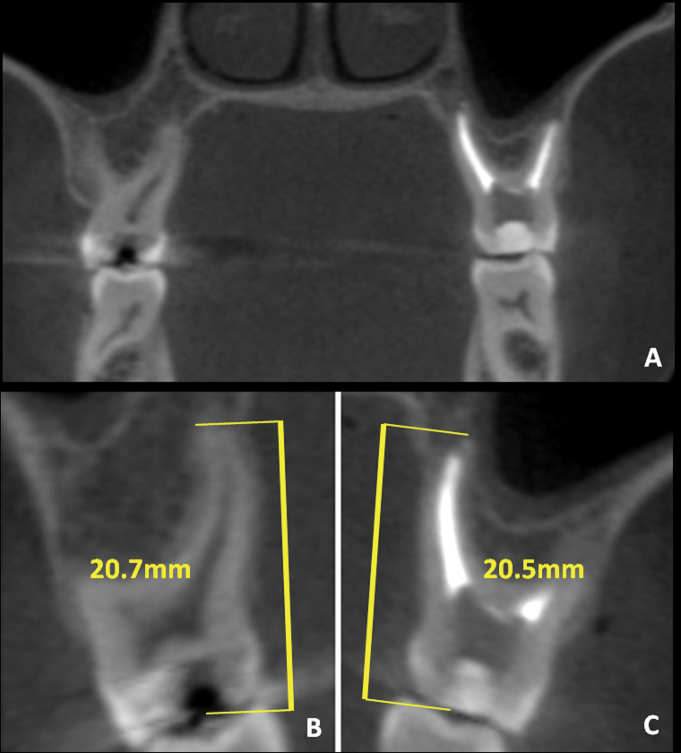
Root-filled teeth were measured in their maximum long axial length from root apex to corresponding cusp tip. These measurements were done before and after orthodontic treatment using the axial guided navigation method.2 All measurements were performed by a single calibrated orthodontist with the aid of specific tomograph software (version 3.1.62 Xoran, Xoran Technologies). Measurements were obtained in tenths of millimeters, and the data were recorded using Microsoft Office Excel, version 2007. The vital contralateral teeth (homologs) were used as a control group (Figure 1).
Figure 1.
Coronal view of contralateral upper molars (A). Vital (B) and filled (C) tooth measurements.
Error Analysis
To assess intraexaminer reliability, the total sample was remeasured by the same researcher after a 30-day interval. The random error (S) was calculated according to Dahlberg formula: S = √Σd2/2n, where d is the difference between the two measurements and n is the number of tested scans. Systematic errors were evaluated with the paired t-test at P < .05.24
Statistical Analysis
Data normality was checked using Kolmogorov-Smirnov test after obtaining descriptive statistics. Tooth lengths in the root-filled and vital pulp groups before treatment were compared using the paired t-test. In addition, the mean difference between root lengths of filled and vital teeth, before and after orthodontic treatment, were compared using the paired t-test. Statistical analysis was performed using two-tailed distribution at P < .05 using the BioEstat statistical software (version 5.0, Mamirauá Maintainable Development Institute, Belém, PA, Brazil).
RESULTS
Random and systematic errors in the first and repeated measurements are presented in Table 1. The random errors ranged from 0.2795 mm (vital-root group, T2) to 0.3557 mm (vital-root group, T1), which are within the acceptable range. There was a systematic error only in the root-filled group (T2), but the mean difference between the measurements was 0.19 mm.
Table 1.
Random and Systematic Errors Between the First and Second Tooth Measurement
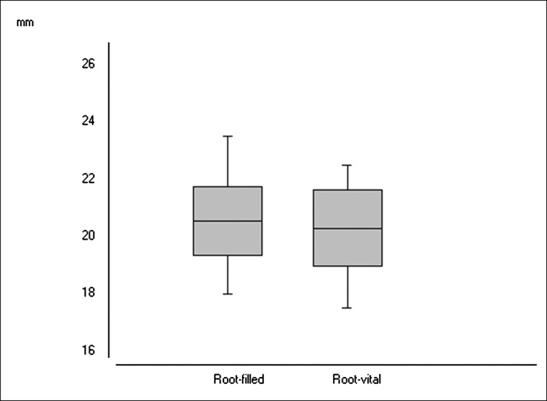
The sample was composed of 20 root-filled teeth from six adolescents (two boys and four girls; mean [SD] age 12.8 [1.8] years, range 11 to 15 years) originally selected after analyzing 1256 roots from 30 orthodontic patients. The teeth selected were first upper premolars (n = 1), second upper premolars (n = 1), first upper molars (n = 4), and first lower molars (n = 2). These root-filled teeth were similar in root length to their contralateral vital teeth before orthodontic treatment (P = .4364; Figure 2). Root-filled incisors were not included in the sample because of the exclusion criteria in relation to dental trauma.
Figure 2.
Comparison of tooth length in filled and vital groups before treatment (P = .4364, paired t-test).
The degree of root shortening after orthodontic treatment was minimal, with no statistical difference between root-filled (P = .0552) and vital teeth (P = .3178; Table 2). The orthodontic treatment response (mean difference in root length before and after treatment) induced a root shortening of 0.30 mm and 0.16 mm, respectively, for filled and vital teeth, without any statistical difference between them (P = .4197; Table 3).
Table 2.
Comparison of Tooth Length Before (T1) and After (T2) Orthodontic Treatmenta
Table 3.
Orthodontic Treatment Response (Mean Difference in Tooth Length Before and After Treatment)a
A post hoc power analysis was carried out by using G*Power software (version 3.1.3).25 The comparison of treatment response with t-test had a power of .4576 (alpha .05; effect size .19).
DISCUSSION
This study showed that after evaluation by CBCT root-filled and vital teeth induced ARR to a similar degree in the wake of orthodontic treatment. The sample was composed of posterior teeth in a split-mouth design study with no difference in root length before treatment, which presupposes no presence of external ARR prior to treatment. The selection criteria excluded teeth with any previous history of trauma, ankylosis, or presence of periapical lesions. Thus, the influence of these confounding variables was controlled, and the isolated effect of endodontic treatment was also considered. Furthermore, no previous study used CBCT methodology for the analysis of the issue in question.
CBCT is a reliable diagnostic tool that provides a more accurate image of root resorption, including posterior teeth measurements,2 than conventional radiographs.23 For this reason, CBCT scans have been recommended to identify different types of resorption along the root surface.26
In our study, the tooth measurements were carried out using a dynamic method, called axial guided navigation.2 The teeth were measured in their maximum long axial length from root apex to its corresponding cusp tip. It is expected that the cusp tip would be preserved after orthodontic treatment and that no significant occlusal abrasion would occur during the orthodontic treatment interval between T1 and T2. Another study18 based the most occlusal landmark on the cementoenamel junction. However, this anatomic structure varies throughout the cervical region; and the axial guided navigation method based on this landmark could provide different sizes of root length.
The research showed that the level of ARR was mild and that there was no statistical significant difference in root lengths before or after orthodontic treatment between root-filled and vital teeth. This contrasts with results of studies by Spurrier et al.15 and Esteves et al.,17 who found significant differences in the occurrence of ARR due to orthodontic treatment. Greater root resorption is normally expected to occur in incisors after orthodontic treatment,1,18 but the teeth we analyzed were premolars and molars, which demonstrate less sensitivity to ARR. Although the mean resorption in root-filled teeth (−0.30 mm) was almost twice that of the vital teeth (−0.16 mm), there was no statistical difference between them. In conclusion, the shortening of both filled and vital teeth was similar and mild, and could be considered of no clinical significance.
The majority of earlier studies corroborate the findings that root-filled teeth do not increase, or may even reduce resorption as a result of orthodontic treatment.21,22 Paradoxically, the inclusion of incisors in clinical studies raises the following question: is the amount of ARR in root-filled teeth following orthodontic treatment due to traumatic injury or to the endodontic treatment itself? It is known that trauma can be subclinical and is not always remembered or mentioned by patients or parents, and this can enhance the bias. It has also been suggested that orthodontic treatment can increase ARR in traumatized teeth, which have previously shown root resorption.5
It is also certain that split-mouth design studies do not guarantee similar conditions in relation to orthodontic tooth movement. There can be differences between the positions of the contralateral teeth themselves, and this requires asymmetric orthodontic biomechanics, which would contribute to the variation in the level of ARR.
The low occurrence of a need for endodontic treatment found in our original sample can be explained by the fluoridated public water supply and the tender age of the patients. Furthermore, the rigid criteria used in sample selection resulted in a small sample size with a power of .4576 (alpha .05; effect size .19). This heightens the risk of a type II error, so a larger sample would be recommended to confirm these results. On the other hand, a small sample size was used for ethical reasons, as a higher radiation dose is questionable in routine orthodontic treatment. However, an extremely homogeneous sample was obtained, which decreased the bias.
Our results support the finding that orthodontic force can be applied both to filled and vital teeth in a similar way. It can be hypothesized that orthodontic tooth movement occurs using the same biological mechanisms in endodontically treated or vital teeth, except when a concomitant ankylosis is present in the periodontal ligament.27 Based on this result, root-filled teeth will not demand more time between adjustments or modification in the biomechanical orthodontic planning unless earlier ARR is detected as a result of trauma, the ectopical eruption of an adjacent tooth, a persistent periapical lesion, or unknown factors.5–8
CONCLUSIONS
Root-filled posterior teeth do not seem to increase ARR after orthodontic treatment.
No change in orthodontic planning is required to move root-filled teeth unless earlier ARR, attributed to other etiologic factors, is detected.
REFERENCES
- 1.Levander E, Malmgren O. Evaluation of the risk of root resorption during orthodontic treatment: a study of upper incisors. Eur J Orthod. 1988;10:30–38. doi: 10.1093/ejo/10.1.30. [DOI] [PubMed] [Google Scholar]
- 2.Castro IO, Alencar AHG, Valladares-Neto J, Estrela C. Apical root resorption due to orthodontic treatment detected by cone beam computed tomography. Angle Orthod. 2013;83:196–203. doi: 10.2319/032112-240.1. [DOI] [PMC free article] [PubMed] [Google Scholar]
- 3.Brudvik P, Rygh P. Multi-nucleated cells remove the main hyalinized tissue and start resorption of adjacent root surfaces. Eur J Orthod. 1994;16:265–273. doi: 10.1093/ejo/16.4.265. [DOI] [PubMed] [Google Scholar]
- 4.Sameshima GT, Sinclair PM. Predicting and preventing root resorption: Part II. Treatment factors. Am J Orthod Dentofacial Orthop. 2001;119:511–515. doi: 10.1067/mod.2001.113410. [DOI] [PubMed] [Google Scholar]
- 5.Malmgren O, Goldson L, Hill C, Orwin A, Petrini L, Lundberg M. Root resorption after orthodontic treatment of traumatized teeth. Am J Orthod Dentofacial Orthop. 1982;82:487–491. doi: 10.1016/0002-9416(82)90317-7. [DOI] [PubMed] [Google Scholar]
- 6.Ericson S, Kurol PJ. Resorption of incisors after ectopic eruption of maxillary canines: a CT study. Angle Orthod. 2000;70:415–423. doi: 10.1043/0003-3219(2000)070<0415:ROIAEE>2.0.CO;2. [DOI] [PubMed] [Google Scholar]
- 7.Felippe WT, Ruschel MF, Felippe GS, Pozzobon MH, Felippe MC. SEM evaluation of the apical external root surface of teeth with chronic periapical lesion. Aust Endod J. 2009;35:153–157. doi: 10.1111/j.1747-4477.2009.00165.x. [DOI] [PubMed] [Google Scholar]
- 8.Brezniak N, Wasserstein A. Root resorption after orthodontic treatment: Part 2. Literature review. Am J Orthod Dentofacial Orthop. 1993;103:138–146. doi: 10.1016/S0889-5406(05)81763-9. [DOI] [PubMed] [Google Scholar]
- 9.Sameshima GT, Sinclair PM. Predicting and preventing root resorption: Part I. Diagnostic factors. Am J Orthod Dentofacial Orthop. 2001;119:505–510. doi: 10.1067/mod.2001.113409. [DOI] [PubMed] [Google Scholar]
- 10.Al-Qawasmi RA, Hartsfield JK, Jr, Everett ET, et al. Genetic predisposition to external apical root resorption. Am J Orthod Dentofacial Orthop. 2003;123:242–252. doi: 10.1067/mod.2003.42. [DOI] [PubMed] [Google Scholar]
- 11.Huettner RJ, Young RW. The movability of vital and devitalized teeth in the Macacus rhesus monkey. Oral Surg Oral Med Oral Pathol. 1955;8:189–197. doi: 10.1016/0030-4220(55)90192-4. [DOI] [PubMed] [Google Scholar]
- 12.Mattison GD, Delivanis HP, Delivanis PD, Johns PI. Orthodontic root resorption of vital and endodontically treated teeth. J Endod. 1984;10:354–358. doi: 10.1016/S0099-2399(84)80154-5. [DOI] [PubMed] [Google Scholar]
- 13.Mah R, Holland GR, Pehowich E. Periapical changes after orthodontic movement of root-filled ferret canines. J Endod. 1996;22:298–303. doi: 10.1016/S0099-2399(96)80263-9. [DOI] [PubMed] [Google Scholar]
- 14.Satoh I. Root resorption of vital and endodontically treated teeth in orthodontic movement [in Japanese] Kanagawa Shigaku. 1990;24:601–617. [PubMed] [Google Scholar]
- 15.Spurrier SW, Hall SH, Joondeph DR, Shapiro PA, Riedel RA. A comparison of apical root resorption during orthodontic treatment in endodontically treated and vital teeth. Am J Orthod Dentofacial Orthop. 1990;97:130–134. doi: 10.1016/0889-5406(90)70086-R. [DOI] [PubMed] [Google Scholar]
- 16.Mirabella AD, Årtun J. Prevalence and severity of apical root resorption of maxillary anterior teeth in adult orthodontic patients. Eur J Orthod. 1995;17:93–99. doi: 10.1093/ejo/17.2.93. [DOI] [PubMed] [Google Scholar]
- 17.Esteves T, Ramos AL, Pereira CM, Hidalgo MM. Orthodontic root resorption of endodontically treated teeth. J Endod. 2007;33:119–122. doi: 10.1016/j.joen.2006.09.007. [DOI] [PubMed] [Google Scholar]
- 18.Llamas-Carreras JM, Amarilla A, Solano E, Velasco-Ortega E, Rodríguez-Varo L, Segura-Egea JJ. Study of external root resorption during orthodontic treatment in root filled teeth compared with their contralateral teeth with vital pulps. Int Endod J. 2010;43:654–662. doi: 10.1111/j.1365-2591.2010.01722.x. [DOI] [PubMed] [Google Scholar]
- 19.Llamas-Carreras JM, Amarilla A, Espinar-Escalona E, et al. External apical root resorption in maxillary root-filled incisors after orthodontic treatment: a split-mouth design study. Med Oral Patol Oral Cir Bucal. 2012;17:e523–e527. doi: 10.4317/medoral.17586. [DOI] [PMC free article] [PubMed] [Google Scholar]
- 20.Wickwire NA, McNeil MH, Norton LA, Duell RC. The effects of tooth movement upon endodontically treated teeth. Angle Orthod. 1974;44:235–242. doi: 10.1043/0003-3219(1974)044<0235:TEOTMU>2.0.CO;2. [DOI] [PubMed] [Google Scholar]
- 21.Ioannidou-Marathiotou I, Zafeiriadis AA, Papadopoulos MA. Root resorption of endodontically treated teeth following orthodontic treatment: a meta-analysis. Clin Oral Investig. 2013;17:1733–1744. doi: 10.1007/s00784-012-0860-8. [DOI] [PubMed] [Google Scholar]
- 22.Walker SL, Tieu LD, Flores-Mir C. Radiographic comparison of the extent of orthodontically induced external apical root resorption in vital and root-filled teeth: a systematic review. Eur J Orthod. 2013;35:796–802. doi: 10.1093/ejo/cjs101. [DOI] [PubMed] [Google Scholar]
- 23.Ren H, Chen J, Deng F, Zheng L, Liu X, Dong Y. Comparison of cone-beam computed tomography and periapical radiography for detecting simulated apical root resorption. Angle Orthod. 2013;83:189–195. doi: 10.2319/050512-372.1. [DOI] [PMC free article] [PubMed] [Google Scholar]
- 24.Houston WJ. The analysis of errors in orthodontic measurements. Am J Orthod. 1983;83:382–390. doi: 10.1016/0002-9416(83)90322-6. [DOI] [PubMed] [Google Scholar]
- 25.Faul F, Erdfelder E, Lang AG, Buchner A. G*Power 3: a flexible statistical power analysis program for the social, behavioral, and biomedical sciences. Behav Res Methods. 2007;39:175–191. doi: 10.3758/bf03193146. [DOI] [PubMed] [Google Scholar]
- 26.Estrela C, Bueno MR, Alencar AHG, et al. Method to evaluate inflammatory root resorption by using cone beam computed tomography. J Endod. 2009;35:1491–1497. doi: 10.1016/j.joen.2009.08.009. [DOI] [PubMed] [Google Scholar]
- 27.Kim Y, Park S, Son W, Kim S, Kim Y, Mah J. Treatment of an ankylosed maxillary incisor by intraoral alveolar bone distraction osteogenesis. Am J Orthod Dentofacial Orthop. 2010;138:215–220. doi: 10.1016/j.ajodo.2008.07.024. [DOI] [PubMed] [Google Scholar]


