



Case description
A 79-year-old Caucasian man and his wife were hospitalized in a peripheral hospital because of nausea, vomiting, watery diarrhea, and oliguria. Symptoms appeared 6 h after ingestion of about 200 g of cooked mushrooms (mistaken by the patient for Amanita caesarea and Boletus impolitus), foraged days before. In the emergency room, the standard protocol for mushroom poisoning was performed (gastric lavage, intravenous acetylcysteine: 150 mg/kg in 60 min, and electrolyte replacement). Twenty-four hours after admission, he showed progressive decline of urine output and an abrupt increase of transaminases. He was therefore referred to our Division the following day, while his wife was discharged after 72 h of clinical observation. His past medical history showed type 2 diabetes with microvascular complications (diabetic retinopathy stage II), and hypertension being treated with angiotensin converting enzyme (ACE) inhibitors.
At admission, his vital parameters were normal (blood pressure [BP] 130/90 mmHg, heart rate [HR] 82/m, body temperature 36.5 °C). Serum creatinine was 7.6 mg/dl, blood urea nitrogen (BUN) 160 mg/dl, with signs of acute liver and pancreas injury (aspartate aminotransferase [AST] 509 U/ml, alanine aminotransferase [ALT] 1,013 U/ml, total bilirubin 2.4 mg/dl, amylase 566 U/l, lipase 1402 U/l, pancreatic amylase 525 U/l). Urinalysis showed normal specific gravity (1.012), glycosuria (50 mg/dl), and mild proteinuria (50 mg/dl), while urinary sediment displayed several granular casts with mild hematuria (16 red cells/HPF). Urinary amatoxin (alpha amanitin) was positive, confirming the diagnosis; interestingly, the toxin was still detected in the urine as late as 14 days after poisoning. Renal ultrasound showed kidneys of normal size, with hyperechoic parenchyma.
Intravenous acetylcysteine was continued until his liver parameters improved; dialysis was started with sustained low-efficiency daily diafiltration (SLEDD-f) in an attempt to reduce amatoxin concentration. In view of the lack of kidney function recovery, the patient was later switched to standard hemodialysis. To explore the causes of this persistent kidney failure, we performed a kidney biopsy that showed severe, diffuse, acute tubular necrosis, consistent with the clinical suspicion of amatoxin toxicity superimposed on diabetic nephropathy (Figs. 1 and 2).
Fig. 1.
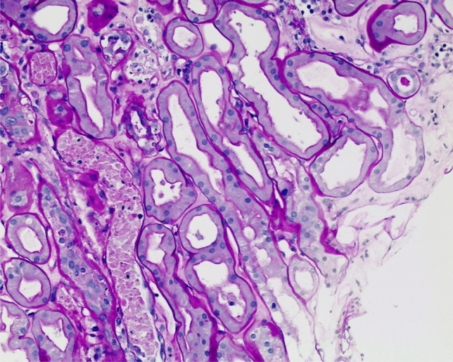
Acute tubular necrosis with granular casts containing nuclear fragments and eosinophilic cytoplasmic debris derived from necrotic tubular epithelial cells (PAS, 100×)
Fig. 2.

Marked global expansion of the mesangial matrix and cellularity with segmental capillary loop adhesions to Bowman’s capsule. (PAS, 200×)
The patient was discharged on chronic hemodialysis and five years later he is still undergoing dialysis with no signs of chronic liver disease.
Lesson for the clinical nephrologist
Amanita phalloides (also called “deathcap”) is a basidiomycete fungus of the genus Amanita known by foragers and clinicians to be a potential cause of fatal acute liver injury. Among food poisoning cases in the US, Amanita phalloides accounts for more than 90% of fatalities, with an estimated mortality of 50% in adults and 33% in children in a dated series [1]. In the absence of up-to-date, high quality evidence, it is legitimate to assume that survival has considerably improved thanks to progress in supportive care and liver transplant [2, 3].
Amanita phalloides produces two types of toxins that cause the phalloideal syndrome: amatoxins and phallotoxins. In humans, phallotoxins exert a direct influence on the cytoskeleton, thus causing a mild and transient enteric syndrome within 6–24 h from ingestion, while amatoxins (in particular alpha amanitin) tightly inhibit RNA polymerase II in the hepatocytes. The impairment of synthetic activity and the loss of structural proteins cause hepatocyte necrosis, which manifests as severe, often fatal, acute liver failure. Kidney failure and anuria are initially the result of dehydration but subsequently kidney damage may be the expression of direct alpha amanitin toxicity to the proximal tubular cells.
As a general rule, once the liver injury is resolved, the renal tubular epithelium progressively regenerates, leading to complete or partial recovery of kidney function [3, 4]. End-stage kidney disease with the need for definitive renal replacement therapy has been described [5]; however, in this condition, renal histology has seldom been reported [6, 7].
Why this patient developed end-stage kidney disease is unclear. Our patient had additional risk factors: type 2 diabetes, hypertension, and a previously known chronic kidney disease. Indeed, although not quantitated, we observed traces of alpha amanitin in the urine 14 days after ingestion, suggesting a relevant toxin load; whether the loss of kidney function is dose-related or independent is not known. It is also reasonable to assume that age, which increases the risk of developing comorbidities, has an impact on the renal and overall outcomes.
In animal models of poisoning by Amanita phalloides, the histological pattern is that of acute tubular necrosis and reactive interstitial nephritis [8]. In our patient, interstitial inflammation was mild, with focal tubulitis and mild interstitial edema, and there were foci of regenerating tubular epithelium, which led us to assume there would be prompt recovery. We were unable to precisely determine the prognosis because (1) how to determine the extent to which tissue has undergone irreversible functional injury after severe acute tubular toxicity is not known; (2) the prognostic value of tissue regeneration on kidney histology is misleading; (3) we do not know what the real proliferating capacity of tubular cells with chronic kidney disease in the background is.
Despite all the measures undertaken to reduce the toxic load as per the current literature data (Table 1), and taking into consideration the pre-existing CKD, prolonged toxicity may explain the dissociation between liver recovery and kidney failure.
Table 1.
Current literature data
| Category | Intervention | Rationale | Dose (or target) | Evidence |
|---|---|---|---|---|
| Prompt consultation with a regional poison center | Provide the best-known clinical approach in rare toxic syndromes | High | ||
| Supportive care | Volume repletion; electrolyte correction; bicarbonate repletion | Avoid hypovolemic shock and reduce the risk of tubular necrosis | Personalized | High |
| Gastrointestinal decontamination | Gastric Lavage | Remove the contaminated meal and the amatoxin concentration | Gastric emptying, synergic with activated charcoal | High |
| Multiple-dose activated charcoal | Absorb amatoxin in the enteric tract to reduce amatoxin concentration | 50 g every 4 h or 25 g every 2 h. Some use it days after the meal to interrupt the enterohepatic reabsorption of amatoxin | High | |
| Enhance clearance | Biliary drainage | Interrupt the enterohepatic reabsorption of amatoxin (observed after liver transplant following amatoxin exposure) | Rapid decrease of concentrations after a mean of 4 days from the procedure | Low |
| Therapeutic plasma exchange | Removes amatoxins and support the insufficient synthetic activity of the liver | Useful when significant concentrations of amatoxin are present (first 36–48 h). Can be repeated up to 84 h following the meal | Low | |
| Intermittent and continuous hemodialysis | Useful for removing amatoxins and replacing renal function when needed | Useful when significant concentrations of amatoxin are present (first 36–48 h), otherwise use it if indicated as renal replacement therapy | Low | |
| Molecular absorbent recirculating system (MARS) | May remove protein-bound substances and water-soluble toxins | Use it as a bridge for liver transplantation | Low | |
| Fractionated plasma separation and adsorption system (FPSA) | May remove protein-bound substances and water-soluble toxins | Use it as a bridge for liver transplantation | Low | |
| Amatoxin uptake inhibitors | Silibinin dihemisuccinate (intravenous) | Strong inhibitor of Organic Anion Transporting Polypeptide 1B3 (OATP1B3) transporter. Blocks α-amanitin uptake in hepatocytes | Effective when given within 24 h of ingestion. 5 mg/kg intravenously followed by a continuous dose of 20 mg/kg per day for 6 days or until recovery | High |
| Silymarin (oral) | Strong inhibitor of Organic Anion Transporting Polypeptide 1B3 (OATP1B3) transporter. Blocks α-amanitin uptake in hepatocytes | Start with 50–100 mg/kg (max 2 g) of oral silymarin every 8 h, then increase to a maximum of 200 mg/kg per dose (maximum single dose; 3 g) if tolerated, for 6 days | Low | |
| Benzylpenicillin | Strong inhibitor of Organic Anion Transporting Polypeptide 1B3 (OATP1B3) transporter. Blocks α-amanitin uptake in hepatocytes | May be used within 36 h after mushroom ingestion. Suggested 1 MU/kg/day and 0.5 MU/kg/day | High | |
| Antioxidant therapy | N-acetylcysteine | Inactivate free radicals and support glutathione depletion | 150 mg/kg intravenously over 15 min followed by 50 mg/kg over 4 h followed by 100 mg/kg over 16 h | Low |
| Cimetidine and vitamin C | Antioxidant effects in animal models of amatoxin-containing mushroom poisoning | Cimetidine: 300 mg i.v. every 8 h until clinical improvement; Vitamin C: 3 g i.v. daily until clinical improvement | Low | |
| Experimental therapies | Polymyxin B | Bind RNA Polymerase II preventing α-amanitin from binding | 1 mg/kg/day, 4, 8, and 12 h after the meal | Low |
| Amatoxin uptake inhibitors | Significantly inhibit amatoxin uptake into liver cells | Not known | Low | |
| Aucubin | Antioxidant activity in animal models of alpha-amanitin toxicity | Not known | Low | |
| Liver transplantation | Liver replacement therapy | Clinical signs of hepatic injury are moderate to severe | High | |
AKI acute kidney injury
Our case points out the importance of considering mushroom poisoning as a potential cause of acute liver and kidney failure and highlights the fact that, especially in subjects with a pre-existing kidney disease (hypertension, diabetes), renal failure may be irreversible.
Acknowledgements
Andrea Angioi, Matteo Floris and Nicola Lepori contributed equally to the preparation of this paper
Compliance with ethical standard
Conflict of interest
On behalf of all authors, the corresponding author states that there is no conflict of interest.
Ethical approval
This article does not contain any studies with human participants performed by any of the authors.
Footnotes
Publisher's Note
Springer Nature remains neutral with regard to jurisdictional claims in published maps and institutional affiliations.
References
- 1.Jaeger A, Jehl F, Flesch F, Sauder P, Kopferschmitt J. Kinetics of amatoxins in human poisoning: therapeutic implications. J Toxicol Clin Toxicol. 1993;31(1):63–80. doi: 10.3109/15563659309000374. [DOI] [PubMed] [Google Scholar]
- 2.Pond SM, Olson KR, Woo OF, Osterloh JD, Ward RE, Kaufman DA, Moody RR. Amatoxin poisoning in northern California, 1982–1983. West J Med. 1986;145(2):204–209. [PMC free article] [PubMed] [Google Scholar]
- 3.Krenova M, Pelclova D, Navratil T. Survey of Amanita phalloides poisoning: clinical findings and follow-up evaluation. Hum Exp Toxicol. 2007;26(12):955–961. doi: 10.1177/0960327107085832. [DOI] [PubMed] [Google Scholar]
- 4.Leathem AM, Purssell RA, Chan VR, Kroeger PD. Renal failure caused by mushroom poisoning. J Toxicol Clin Toxicol. 1997;35(1):67–75. doi: 10.3109/15563659709001168. [DOI] [PubMed] [Google Scholar]
- 5.Unverir P, Soner BC, Dedeoglu E, Karcioglu O, Boztok K, Tuncok Y. Renal and hepatic injury with elevated cardiac enzymes in Amanita phalloides poisoning: a case report. Hum Exp Toxicol. 2007;26(9):757–761. doi: 10.1177/0960327107083972. [DOI] [PubMed] [Google Scholar]
- 6.Fineschi V, Di Paolo M, Centini F. Histological criteria for diagnosis of amanita phalloides poisoning. J Forensic Sci. 1996;41(3):429–432. doi: 10.1520/JFS13929J. [DOI] [PubMed] [Google Scholar]
- 7.Garrouste C, Hemery M, Boudat AM, Kamar N. Amanita phalloides poisoning-induced end-stage renal failure. Clin Nephrol. 2009;71(5):571–574. doi: 10.5414/CNP71571. [DOI] [PubMed] [Google Scholar]
- 8.Garcia J, Costa VM, Carvalho AT, Silvestre R, Duarte JA, Dourado DF, Arbo MD, Baltazar T, Dinis-Oliveira RJ, Baptista P, et al. A breakthrough on Amanita phalloides poisoning: an effective antidotal effect by polymyxin B. Arch Toxicol. 2015;89(12):2305–2323. doi: 10.1007/s00204-015-1582-x. [DOI] [PubMed] [Google Scholar]