Abstract
Objective:
To compare the periodontal health of maxillary and mandibular anterior teeth retained with two types of fixed retainers.
Materials and Methods:
A fixed straight retainer (SR) group had 39 subjects, and a fixed wave retainer (WR) group had 35 subjects. Subjects were between the ages of 13 and 22 years and had been in fixed retention for 2 to 4 years. Pocket probing depths, bleeding on probing, plaque index, calculus index, recession, and gingival crevicular fluid volume were compared between the two retainer groups. A four-question oral hygiene survey was given to each subject. The Mann-Whitney U-test and Fisher exact test was used to analyze the data.
Results:
There was no clinically significant difference between the retainer groups regarding plaque index, gingival crevicular fluid volume, calculus index, recession, bleeding on probing, and pocket probing depths. A statistically significant increase in the reported frequency of flossing (P = .006) and ease of flossing (P < .001) was associated with the WR group. There was no significant difference between the groups in reported frequency of brushing and comfort of the retainer.
Conclusions:
Under the conditions of this study, no clinical difference was found in the periodontal health of anterior teeth retained with a SR or WR for a period of 2 to 4 years. Subjects in the WR group reported an increase in frequency and ease of flossing.
Keywords: Fixed retainer, Periodontal health, Wave retainer
INTRODUCTION
The use of fixed retention in orthodontic practice has been increasing. In 2002, a survey found that one third of orthodontic practitioners used mandibular fixed retainers, and 5% used maxillary fixed retainers.1 By 2011, those numbers had increased to 42% in the mandibular arch, and 11% in the maxillary arch.2 The expanding use of fixed retainers has raised concerns among practitioners about the potential increase in dental caries or decline in periodontal health.
No association between dental caries and fixed retention has been observed, despite greater plaque accumulations along the wires.3,4 While it seems clear that dental caries are not related to the presence of a fixed straight retainer, the presence of a fixed retainer may cause periodontal decline. For example, Levin et al.5 found that fixed retainers have been associated with increased gingival recession, plaque retention, and bleeding on probing.
It has also been suggested that fixed retainers have some influence on other aspects of periodontal health.6 Pandis et al.7 found that long-term fixed retainer wear causes greater calculus accumulations, marginal recession, and increased probing depths. All of these are likely associated with long-term irritation of the tissue induced by the fixed retainer (or by bacteria around the fixed retainer).7 It also seems that plaque and calculus accumulation is more related to the length of time the bonded retainer is in place, than to the type or size of wire.3 Interproximal and areas gingival to the wire have been shown to accumulate deposits of plaque and calculus. This is probably because as the wire crosses the interdental region, it creates an area that is difficult to clean.1,7,8
Other studies have shown no apparent damage to hard tissues, including bone levels, even though there was some evidence for soft tissue effects.7,9 Booth et al.10 found that long-term retention of mandibular incisors with fixed retention appears to be compatible with periodontal health.10 A more recent study by Rody et al.9 found that the clinical periodontal health of subjects was not affected by bonded lingual retainers despite increased plaque accumulations in the lower incisor region.9
While the periodontal effects of long-term fixed retainer wear are not clearly understood, there is general agreement that fixed retainers make oral hygiene procedures more difficult.4 When bonded retainers are placed, patients must be educated on maintenance that includes some form of interdental cleaning aid.8 This added cleaning process complicates oral hygiene and suggests that a patient's motivation level should be an important factor in deciding whether or not to place a fixed retainer.9 Bonding to each tooth may also restrict access of the toothbrush to interdental areas, may limit the ability of floss to slide freely from canine to canine, and may lead to an overall decline in maintenance and compliance.4
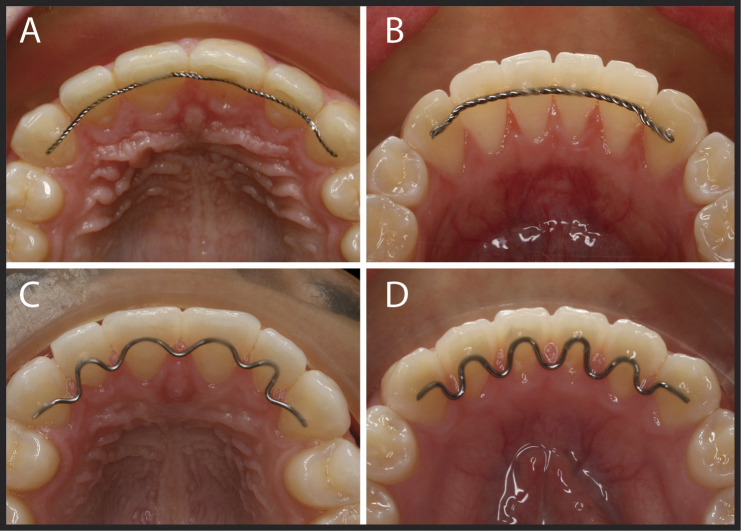
Most fixed retainers are made of a straight, single stranded, or braided stainless steel wire intimately adapted to the lingual surface of the teeth and placed at or slightly above the cingulum (Figure 1).11 The V-Loop retainer4 and the more recently modified “wave” retainer are fixed retainers in which the wire is scalloped toward the soft tissues around the retained teeth in order to make oral hygiene less complicated for patients (Figure 1). The position of the lower loop of the retainer is just slightly above the lingual interdental papilla to allow for normal flossing technique to be used during routine oral hygiene. The wave retainer was designed with the idea that flossing would be easier, and this might lead to improved periodontal health.4
Figure 1.
Fixed bonded retainers. (A) Maxillary straight retainer. (B) Mandibular straight retainer. (C) Maxillary wave retainer. (D) Mandibular wave retainer.
Since the use of fixed bonded retainers is increasing in orthodontic practice, and because the wave retainer may confer periodontal health benefits, this study was designed to compare the periodontal health of anterior teeth retained with fixed straight retainers with the periodontal health of anterior teeth retained with fixed wave retainers. The null hypothesis was that there is no significant difference in periodontal health in anterior teeth retained with the two types of fixed retainers.
MATERIALS AND METHODS
This research project was an observational cross-sectional study that was reviewed and approved by the Institutional Review Board at Loma Linda University (OSR 5120106). The study sample included 35 subjects with a straight twisted wire retainer (SR) and 39 subjects with a wave-type retainer (WR) (sometimes called the V-loop retainer). All subjects in this study were selected from a single orthodontic practice. SR and WR groups were available in the single practice because the practitioner changed his fixed retention strategy from only SR to predominately WR. The study included the collection of data commonly recorded during routine dental prophylaxis appointments, an intraoral photograph of the maxillary and mandibular anterior teeth, and a brief survey of oral hygiene habits. The periodontal data on SR was collected on maxillary anterior teeth retained with a 0.546-mm (0.0215-inch) Tri-Flex stainless steel twisted three-strand orthodontic wire (RMO, Denver, Colo), and mandibular anterior teeth retained with a 0.8-mm twisted stainless steel wire (3M Unitek, Monrovia, Calif) (Figure 1). The WR periodontal data were collected on maxillary and mandibular anterior teeth retained with a 0.569-mm (0.022-inch) Blue Elgiloy (soft) round wire (RMO) (Figure 1).
Male and female patients between the ages of 13 and 22 years and in postorthodontic treatment with continuous fixed retention lasting between 24 and 48 months were included in this study. A list of potential subjects who had been debanded within the time period of interest and met the inclusion criteria were consecutively called until the sample size for both groups had been met.
Exclusion criteria included: (1) a professional dental cleaning within the last 4 months, (2) a history of diabetes, (3) a habit of smoking, (4) preexisting periodontal disease, (5) postorthodontic periodontal disease, (6) antibiotic prophylaxis prior to periodontal data collection, (7) current use of antibiotics, and (8) pregnancy.
The FDI World Dental Federation notation system was used to identify teeth. A dental hygiene survey was collected on each subject. The six measures of periodontal health included in the study are shown below.
The Löe plaque index (PI). The following scores were used for plaque accumulation measurements: 0, no plaque in the gingival area; 1, no plaque visible by the unaided eye, but plaque is made visible on the point of the probe after it has been moved across surface at entrance of the gingival crevice; 2, gingival area is covered with a thin to moderately thick layer of plaque and deposit is visible to the naked eye; and 3, heavy accumulation of soft matter, the thickness of which fills out niche produced by the gingival margin and tooth surface, and interdental area is stuffed with soft debris.12
Gingival crevicular fluid volume (GCFV), which was measured with the Periotron 8000 (Oraflow, Smithtown, NY). PerioPaper (Oraflow) gingival fluid collection strips were used for instrument calibration and crevicular fluid collection from each subject. A calibration curve was constructed using known volumes of distilled water at 0.25 µL, 0.50 µL, 0.75 µL, 1.00 µL, and 1.25 µL dispensed with a fixed-volume pipette. The computer software on which the analysis was completed was the Periotron Professional (v3.0a) (Oraflow). Plaque and/or calculus accumulations that interfered with the collection of crevicular fluid were removed before each sample was collected. Four sites were chosen for fluid collection: the direct facial and lingual sulcus of an upper and lower right central incisor. Each site was gently air dried for approximately 5 seconds and isolated from saliva with cotton rolls as necessary. Two strips of PerioPaper were individually inserted into the gingival sulcus for 5 seconds with 30 seconds between samplings. Two samples per site were taken for a total of eight samples. Each PerioPaper strip was immediately placed between the counterparts of the Peritron 8000 and the Periotron score recorded. The Periotron score for each collection site was averaged and entered into the Periotron Professional software, from which a volume of fluid was determined by the Periotron computer program using interpolation from the standard curve developed from the instrument calibration.
The Greene and Vermillion calculus index (CI). The associate scale of 0–3 included: 0, no calculus; 1, supragingival calculus covering not more than one third of the tooth surface; 2, supragingival calculus covering between one third and two thirds of the tooth surface or scattered subgingival calculus; and 3, supragingival calculus covering more than two thirds of the tooth surface or a continuous ring of subgingival calculus.13
Gingival pocket probing depths (PPD), which were measured with a standard periodontal probe with 2-mm increments. Sulcular pocket depths were measured at six locations around each study tooth: mesial buccal (MB), direct facial (F), distal buccal (DB), distal lingual (DL), direct lingual (L), and mesial lingual (ML). The PPD was recorded to the nearest millimeter for each site and entered into the research record.
Gingival recession (REC), which was recorded to the nearest millimeter from the cementoenamel junction to the free gingival margin for the direct facial and direct lingual surfaces of each anterior tooth using the same periodontal probe as used for PPD.
Bleeding on probing (BOP) that occurred within 30 seconds of making a PPD measurement anywhere along the gingival sulcus. Data were recorded as a yes (Y) or no (N).
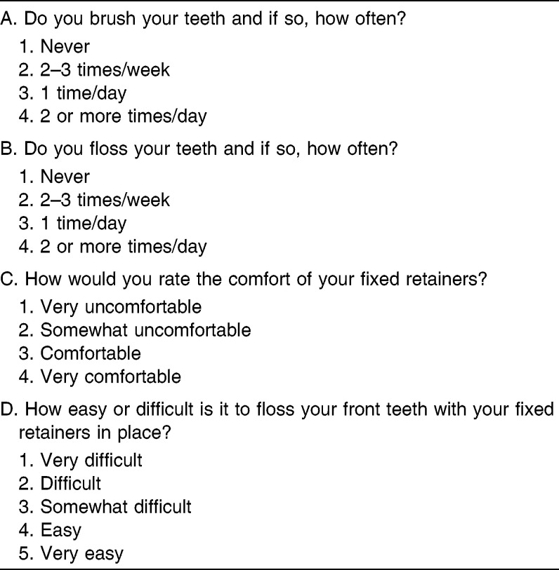
An oral hygiene questionnaire with four questions was given to each subject at the time of the clinical exam that asked for the subject's frequency of brushing, flossing, ease of flossing, and comfort of retainers (Table 1).
Table 1.
Oral Hygiene Questionnaire and Scoring Scales
One examiner collected the research data on all subjects during a 1-week period. The sequence of data collection was PI, GCFV, CI, PPD, BOP, and REC. The examiner (an orthodontic resident) was calibrated with a periodontist prior to the collection of research data (ICC = 0.907). When any pathologic condition was discovered during the data collection, the patient was informed of the finding and referred to the appropriate dental professional for follow-up care.
Statistical Analysis
The Mann-Whitney U-test (MWU) was used to compare the retainer groups with respect to plaque index, gingival crevicular fluid volume, calculus index, pocket probing depths, gingival recession, and responses to the oral hygiene questionnaire between the groups. Fisher exact test for categorical data was used to analyze the bleeding on probing scores.
RESULTS
A summary of demographic data is presented in Table 2. The SR retainer group showed a statistically greater age and retention time than the WR retainer group.
Table 2.
Demographic Data
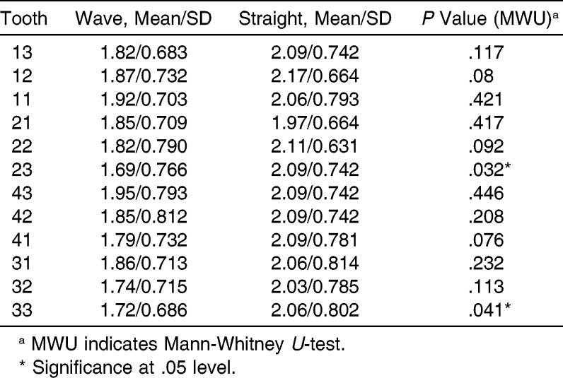
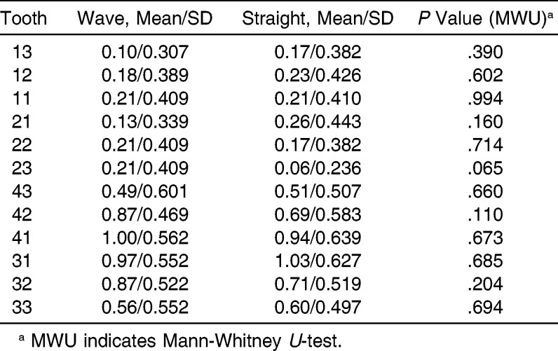
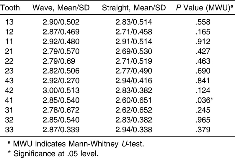
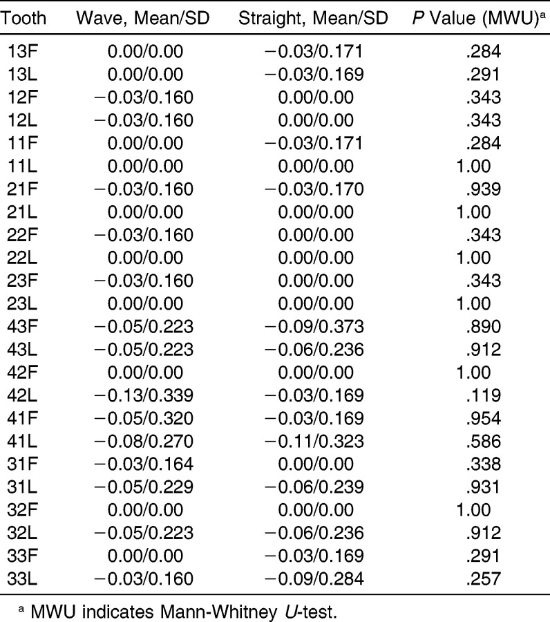
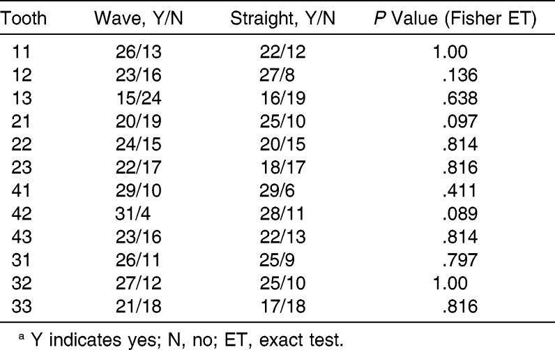
An analysis of the plaque index using the MWU test indicated statistical significance for tooth numbers 23 and 33 with P values of .032 and .041, respectively. The remaining P values ranged from .08 to .446 (Table 3). Analysis of the gingival crevicular fluid volume using the MWU test indicated no significant difference between the two retainer groups. P values ranged from .303 to .914 (Table 4). The MWU test for the calculus index indicated there was no significant difference between the two retainer groups in terms of calculus accumulation. P values ranged from .110 to .994 (Table 5). The MWU test for gingival pocket probing depths indicated a statistically significant difference for tooth number 41 with a P value of .036. The remaining P values ranged from .124 to .965 (Table 6). No significant difference was found for gingival recession between the two groups using the MWU test. P values ranged from .119 to 1.00 (Table 7). Bleeding on probing along the gingival sulcus was recorded as “yes” or “no.” Fisher exact test for categorical data indicated no significant difference between the two groups. P values ranged from .089 to 1.00 (Table 8).
Table 3.
Plaque Index
Table 4.
Gingival Crevicular Fluid Volume (µL)
Table 5.
Calculus Index
Table 6.
Pocket Probing Depth (mm)
Table 7.
Recession (mm)
Table 8.
Bleeding On Probinga
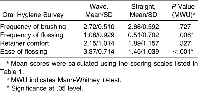
The self-reported oral hygiene survey (Table 1) indicated a signifcant difference in frequency of flossing and ease of flossing, P = .006 and P < .001, respectively, using the MWU test (Table 9). Self-reported retainer comfort and frequency of brushing was found to have no significant difference between the groups. Mann Whitney U-test P values were .327 and .727, respectively (Table 9).
Table 9.
Oral Hygiene Survey Resultsa
DISCUSSION
Although the sample groups differed with respect to age (WR, 16.9 ± 0.96 years; SR, 18.3 ± 1.3 years) and retention time (WR, 31.6 ± 3.2 months; SR, 42.3 ± 2.4 months), the mean and standard deviation of each retention group fit within the inclusion criteria of this study (13–22 years old, 2–4 years in retention). Because of this, the retention groups were considered similar in age and retention time. In addition, the SR group was expected to be older and have longer retention times because the orthodontic practitioner changed his retention strategy from SR to predominately WR.
Many of the periodontal factors considered in this study are well grounded in the periodontal literature. The use of GCFV as a diagnostic tool for assessment of changes in periodontal health has been challenged because unknown systemic or environmental factors may influence GCFV measurements.14 Deinzer et al.14 showed that GCFV measurements made 24 hours apart showed low stability (high variability). Despite this challenge, the preponderance of evidence suggests that GCFV can be used as a proxy for periodontal inflammation.15 This study controlled some systemic and environmental factors by eliminating variables related to diabetes, pregnancy, smoking, antibiotic treatment, and antibiotic prophylaxis.
Three statistically significant findings concerning PI (numbers 22 and 33) and PPD (number 41) are probably not clinically important. In context of the other PI and PPD statistical tests, and considering the low magnitude of the differences, it seems more reasonable to conclude that there were no clinically significant differences in PI and PPD between the two types of fixed retainers. The main indicators of potential periodontal inflammation, the BOP and the GCFV were also similar between the groups. When all of the studied proxies for periodontal condition (PI, GCFV, CI, PPD, BOP, REC) are considered as a group, it is clear that SR and WR are equal in terms of periodontal health parameters after 2 to 4 years of fixed retention.
Oral Hygiene Questionnaire
In order to evaluate oral hygiene experience of subjects in the two retainer groups, each subject was asked to answer a four-question survey (Table 1). Two questions asked about the patient experience with flossing (frequency and ease), one question asked about the comfort of the fixed retainer, and one question asked about brushing frequency.
The WR group reported much higher frequencies of flossing and higher ease of flossing than the SR group (Table 9). This seems like a reasonable result since the WR was designed to make flossing simpler. It is interesting to note that the reported frequency of flossing did not appear to make a significant difference in the periodontal parameters of the teeth bonded to the fixed retainer. Since the frequency of brushing was the same for both groups (Table 9), it is interesting that the increased use of dental floss did not lead to better periodontal health for the WR group.
A possible explanation for this result can be found in two recent systematic reviews that evaluated the importance of flossing. A 2008 review concluded that routine instruction to use floss is not supported by scientific evidence.16 Another review downplayed the importance of flossing but concluded that there was some evidence that flossing in addition to tooth brushing reduces gingivitis (compared to simply brushing alone), and that there was weak, unreliable evidence that flossing plus brushing is associated with a small reduction in plaque at 1 and 3 months.17
Another possible explanation is that the gingival loops of the WR might not be placed gingival enough to allow flossing to the bottom of the gingival sulcus. The WR group may not have been able to floss correctly due to wire interference. If the WR group flossing can be characterized as frequent but ineffective, and the SR group flossing can be characterized as infrequent but effective (using a flossing aid), the two groups might end up with similar periodontal health. Finally, the expected improved outcomes of the WR group as compared to the SR subjects may be difficult to detect due to the inherent weakness of standard clinical measurements in the form of a measurement error.
The WR requires a greater length of wire and its gingival position might reduce the perceived comfort by the patient; however, the subjects in this study were equally comfortable with both types of fixed retainers (Table 9).
CONCLUSIONS
The periodontal parameters of anterior teeth retained with SR or WR are similar after 2 to 4 years of retention in adolescent children and young adults (age 13–22 years). The two retainer types appear to be clinically interchangeable in terms of their effect on the associated periodontal tissues.
The WR enables patients to floss deeper into the interproximal contact area (deeper than the SR) and may reinforce flossing compliance because dental floss is not blocked above the interproximal contact area. Even so, self-reported flossing compliance did not improve the periodontal status of the WR group.
ACKNOWLEDGMENTS
This research was partially funded by the Center for Dental Research, Loma Linda University School of Dentistry. Udochukwu Oyoyo provided statistical analysis. Roger Clawson allowed access to his orthodontic practice and patients.
REFERENCES
- 1.Keim RG, Gottlieb EL, Nelson AH, Vogels DS., 3rd JCO study of orthodontic diagnosis and treatment procedures. Part 1. Results and trends. J Clin Orthod. 2002;36:553–568. [PubMed] [Google Scholar]
- 2.Prat MC, Kluemper GT, Hartsfield JK, Jr, Fardo D, Nash DA. Evaluation of retention protocols among members of the American Association of Orthodontists in the United States. Am J Orthod Dentofacial Orthop. 2011;140:520–526. doi: 10.1016/j.ajodo.2010.10.023. [DOI] [PMC free article] [PubMed] [Google Scholar]
- 3.Artun J. Caries and periodontal reactions associated with long-term use of different types of bonded lingual retainers. Am J Orthod. 1984;86:112–118. doi: 10.1016/0002-9416(84)90302-6. [DOI] [PubMed] [Google Scholar]
- 4.Lew K. Direct-bonded lingual retainer. J Clin Orthod. 1989;23:490–491. [PubMed] [Google Scholar]
- 5.Levin L, Samorodnitzky-Naveh GR, Machtei EE. The association of orthodontic treatment and fixed retainers with gingival health. J Periodontol. 2008;79:2087–2092. doi: 10.1902/jop.2008.080128. [DOI] [PubMed] [Google Scholar]
- 6.Ârtun J, Spadafora AT, Shapiro PA, McNeill RW, Chapko MK. Hygiene status associated with different types of bonded, orthodontic canine-to-canine retainers. J Clin Periodontol. 1987;14:89–94. doi: 10.1111/j.1600-051x.1987.tb00948.x. [DOI] [PubMed] [Google Scholar]
- 7.Pandis N, Vlahopoulos K, Madianos P, Eliades T. Long-term periodontal status of patients with mandibular lingual fixed retention. Eur J Orthod. 2007;29:471–476. doi: 10.1093/ejo/cjm042. [DOI] [PubMed] [Google Scholar]
- 8.Butler J, Dowling P. Orthodontic bonded retainers. J Ir Dent Assoc. 2005;51:29–32. [PubMed] [Google Scholar]
- 9.Rody WJ, Jr, Akhlaghi H, Akyalcin S, Wiltshire WA, Wijegunasinghe M, Filho GN. Impact of orthodontic retainers on periodontal health status assessed by biomarkers in gingival crevicular fluid. Angle Orthod. 2011;81:1083–1089. doi: 10.2319/011011-15.1. [DOI] [PMC free article] [PubMed] [Google Scholar]
- 10.Booth FA, Edelman JM, Proffit WR. Twenty-year follow-up of patients with permanently bonded mandibular canine-to-canine retainers. Am J Orthod Dentofacial Orthop. 2008;133:70–76. doi: 10.1016/j.ajodo.2006.10.023. [DOI] [PubMed] [Google Scholar]
- 11.Al-Nimri K, Al Habashneh R, Obeidat M. Gingival health and relapse tendency: a prospective study of two types of lower fixed retainers. Aust Orthod J. 2009;25:142–146. [PubMed] [Google Scholar]
- 12.Löe H. The Gingival Index, the Plaque Index and the Retention Index Systems. J Periodontol. 1967;38:610. doi: 10.1902/jop.1967.38.6.610. [DOI] [PubMed] [Google Scholar]
- 13.Greene JC, Vermilion JR. The simplified oral hygiene index. J Am Dent Assoc. 1964;68:7–13. doi: 10.14219/jada.archive.1964.0034. [DOI] [PubMed] [Google Scholar]
- 14.Deinzer R, Mossanen BS, Herforth A. Methodological considerations in the assessment of gingival crevicular fluid volume. J Clin Periodontol. 2000;27:481–488. doi: 10.1034/j.1600-051x.2000.027007481.x. [DOI] [PubMed] [Google Scholar]
- 15.Goodson JM. Gingival crevice fluid flow. Periodontology 2000. 2003;31:43–54. doi: 10.1034/j.1600-0757.2003.03104.x. [DOI] [PubMed] [Google Scholar]
- 16.Bercheir CE, Slot DE, Haps S, Van der Weijden GA. The efficacy of dental floss in addition to a toothbrush on plaque and parameters of gingival inflammation: a systematic review. Int J Dent Hyg. 2008;6:265–279. doi: 10.1111/j.1601-5037.2008.00336.x. [DOI] [PubMed] [Google Scholar]
- 17.Sambunjak D, Nickerson JW, Poklepovic T, et al. Flossing for the management of periodontal diseases and dental caries in adults. Cochrane Database Syst Rev. 2011;12:CD008829. doi: 10.1002/14651858.CD008829.pub2. [DOI] [PubMed] [Google Scholar]


