Abstract
Objective:
To clinically evaluate the pain intensity during the week following initial placement of three different orthodontic aligning archwires.
Materials and Methods:
A consecutive sample of 75 patients requiring upper and lower fixed orthodontic appliances were alternately allocated into three different archwires (0.014-inch superelastic NiTi, 0.014-inch thermoelastic NiTi or 0.014-inch conventional NiTi). Assessments of pain/discomfort were made on a daily basis over the first 7-day period after bonding by means of visual analog scale and consumption of analgesics. The maximum pain score was recorded. The possible associations between age, gender, degree of crowding, and teeth irregularity and the pain intensity were also examined. Demographic and clinical differences between the three groups were compared with chi-square test or analysis of variance (ANOVA) test.
Results:
No statistically significant differences were found in the pain intensity when the three aligning NiTi archwires were compared (P = .63). No significant differences in pain perception were found in terms of gender, age, lower arch crowding, and incisor irregularity. The intake of analgesics was the least in the superelastic NiTi group.
Conclusion:
The three forms of NiTi wires were similar in terms of pain intensity during the initial aligning stage of orthodontic fixed appliance therapy. Gender, age, and the degree of crowding have no effect on the perceived discomfort experienced by patients undergoing fixed orthodontic treatment.
Keywords: NiTi archwires, Pain intensity
INTRODUCTION
This level of pain and discomfort is considered an important factor in discouraging patients from seeking orthodontic treatment. Lew reported that about 30% of patients discontinue the treatment because of the pain experienced in the initial stages of orthodontic treatment.1
The prevalence and magnitude of pain has been studied by several groups of researchers.2–6 Ninety-one percent of orthodontic patients reported some degree of pain and discomfort at some stage during treatment.1
Patients reported variable degrees of pain, with some patients reporting no pain at all. The majority of patients (95%) reported pain 24 hours following the insertion of a fixed orthodontic appliance.3,6,7 Adults reported higher degree of pain than children.3 Compared to the pain associated with dental extraction, the pain following placement of an archwire was reported to be more intense and of longer duration.2
The variations in individual responses to insertion of orthodontic archwires have led several groups of investigators to look for factors that could be helpful in predicting which patients will experience the most pain. Discomfort may be influenced by a number of factors, including the force generated by the archwire, the ligation technique, soft tissue ulceration, or difficulties with mastication.8 Burstone9 identified an immediate pain response related to the periodontal ligament being compressed immediately after archwire placement, and a latter response “hyperalgesia,” related to changes in the blood flow and correlated with the presence of prostaglandins, substance P, and other substances.5,7,9
Fixed orthodontic appliances include a wide variety of archwires as means of delivering forces upon teeth. Light and continuous forces are desirable to achieve physiologic tooth movement with minimum pathological effect on the teeth and their surrounding structures.10,11 It has been suggested that superelastic nickel-titanium (NiTi) archwires are capable of producing light continuous forces capable of achieving fast tooth movement with minimal patient discomfort and tissue trauma.10,12–15 However, this theoretical advantage of superelastic NiTi wires over other archwires is based solely on in vitro testing, and in order to be validated, this should be assessed clinically. Few studies evaluated the pain intensity experienced by patients during the initial alignment stage of treatment with different archwires.16–18 Bearing these studies in mind, there are no definite conclusions as to which archwire is associated with the least pain.19
Therefore, the aim of this study was to evaluate the pain experience during the initial aligning phase of orthodontic treatment with three types of NiTi wires: superelastic NiTi, thermoelastic NiTi, and Nitinol aligning archwires. Further aims were to examine any possible associations between age, gender, and degree of crowding/teeth irregularity and the pain intensity.
MATERIALS AND METHODS
This study was approved and supported by the institutional research board at Jordan University of Science and Technology.
A prospective double blind clinical trial was conducted in private orthodontic practice clinics and graduate dental clinics in Jordan University of Science and Technology to clinically evaluate the effects of three orthodontic tooth-aligning archwires—conventional NiTi, superelastic NiTi and thermoelastic NiTi—in relation to pain intensity experienced by patients during the initial alignment stage of treatment. All patients received 0.022 × 0.028-inch slot Gemini 3M Unitek (Monrovia, Calif) Roth Rx brackets, and a supply of relief wax was provided. All archwires were from 3M Unitek. The method of ligation was standardized as archwires were tied with figure-of-eight elastomeric modules to achieve complete engagement where clinically possible.
The overall study sample size consisted of 81 patients requiring upper and lower fixed orthodontic appliance therapy. Sample size calculation revealed that at least 75 subjects would provide adequate statistical power (80%) to detect a significant difference between the three types of archwires (P < .05). To compensate for nonresponsive and incomplete data, six additional patients were recruited. The power and sample size calculation was carried out with Stata software (StataCorp, College Station, Tex).
Inclusion criteria for participants' selection were:
patients requiring full upper and lower fixed orthodontic appliance with no additional appliances (eg, Quadhelix, TPA, HG) that can cause discomfort;
medically fit patients with no medical or mental problems;
patients with crowding in the lower labial segment;
patients with adequate oral hygiene (OH) and no periodontal diseases;
patients without caries who did not receive any dental treatment nor had any sort of dental pain in the past 3 weeks; and
patients who agreed to participate in the study.
Exclusion criteria for participants' selection were:
previous active orthodontic treatment;
blocked out tooth that did not allow for placement of the bracket at the initial bonding appointment;
relevant medical history such as neuralgias, migraine, or any condition requiring daily intake of analgesics; and
Following informed consent, consecutive patients were alternately allocated for treatment with three different archwires:
the first group (27 patients) used 3M Unitek 0.014-inch superelastic NiTi aligning archwire;
the second group (27 patients) used 3M Unitek 0.014-inch thermoelastic NiTi aligning archwire; and
the third group (27 patients) used 3M Unitek 0.014-inch conventional Nitinol aligning archwire.
Patients were matched according to age, gender, degree of initial crowding, malocclusion (incisors classification), and type of treatment (extraction vs nonextraction). Teeth extraction, if required, was to be done at least 3 weeks before bonding. The patients and the investigator who carried out all of the measurements were blinded to the allocated groups.
Data Collection
The pretreatment lower anterior crowding was assessed to determine pretreatment equivalence between the three groups. This was calculated as the difference between the available and the required arch lengths. Lower incisor irregularity was measured using Little's irregularity index,20 with Vernier caliper that is accurate to 0.05 mm.
Measurements
Assessments of pain/discomfort were made at night on a daily basis over the first 7-day period after bonding by means of a 10-point visual analog scale (VAS) of 10 cm length. The maximum pain experienced by each patient was recorded.
All of the patients received a recording sheet with seven visual analog scales and were given oral instructions on how to complete the VAS questionnaire by marking the point on the line which they believed to best represent the maximum pain they experienced per day, with 0 indicating no pain and 10 indicating unbearable pain. Patients were reminded daily by a phone call or a text message to fill in the recording sheet and to bring it on their next visit. Patients were free to take any nonprescription analgesic as required. They were asked to report whether they had taken an analgesic during the recording period, and if so, when.
Statistics
Data analysis included descriptive and analytic statistics obtained with Statistical Package for the Social Sciences (SPSS) software, version 21.0 (Chicago, Ill). Descriptive statistics were calculated, and the three archwire groups were compared for pretreatment characteristics including gender, age, treatment modality (extraction vs nonextraction), lower anterior crowding, malocclusion, and Little's irregularity index. Data were checked for normality. Comparisons in the mean highest pain score between the three groups were investigated using one-way analysis of variance (ANOVA) test. Repeated measures ANOVA was used to compare the wire groups for differences in perceived pain level over time. A significance level of P < .05 was used for all tests.
RESULTS
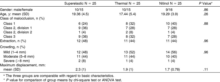
Eighty-one participants met the inclusion criteria and were enrolled in this trial. Two participants were lost to follow-up, and four were excluded due to a lost or incomplete questionnaire. In total, the sample consisted of 29 male and 46 female patients, with a mean age of 18.6 years (SD 4.6 years). The baseline demographic and clinical characteristics for the three groups are shown in Table 1. No variable was identified to discriminate the three groups.
Table 1.
Basic Thermoelastic Characteristics of the Three Groupsa
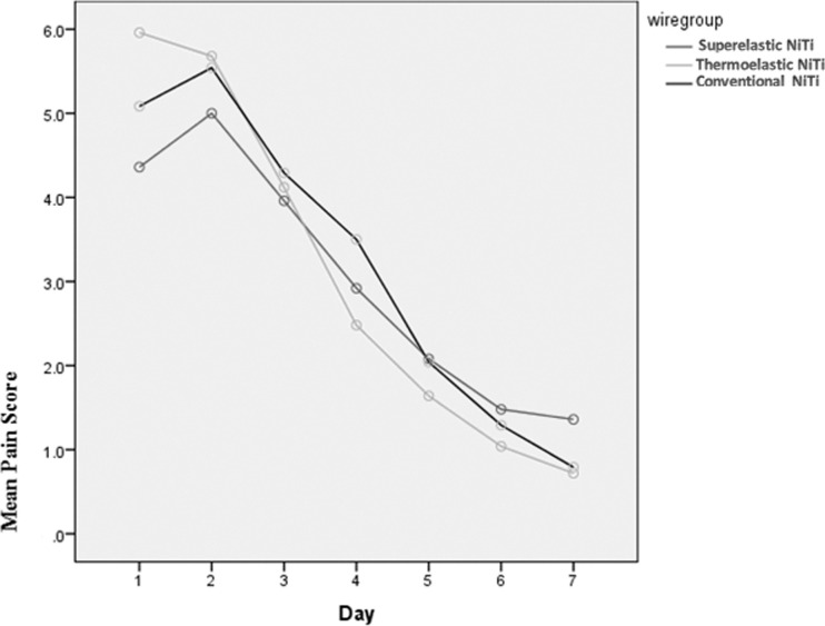
No significant differences were detected in the mean highest pain score between the three groups (P = .63). Eighty-seven percent of patients experienced the maximum pain within the first two days after archwire placement (Table 2). The pain intensity decreased as a function of time for all wires over the observation period (Figure 1). Repeated measures ANOVA confirmed that wire type had no significant effect on the perceived pain over time (P = .155). Time had a significant effect on pain (P < .0001). Subsequent analysis using contrasts showed that there were no significant difference in the pain scores between day 1 and 2, but pain scores were significantly different at the following days. A high percentage (67%) of patients relied on analgesics for symptomatic relief in the week following orthodontic appliance placement. Two patients reported no pain at all. The need for analgesics was significantly different between the three groups (P = .048) (Table 2). Multiple regression analysis showed no significant effect of gender (P = .22), age (P = .24), crowding severity (P = .91), or teeth irregularity (P = .2) on the highest perceived discomfort.
Table 2.
Thermoelastic Side Effects of Managementa
Figure 1.
Plot of mean VAS as a function of time for the three NiTi archwires.
DISCUSSION
Most clinicians believe that discomfort is related to high forces applied to the teeth. This suggestion is derived from the early classic histologic studies that promoted the idea of light forces being more efficient, more biologic, and less painful.12,15 However, some investigators failed to prove such an association between the force applied to the teeth and the resultant pain.4,21 On the other hand, a recent study concluded that heavy forces produce significantly greater pain than light forces 24 hours after force application.22
The introduction of nickel-titanium archwires has revolutionized the field of orthodontics because of the ability of these archwires to deliver light continuous forces, thus increasing the intervals between appointments. There are three nickel-titanium archwires currently available commercially. The first nickel-titanium archwire “Nitinol” was introduced to orthodontics by Andreasen and Hilleman23 in 1971 and later produced for clinical use by Unitek Corporation. Nitinol (martensitic stable) archwires have a stress-strain curve similar to stainless steel wires. Austenitic nickel-titanium alloys (superelastic and thermoelastic) were introduced later, and these were widely accepted for initial alignment of malocclusions mainly because of their unique properties of superelasticity and shape memory.
This is the first clinical trial to compare pain intensity between the three types of NiTi archwires. Although in vitro studies demonstrated that superelastic wires are able to deliver almost continuous light forces with large activations that may generate less pain,10,12–15 the present clinical study found no evidence of significant difference in the pain intensity when the three types of NiTi aligning archwires (martensitic stable, austenitic active, and martensitic active) were compared. Jones and Chan16 failed to demonstrate a difference in the pain experience between multistranded stainless steel and superelastic NiTi archwires during the first 2 weeks after archwire placement.16 A similar finding was reported in another study; however, superelastic wires had a significantly higher pain at peak level.18 When comparing conventional Nitinol wires to superelastic Sentalloy wires over 1 week following archwire placement, a significant difference in the overall pain response could not be found.17 However, Fernandes et al.17 found that conventional Nitinol wires induced significantly higher pain levels than superelastic Sentalloy wires at 4 hours.
Similar to our study, Jones and Chan16 and Fernandes et al.17 used a visual analog scale to evaluate the pain intensity. VAS is one of the most commonly used tools in the measurement of the perceived discomfort during orthodontic treatment.5,16,17,24 This scale is simple to use, reliable, reproducible, and readily understood by most patients.25,26 When compared to other pain/discomfort assessment methods like the verbal rating scales, VAS is more precise and demonstrates better sensitivity between small changes in pain intensity.27,28
The general time-course of pain intensity concurs with previous studies as the pain level peaked within the first 2 days after archwire insertion, and then gradually declined to near baseline levels 6 to 7 days postoperatively,4,5,16,17 which indicates that any differences in pain/discomfort are likely to be minimal after 7 days. This observed pain time-course correlates well with the underlying biologic response to orthodontic forces. An increased concentration of interleukin-1β (inflammatory mediator that induces the secretion of pain-producing substances) in human gingival crevicular fluid was found after 1 hour of orthodontic force application that reaches its peak level after 24 hours and subsequently declines to normal level in 1 week to 1 month.22
As a second form of pain intensity assessment, patients were asked to report the use of self-prescribed analgesics. A high percentage (67%) of patients relied on analgesics for symptomatic relief in the week following orthodontic appliance placement, which underlines the severity of orthodontic pain. In agreement with a previous study,17 no significant difference was found in the amount of consumed analgesics between superelastic and conventional NiTi archwires. However, the need for analgesics was significantly different between superelastic and thermoelastic wire groups. Although this finding is inconsistent with the results from the VAS, analgesic requirements provide only a rough assessment of pain response since it is correlated with personality factors such as anxiety and depression.29
Since pain is a subjective experience, it can be influenced by a number of factors other than the magnitude of the applied force, such as age, gender, degree of teeth irregularity, and psychologic factors. In agreement with previous studies,5,16,17,24,29 there were no statistically significant differences in pain scores between female and male patients with regard to VAS and consumption of analgesics. However, Scheurer et al.6 reported that female patients experienced greater pain and consumed more analgesics than male patients.
As reported previously and confirmed in the present study, neither the degree of initial crowding4 nor the amount of incisor irregularity was found to be a statistically significant variable in the pain response. This suggests that the degree of incisor irregularity and the related interbracket span may not significantly influence the forces applied to the teeth.
In addition, no significant association between age and the level of pain/discomfort experienced by patients following archwire placement was found. This is in disagreement with previous research that has shown patients over the age 16 years to have higher pain scores3 and those under the age 13 years to experience less pain.6
CONCLUSIONS
No significant difference between the three types of NiTi archwires (conventional, superelastic and thermoelastic) was found in pain intensity experienced by patients during initial tooth alignment.
Gender, age, and the degree of crowding have no effect on the perceived discomfort experienced by patients undergoing fixed orthodontic treatment.
ACKNOWLEDGMENTS
Our thanks go to all clinicians, dental assistants, and laboratory technicians involved in this project. We would also like to thank Dr Fares Chedid for his assistance in the statistical analysis.
REFERENCES
- 1.Lew KK. Attitudes and perceptions of adults towards orthodontic treatment in an Asian community. Community Dent Oral Epidemiol. 1993;21:31–35. doi: 10.1111/j.1600-0528.1993.tb00715.x. [DOI] [PubMed] [Google Scholar]
- 2.Jones M. An investigation into the initial discomfort caused by placement of an archwire. Eur J Orthod. 1984;6:48–54. doi: 10.1093/ejo/6.1.48. [DOI] [PubMed] [Google Scholar]
- 3.Jones M, Richmond S. Initial tooth movement: force application and pain—a relationship. Am J Orthod. 1985;88:111–116. doi: 10.1016/0002-9416(85)90234-9. [DOI] [PubMed] [Google Scholar]
- 4.Jones ML, Chan C. Pain in the early stages of orthodontic treatment. J Clin Orthod. 1992;26:311–313. [PubMed] [Google Scholar]
- 5.Ngan P, Kess B, Wilson S. Perception of discomfort by patients undergoing orthodontic treatment. Am J Orthod Dentofacial Orthop. 1989;96:47–53. doi: 10.1016/0889-5406(89)90228-x. [DOI] [PubMed] [Google Scholar]
- 6.Scheurer PA, Firestone AR, Burbin WB. Perception of pain as a result of orthodontic treatment with fixed appliances. Eur J Orthod. 1996;18:349–357. doi: 10.1093/ejo/18.4.349. [DOI] [PubMed] [Google Scholar]
- 7.Kvam E, Gjerdet N, Bondevik O. Traumatic ulcers and pain during orthodontic treatment. Community Dent Oral Epidemiol. 1987;15:104–107. doi: 10.1111/j.1600-0528.1987.tb00493.x. [DOI] [PubMed] [Google Scholar]
- 8.Rock WP, Wilson HJ. Forces exerted by orthodontic aligning archwires. Br J Orthod. 1988;15:255–259. doi: 10.1179/bjo.15.4.255. [DOI] [PubMed] [Google Scholar]
- 9.Burstone C. Biomechanics of tooth movement. In: Kraus BS, Riedel RA, editors. Vistas in Orthodontics. Philadelphia, Pa: Lea & Febiger; 1964. pp. 197–213. [Google Scholar]
- 10.Burstone CJ. Variable-modulus orthodontics. Am J Orthod. 1981;80:1–16. doi: 10.1016/0002-9416(81)90192-5. [DOI] [PubMed] [Google Scholar]
- 11.Linge L, Linge BO. Patient characteristics and treatment variables associated with apical root resorption during orthodontic treatment. Am J Orthod Dentofacial Orthop. 1991;99:35–43. doi: 10.1016/S0889-5406(05)81678-6. [DOI] [PubMed] [Google Scholar]
- 12.Storey E, Smith R. Force in orthodontics and its relation to tooth movement. Aust J Dent. 1952;56:11–18. [Google Scholar]
- 13.Kapila S, Haugen JW, Watanabe LG. Load-deflection characteristics of nickel-titanium alloy wires after clinical recycling and dry heat sterilization. Am J Orthod Dentofacial Orthop. 1992;102:120–126. doi: 10.1016/0889-5406(92)70023-4. [DOI] [PubMed] [Google Scholar]
- 14.Miura F, Mogi M, Ohura Y, Hamanaka H. The super-elastic property of the Japanese NiTi alloy wire for use in orthodontics. Am J Orthod Dentofacial Orthop. 1986;90:1–10. doi: 10.1016/0889-5406(86)90021-1. [DOI] [PubMed] [Google Scholar]
- 15.Reitan K. Some factors determining the evaluation of forces in orthodontics. Am J Orthod. 1957;43:32–45. [Google Scholar]
- 16.Jones M, Chan C. The pain and discomfort experienced during orthodontic treatment: a randomized controlled clinical trial of two initial aligning arch wires. Am J Orthod Dentofacial Orthop. 1992;102:373–381. doi: 10.1016/0889-5406(92)70054-e. [DOI] [PubMed] [Google Scholar]
- 17.Fernandes LM, Ogaard B, Skoglund L. Pain and discomfort experienced after placement of a conventional or a superelastic NiTi aligning archwire. A randomized clinical trial. J Orofac Orthop. 1998;59:331–339. doi: 10.1007/BF01299769. [DOI] [PubMed] [Google Scholar]
- 18.Sandhu SS, Sandhu J. A randomized clinical trial investigating pain associated with superelastic nickel-titanium and multistranded stainless steel archwires during the initial leveling and aligning phase of orthodontic treatment. J Orthod. 2013;40:276–285. doi: 10.1179/1465313313Y.0000000072. [DOI] [PMC free article] [PubMed] [Google Scholar]
- 19.Wang Y, Jian F, Lai W, et al. Initial arch wires for alignment of crooked teeth with fixed orthodontic braces. Cochrane Database Syst Rev. 2010 Apr 14;:CD007859. doi: 10.1002/14651858.CD007859.pub2. [DOI] [PubMed] [Google Scholar]
- 20.Little RM. The irregularity index: a quantitative score of mandibular anterior alignment. Am J Orthod. 1975;68:554–563. doi: 10.1016/0002-9416(75)90086-x. [DOI] [PubMed] [Google Scholar]
- 21.Boester CH, Johnston IE. A clinical investigation of the concepts of differential and optimal force in canine retraction. Angle Orthod. 1974;44:113–119. doi: 10.1043/0003-3219(1974)044<0113:ACIOTC>2.0.CO;2. [DOI] [PubMed] [Google Scholar]
- 22.Luppanapornlarp S, Kajii TS, Surarit R, Iida J. Interleukin-1beta levels, pain intensity, and tooth movement using two different magnitudes of continuous orthodontic force. Eur J Orthod. 2010;32:596–601. doi: 10.1093/ejo/cjp158. [DOI] [PubMed] [Google Scholar]
- 23.Andreasen CF, Hilleman TB. An evaluation of 55 cobalt substituted Nitinol wire for use in orthodontics. J Am Dent Assoc. 1971;82:1373–1375. doi: 10.14219/jada.archive.1971.0209. [DOI] [PubMed] [Google Scholar]
- 24.Erdinc E, Aslihan M, Dincer B. Perception of pain during orthodontic treatment with fixed appliances. Eur J Orthod. 2004;26:79–85. doi: 10.1093/ejo/26.1.79. [DOI] [PubMed] [Google Scholar]
- 25.Huskisson EC. Visual analogue scales. In: Melzack R, editor. Pain Measurement and Assessment. New York, NY: Raven Press; 1983. pp. 33–37. [Google Scholar]
- 26.Scott P, Sherriff M, Dibiase AT, Cobourne MT. Perception of discomfort during initial orthodontic tooth alignment using a self-ligating or conventional bracket system: a randomized clinical trial. Eur J Orthod. 2008;30:227–232. doi: 10.1093/ejo/cjm131. [DOI] [PubMed] [Google Scholar]
- 27.Langley GB, Sheppeard H. Problems associated with pain measurement in arthritis: comparison of the visual analogue and verbal rating scales. Clin Exp Rheumatol. 1984;2:231–234. [PubMed] [Google Scholar]
- 28.Deschamps M, Band PR, Coldman AJ. Assessment of adult cancer pain: shortcomings of current methods. Pain. 1988;32:133–139. doi: 10.1016/0304-3959(88)90061-9. [DOI] [PubMed] [Google Scholar]
- 29.Feinmann C, Ong M, Harvey W, Harris M. Psychological factors influencing postoperative pain and analgesic consumption. Br J Oral Maxillofac Surg. 1987;25:285–292. doi: 10.1016/0266-4356(87)90067-2. [DOI] [PubMed] [Google Scholar]


