Abstract
Objective:
To evaluate the effectiveness of rapid maxillary expansion (RME) on the sagittal dental or skeletal parameters of growing children with Class II malocclusion.
Materials and Methods:
A systematic review intended to identify relevant literature was conducted. The search was performed on Medline, Embase, Cochrane Library, and Scopus databases. Reference lists of the included articles were also screened for relevant documents. The qualitative assessment was performed according to the Methodological Index for Non-Randomized Studies (MINORS) tool, and the resultant data were grouped and analyzed concerning dental and skeletal sagittal effects of RME.
Results:
Of 25 screened studies, seven articles met eligibility criteria and were included. Study samples were observed during mixed dentition stage and characterized as having either Class II dental malocclusion or skeletal discrepancy. None of the included studies was a randomized clinical trial. Included controlled studies presented several inadequacies related to control group or lacked appropriate comparative statistical analysis. Besides being frequently based on deficient methodology, dental and skeletal sagittal effects of RME were either controversial or lacked clinical relevance.
Conclusion:
The effect of RME on the sagittal dimension of Class II malocclusions has not been proved yet. Future randomized controlled clinical trials are still needed to definitely address this question.
Keywords: Palatal expansion technique, Malocclusion, Angle Class II
INTRODUCTION
Class II malocclusion is one of the most common orthodontic discrepancies,1–3 and it is likely to produce significant negative esthetic4 and social5 effects on children's lives, affect their dental health,6 or predispose them to dental trauma.7
Plenty of evidence is available to support that Class II, division 1 individuals have smaller transverse maxillary dental or skeletal dimensions.8–11 For this reason, it has been proposed that the treatment of this malocclusion should comprise previous maxillary expansion.12–15 It has been reported that after expansion, a “spontaneous” correction of the Class II malocclusion takes place as a result of a forward posturing of the mandible.13–15 However, such observations have mostly relied upon clinical experience, and the research intended to analyze that question is controversial and presents diverse study methods.16–22
Therefore, the objective of this investigation was to evaluate the effectiveness of rapid maxillary expansion (RME) on the sagittal dental or skeletal parameters of growing children with Class II malocclusion through a systematic review of available clinical trials.
MATERIALS AND METHODS
The Preferred Reporting Item for Systematic Review and Meta-Analysis (PRISMA) checklist23 was used as a guideline for conducting and reporting this review.
Articles were selected if growing individuals who underwent orthodontic treatment with RME for Class II malocclusion were investigated. Studies including adults or growing patients treated with any orthodontic Class II treatment method other than RME were excluded.
In order to be included, the study outcomes should have referred to either dental (molar relationship) or skeletal (cephalometric mandibular parameters) status of Class II malocclusion, and evaluations should have been performed both before and after RME treatment. In addition, included articles should have necessarily presented results derived from inferential statistics.
No language or time restrictions were applied to the searches. Case reports, literature reviews, editorials, interviews, and letters were not considered. Interventional clinical trials, including randomized and nonrandomized controlled studies, as well as case series were accepted.
The following databases were searched: Medline, Embase, Cochrane Library and Scopus. The key words used for this literature search were “maxillary expansion,” “palatal expansion,” and “Class II,” A search strategy was designed first for Medline (Appendix 1) and was applied accordingly to all other databases. The electronic database searching was conducted between September 10 and 15, 2014. A hand search of the reference lists of selected articles was conducted to identify any additional relevant publications that might have been missed by the database search.
Once the search was completed, duplicate results were removed. In the first phase of the selection, two reviewers independently evaluated titles and abstracts, when available. Clinical studies that reported the use of RME for the correction of Class II malocclusion in growing individuals were considered as preselected after phase I screening. Full articles were then retrieved for those preselected publications, as well as for those with inadequate or unavailable abstracts. In the final phase of the study selection, the same reviewers independently evaluated the full-text articles according to critical application of the remaining eligibility criteria. Disagreements between reviewers in any selection phase were resolved through consensus.
The following variables were extracted from the final selected studies: sample and treatment characteristics; examination methods and parameters used to evaluate treatment effects, as well as main results, including baseline status; treatment effect (Δ) observed in the experimental group; net difference (Net Δ) between values observed in the treatment and control groups after observation period; and inferential statistics, which referred to either treatment vs control analysis if a control group was present, or initial vs final analysis in the case of noncontrolled studies. Two reviewers extracted the data independently, and all of the authors reviewed them afterwards. The extracted data were then combined and compared for accuracy, and discrepancies were resolved by reexamination of the literature. Authors were eventually contacted if any information appeared to be unclear.
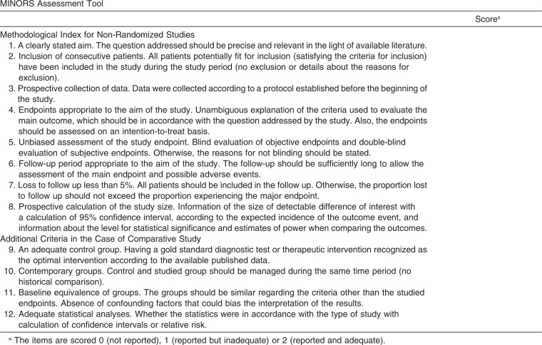
Two reviewers appraised the selected studies according to the Methodological Index for Non-Randomized Studies (MINORS) (Appendix 2).24 This tool was conceived and validated to assess methodological quality for nonrandomized studies, whether comparative or noncomparative.
RESULTS
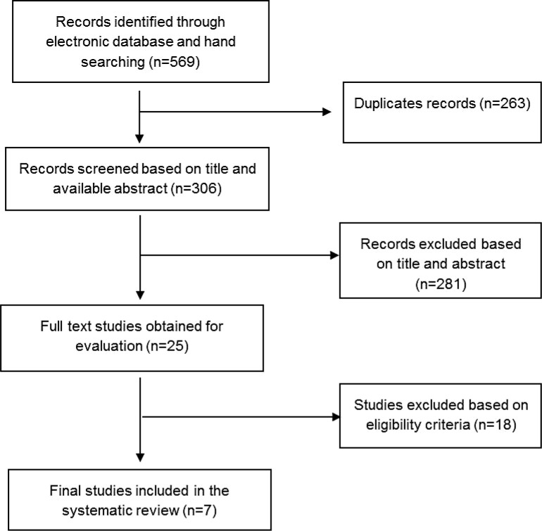
A flowchart illustrating the selection of studies for this systematic review is presented in Figure 1. After the first phase selection, 25 full texts were obtained for the second phase evaluation, of which 18 articles13–15,25–39 were excluded. The reasons for exclusion are listed in Table 1. Finally, seven clinical trials met the eligibility criteria and were considered for this systematic review.16–22
Figure 1.
Flowchart of study selection process.
Table 1.
Articles Excluded After Full Text Evaluation Based on Eligibility Criteria
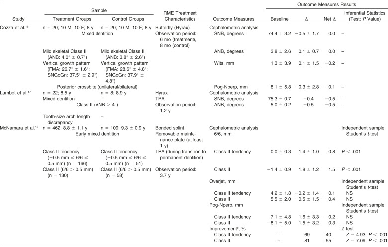
A summary of the key methodological data and study characteristics can be found in Table 2. Study samples were observed during the mixed dentition phase and presented a wide spectrum of Class II malocclusions. Samples were characterized as having either dental18–20 or skeletal discrepancies.16,17,21,22 Transverse maxillary deficiency was reportedly present in the sample of part of the studies.16,19,21,22
Table 2.
Summary of Study Characteristics and Results of the Included Studiesa
Table 2.
Continued
Table 2.
Continued
All subjects in the included studies were treated with RME, either as a sole treatment approach,16,21,22 or associated with other appliances, such as passive transpalatal arches.17–19 The subjects were observed for varying periods of time, according to different outcomes, which related to either skeletal16–19,21,22 or dental characteristics.18–20
Methodological appraisal of the selected studies is presented in Table 3. None of the included studies was a randomized clinical trial. All of the researches16–22 clearly stated the aim of the investigation, presented an appropriate period of observation, and reported no sample loss during follow-up. However, limitations were identified for most of the studies, such as the retrospective enrollment of the sample and collection of the data20,22 or unclear report of these features.16,17 None of the studies16–22 reported if the outcome examiners were blinded during the end-point evaluation, and two trials18,19 utilized a critical examination method (cephalometry) to assess the molar relationship outcome. These two studies,18,19 however, were the only ones that demonstrated that their samples were appropriate in size.
Table 3.
Methodological Appraisal of the Selected Studies, According to MINORS Assessment Tool
Three20–22 of the seven included articles were noncontrolled studies. The other controlled studies16–19 presented adequate paired control groups, although none of them reported if control and experimental groups were contemporary. Among the controlled studies,16–19 two16,17 lacked statistical analysis for comparison between the experimental and control groups changes. Of the remaining two controlled studies,18,19 one of them19 had a baseline difference between the experimental and control groups.
Due to the large variability among the included studies, this systematic review was not designed in a meta-analysis format. Nonetheless, a comprehensive extraction of the study data can be found in Table 2.
Molar Relationship
The dental changes after RME treatment were investigated in three18–20 of the included studies. The first clinical trial18 demonstrated that molar occlusal relationship significantly improved after RME, for both Class II tendency (end-to-end) and Class II (more severe) treated groups. Another included study19 also presented significant positive molar changes following RME treatment. The Class II treatment group demonstrated significant improvement of the molar relationship in comparison with the matched control.19 However, another study20 performed a noncontrolled trial in which Class II patients showed no significant differences regarding molar relationship after treatment, neither in centric occlusion nor maximum intercuspation positions.
Skeletal Mandibular Effects
Among the included studies, six16–19,21,22 evaluated mandibular changes following RME therapy. According to the first one,16 RME did not produce any significant difference in Class II individuals. Among the anteroposterior mandibular parameters investigated by Lambot et al.,17 however, a statistically significant increase in SNB measurement was found for the treated group. In both studies,16,17 when the experimental and control group changes were compared, differences did not reach clinical relevance for all the parameters.
McNamara et al.18 did not observe skeletal differences between treated and control groups in relation to the amount of the mandibular displacement. However, Guest et al.19 demonstrated statistically significant increases in mandibular length and advancement of the symphysis when Class II patients were compared to untreated controls. The RME also produced significant effects on the anteroposterior relationship of the maxillary and the mandibular bones of the treated group, as compared to matched controls.19
According to Baratieri et al.,21 significant anterior displacement of the mandibular symphysis was observed during the RME retention period. In another study, Farronato et al.22 also reported that in Class II subjects, the mandible moved forward in a significant manner, and the ANB angle statistically decreased, improving the skeletal Class II after RME.
DISCUSSION
The results observed during this systematic review are not only contradictory, but also frequently based on deficient methodology, or lack clinical relevance. Even though important studies have been published,16–22 more solid scientific evidence based on reliable methods of assessment and proper study designs is still lacking in order to thoroughly test whether dental correction or mandibular anterior shift and/or supplementary growth take place after RME in Class II individuals.
According to Volk et al.,20 maxillary expansion did not predictably improve Class II molar relationships. And even though the best evidence available18,19 reported statistically significant occlusal improvements, these changes could be attributed to other reasons, eg, the use of passive transpalatal arches during transition from mixed to permanent dentition. As previously documented,9,41,42 most of the flush terminal planes are naturally converted to solid first molar Class I during transition. This fact had been wisely mentioned before,43 and it indicates that most of the end-to-end Class II cases in mixed dentition are self-corrected, and demand neither RME nor transpalatal arches to assure first molar adjustment.
The transformation into a Class I molar relationship, during transition to permanent dentition, depends on a number of dental and facial skeletal changes.44 However, if one assumes that it is possible to preserve additional space by preventing upper molars mesial drift with transpalatal arches, as suggested,45 even more severe than end-to-end Class II occlusions might be supposedly exempt from RME to attenuate their sagittal occlusal imbalances. Unless space management is critical, or transversal discrepancies are proved to be present, RME for sole attenuation of Class II malocclusion thus seems unnecessary.
As for the mandibular skeletal changes, most of the selected studies16–18,20 indicated no mandibular shift, nor supplementary growth after RME. One may claim that the mandibular changes demonstrated by Guest et al.19 might have been a result of the mandibular shift or growth, but differences still seem clinically irrelevant. Moreover, other Class II standard therapies, such as functional removable appliances,46 headgears,43 or bite-jumping appliances,47 have already proved to be effective for the Class II correction; in addition, these therapies promote maxillary expansion, that reduces the usefulness of RME in skeletal Class II cases with no transverse deficiency.
Even though Baratieri et al.21 and Farronato et al.22 have presented data indicating significant mandibular anterior displacement, these changes were not compared to a control group, which considerably decreases the scientific relevance of these evidences.
In order to have a better predictability of the effectiveness of any therapy, it is advisable to consider not only controlled groups, but also randomization.48 Unfortunately, no randomized controlled trial has been performed on that matter so far, and those that were carried out with paired control groups still lack methodological accuracy. Because of the absence of randomized trials, the investigators had to choose to include nonrandomized controlled trials and case series in this systematic review. Unfortunately, no meta-analysis could be executed because of the many methodological differences among the selected studies and the excessively large variability observed in relation to the sample characteristics, treatment features, follow-up period, and outcome measurements.
RME is considered to be one of the safest and most predictable therapies in orthodontic practice.49 However, the effect of maxillary expansion on the sagittal dimension of Class II is still controversial and has not been substantially proved yet. The demonstration of the induced change theory12 still requires methodological concerns, principally in relation to clinical trials design, which should ideally enroll adequate control subjects, randomization, and blindness during outcome assessment.
During performance of future studies, special emphasis must be directed to objectively identify those Class II patients more likely to present favorable sagittal responses to RME therapy. Moreover, long-term studies would be advisable in order to investigate the stability of effects, if present, as well as to verify if RME therapy is likely to avoid Class II specific therapy in late mixed dentition or diminish comprehensive orthodontic treatment time.
APPENDIX 1
Search Strategy Designed for Medline Database Search
APPENDIX 2
MINORS Assessment Tool
REFERENCES
- 1.Reddy ER, Manjula M, Sreelakshmi N, Rani ST, Aduri R, Patil BD. Prevalence of malocclusion among 6 to 10 year old Nalgonda school children. J Int Oral Health. 2013;5:49–54. [PMC free article] [PubMed] [Google Scholar]
- 2.Urrego-Burbano PA, Jiménez-Arroyave LP, Londoño-Bolívar MÁ, Zapata-Tamayo M, Botero-Mariaca P. Epidemiological profile of dental occlusion in children attending school in Envigado, Colombia [in Spanish] Rev Salud Publica (Bogota) 2011;13:1010–1021. doi: 10.1590/s0124-00642011000600013. [DOI] [PubMed] [Google Scholar]
- 3.Karaiskos N, Wiltshire WA, Odlum O, Brothwell D, Hassard TH. Preventive and interceptive orthodontic treatment needs of an inner-city group of 6- and 9-year-old Canadian children. J Can Dent Assoc. 2005;71:649. [PubMed] [Google Scholar]
- 4.Kiekens RM, Maltha JC, van't Hof MA, Kuijpers-Jagtman AM. Objective measures as indicators for facial esthetics in white adolescents. Angle Orthod. 2006;76:551–556. doi: 10.1043/0003-3219(2006)076[0551:OMAIFF]2.0.CO;2. [DOI] [PubMed] [Google Scholar]
- 5.Seehra J, Fleming PS, Newton T, DiBiase AT. Bullying in orthodontic patients and its relationship to malocclusion, self-esteem and oral health-related quality of life. J Orthod. 2011;38:247–256. doi: 10.1179/14653121141641. [DOI] [PubMed] [Google Scholar]
- 6.de Almeida AB, Leite IC. Orthodontic treatment need for Brazilian schoolchildren: a study using the Dental Aesthetic Index. Dental Press J Orthod. 2013;18:103–109. doi: 10.1590/s2176-94512013000100021. [DOI] [PubMed] [Google Scholar]
- 7.Kalha AS. Early orthodontic treatment reduced incisal trauma in children with class II malocclusions. Evid Based Dent. 2014;15:18–20. doi: 10.1038/sj.ebd.6400986. [DOI] [PubMed] [Google Scholar]
- 8.Lux CJ, Conradt C, Burden D, Komposch G. Dental arch widths and mandibular-maxillary base widths in Class II malocclusions between early mixed and permanent dentitions. Angle Orthod. 2003;73:674–685. doi: 10.1043/0003-3219(2003)073<0674:DAWAMB>2.0.CO;2. [DOI] [PubMed] [Google Scholar]
- 9.da Silva LP, Gleiser R. Occlusal development between primary and mixed dentitions: a 5-year longitudinal study. J Dent Child (Chic) 2008;75:287–294. [PubMed] [Google Scholar]
- 10.Marinelli A, Mariotti M, Defraia E. Transverse dimensions of dental arches in subjects with Class II malocclusion in the early mixed dentition. Prog Orthod. 2011;12:31–37. doi: 10.1016/j.pio.2011.02.006. [DOI] [PubMed] [Google Scholar]
- 11.Shu R, Han X, Wang Y, et al. Comparison of arch width, alveolar width and buccolingual inclination of teeth between Class II division 1 malocclusion and Class I occlusion. Angle Orthod. 2013;83:246–252. doi: 10.2319/052412-427.2. [DOI] [PMC free article] [PubMed] [Google Scholar]
- 12.Kingsley NW. A Treatise on Oral Deformities as a Branch of Mechanical Surgery. New York, NY: Appleton; 1880. [Google Scholar]
- 13.McNamara JA., Jr Maxillary transverse deficiency. Am J Orthod Dentofacial Orthop. 2000;117:567–570. doi: 10.1016/s0889-5406(00)70202-2. [DOI] [PubMed] [Google Scholar]
- 14.McNamara JA., Jr Early intervention in the transverse dimension: is it worth the effort. Am J Orthod Dentofacial Orthop. 2002;121:572–574. doi: 10.1067/mod.2002.124167. [DOI] [PubMed] [Google Scholar]
- 15.McNamara JA., Jr Long-term adaptations to changes in the transverse dimension in children and adolescents: an overview. Am J Orthod Dentofacial Orthop. 2006;129(4 suppl):S71–S74. doi: 10.1016/j.ajodo.2005.09.020. [DOI] [PubMed] [Google Scholar]
- 16.Cozza P, Giancotti A, Petrosino A. Rapid palatal expansion in mixed dentition using a modified expander: a cephalometric investigation. J Orthod. 2001;28:129–134. doi: 10.1093/ortho/28.2.129. [DOI] [PubMed] [Google Scholar]
- 17.Lambot T, Van Steenberghe PR, Vanmuylder N, de Maertelaer V, Glineur R. Early treatment with rapid palatal expander and 3D Quad Action mandibular appliance: evaluation of a comprehensive approach in 22 patients [in French] Orthod Fr. 2008;79:107–114. doi: 10.1051/orthodfr:2008005. [DOI] [PubMed] [Google Scholar]
- 18.McNamara JA, Jr, Sigler LM, Franchi L, Guest SS, Baccetti T. Changes in occlusal relationships in mixed dentition patients treated with rapid maxillary expansion. A prospective clinical study. Angle Orthod. 2010;80:230–238. doi: 10.2319/040309-192.1. [DOI] [PMC free article] [PubMed] [Google Scholar]
- 19.Guest SS, McNamara JA, Jr, Baccetti T, Franchi L. Improving Class II malocclusion as a side-effect of rapid maxillary expansion: a prospective clinical study. Am J Orthod Dentofacial Orthop. 2010;138:582–591. doi: 10.1016/j.ajodo.2008.12.026. [DOI] [PubMed] [Google Scholar]
- 20.Volk T, Sadowsky C, Begole EA, Boice P. Rapid palatal expansion for spontaneous Class II correction. Am J Orthod Dentofacial Orthop. 2010;137:310–315. doi: 10.1016/j.ajodo.2008.05.017. [DOI] [PubMed] [Google Scholar]
- 21.Baratieri C, Alves M, Jr, Sant'anna EF, Nojima Mda C, Nojima LI. 3D mandibular positioning after rapid maxillary expansion in Class II malocclusion. Braz Dent J. 2011;22:428–434. doi: 10.1590/s0103-64402011000500014. [DOI] [PubMed] [Google Scholar]
- 22.Farronato G, Giannini L, Galbiati G, Maspero C. Sagittal and vertical effects of rapid maxillary expansion in Class I, II, and III occlusions. Angle Orthod. 2011;81:298–303. doi: 10.2319/050410-241.1. [DOI] [PMC free article] [PubMed] [Google Scholar]
- 23.Preferred Reporting Items for Systematic Reviews and Meta-Analyses (PRISMA) Checklist (2009) Available at: http://www.prisma-statement.org Accessed October 10, 2014. [DOI] [PubMed] [Google Scholar]
- 24.Slim K, Nini E, Forestier D, Kwiatkowski F, Panis Y, Chipponi J. Methodological index for non-randomized studies (minors): development and validation of a new instrument. ANZ J Surg. 2003;73:712–716. doi: 10.1046/j.1445-2197.2003.02748.x. [DOI] [PubMed] [Google Scholar]
- 25.Gray LP. Results of 310 cases of rapid maxillary expansion selected for medical reasons. J Laryngol Otol. 1975;89:601–614. doi: 10.1017/s0022215100080804. [DOI] [PubMed] [Google Scholar]
- 26.Hesse KL, Artun J, Joondeph DR, Kennedy DB. Changes in condylar position and occlusion associated with maxillary expansion for correction of functional unilateral posterior crossbite. Am J Orthod Dentofacial Orthop. 1997;111:410–418. doi: 10.1016/s0889-5406(97)80023-6. [DOI] [PubMed] [Google Scholar]
- 27.Basciftci FA, Karaman AI. Effects of a modified acrylic bonded rapid maxillary expansion appliance and vertical chin cap on dentofacial structures. Angle Orthod. 2002;72:61–71. doi: 10.1043/0003-3219(2002)072<0061:EOAMAB>2.0.CO;2. [DOI] [PubMed] [Google Scholar]
- 28.Garib DG, Henriques JF, Janson G, Freitas MR, Coelho RA. Rapid maxillary expansion—tooth tissue-borne versus tooth-borne expanders: a computed tomography evaluation of dentoskeletal effects. Angle Orthod. 2005;75:548–557. doi: 10.1043/0003-3219(2005)75[548:RMETVT]2.0.CO;2. [DOI] [PubMed] [Google Scholar]
- 29.Wendling LK, McNamara JA, Jr, Franchi L, Baccetti T. A prospective study of the short-term treatment effects of the acrylic-splint rapid maxillary expander combined with the lower Schwarz appliance. Angle Orthod. 2005;75:7–14. doi: 10.1043/0003-3219(2005)075<0007:APSOTS>2.0.CO;2. [DOI] [PubMed] [Google Scholar]
- 30.Lima Filho RM, de Oliveira Ruellas AC. Mandibular behavior with slow and rapid maxillary expansion in skeletal Class II patients: a long-term study. Angle Orthod. 2007;77:625–631. doi: 10.2319/071406-294. [DOI] [PubMed] [Google Scholar]
- 31.Lima Filho RM, Ruellas AC. Long-term anteroposterior and vertical maxillary changes in skeletal class II patients treated with slow and rapid maxillary expansion. Angle Orthod. 2007;77:870–874. doi: 10.2319/071406-293.1. [DOI] [PubMed] [Google Scholar]
- 32.Marini I, Bonetti GA, Achilli V, Salemi G. A photogrammetric technique for the analysis of palatal three-dimensional changes during rapid maxillary expansion. Eur J Orthod. 2007;29:26–30. doi: 10.1093/ejo/cji069. [DOI] [PubMed] [Google Scholar]
- 33.Lima Filho RM, de Oliveira Ruellas AC. Long-term maxillary changes in patients with skeletal Class II malocclusion treated with slow and rapid palatal expansion. Am J Orthod Dentofacial Orthop. 2008;134:383–388. doi: 10.1016/j.ajodo.2006.09.071. [DOI] [PubMed] [Google Scholar]
- 34.Lima Filho RMA. Transverse dimension alterations using rapid maxillary expansion [in Portuguese] R Dental Press Ortodon Ortop Facial. 2009;14:146–157. [Google Scholar]
- 35.Baratieri C, Nojima LI, Alves M, Jr, Souza MMG, Nojima MG. Transverse effects of rapid maxillary expansion in Class II malocclusion patients: a cone-beam computed tomography study. Dental Press J Orthod. 2010;15:89–97. [Google Scholar]
- 36.Kolokitha OE, Papadopoulou AK. Rapid palatal expander: an anchor unit for second molar distalization in Angle Class II treatment. World J Orthod. 2010;11:75–84. [PubMed] [Google Scholar]
- 37.Farronato G, Maspero C, Esposito L, Briguglio E, Farronato D, Giannini L. Rapid maxillary expansion in growing patients. Hyrax versus transverse sagittal maxillary expander: a cephalometric investigation. Eur J Orthod. 2011;33:185–189. doi: 10.1093/ejo/cjq051. [DOI] [PubMed] [Google Scholar]
- 38.Hilgers JJ. A palatal expansion appliance for non-compliance therapy. J Clin Orthod. 1991;25:491–497. [PubMed] [Google Scholar]
- 39.Silvestrini-Biavati A, Angiero F, Gambino A, Ugolini A. Do changes in spheno-occipital synchondrosis after rapid maxillary expansion affect the maxillomandibular complex. Eur J Paediatr Dent. 2013;14:63–67. [PubMed] [Google Scholar]
- 40.Ricketts RM. Perspectives in the clinical application of cephalometrics. The first fifty years. Angle Orthod. 1981;51:115–150. doi: 10.1043/0003-3219(1981)051<0115:PITCAO>2.0.CO;2. [DOI] [PubMed] [Google Scholar]
- 41.Arya BS, Savara BS, Thomas DR. Prediction of first molar occlusion. Am J Orthod. 1973;63:610–621. doi: 10.1016/0002-9416(73)90186-3. [DOI] [PubMed] [Google Scholar]
- 42.Bishara SE, Hoppens BJ, Jakobsen JR, Kohout FJ. Changes in the molar relationship between the deciduous and permanent dentitions: a longitudinal study. Am J Orthod Dentofacial Orthop. 1988;93:19–28. doi: 10.1016/0889-5406(88)90189-8. [DOI] [PubMed] [Google Scholar]
- 43.Gianelly AA. Rapid palatal expansion in the absence of crossbites: added value. Am J Orthod Dentofacial Orthop. 2003;124:362–365. doi: 10.1016/s0889-5406(03)00568-7. [DOI] [PubMed] [Google Scholar]
- 44.Ngan P, Alkire RG, Fields H., Jr Management of space problems in the primary and mixed dentitions. J Am Dent Assoc. 1999;130:1330–1339. doi: 10.14219/jada.archive.1999.0403. [DOI] [PubMed] [Google Scholar]
- 45.Hill CJ, Sorenson HW, Mink JR. Space maintenance in a child dental care program. J Am Dent Assoc. 1975;90:811–815. doi: 10.14219/jada.archive.1975.0178. [DOI] [PubMed] [Google Scholar]
- 46.McWade RA, Mamandras AH, Hunter WS. The effects of Fränkel II treatment on arch width and arch perimeter. Am J Orthod Dentofacial Orthop. 1987;92:313–320. doi: 10.1016/0889-5406(87)90332-5. [DOI] [PubMed] [Google Scholar]
- 47.Hansen K, Iemamnueisuk P, Pancherz H. Long-term effects of the Herbst appliance on the dental arches and arch relationships: a biometric study. Br J Orthod. 1995;22:123–134. doi: 10.1179/bjo.22.2.123. [DOI] [PubMed] [Google Scholar]
- 48.Pandis N, Cobourne MT. Clinical trial design for orthodontists. J Orthod. 2013;40:93–103. doi: 10.1179/1465313313Y.0000000048. [DOI] [PubMed] [Google Scholar]
- 49.Zhou Y, Long H, Ye N, et al. The effectiveness of non-surgical maxillary expansion: a meta-analysis. Eur J Orthod. 2014;36:233–242. doi: 10.1093/ejo/cjt044. [DOI] [PubMed] [Google Scholar]


