Significance
Vitality forms represent the different ways in which actions are performed (e.g., gentle, rude). They express the agent’s attitudes toward others. Previous data indicated that vitality forms of hand actions depend on the dorso-central insula. In the present study, we show that in addition to the insula, the middle cingulate cortex is also involved in hand action modulation. A voxel-based analysis highlighted that voxels showing a similar BOLD signal trend in both action observation and execution are present in both regions. Using a multifiber tractography investigation, we demonstrated that the dorso-central insula and middle cingulate cortex are anatomically connected. These data indicate that the modulation of the parieto-frontal circuit controlling hand actions relies on both the insula and cingulate sectors.
Keywords: vitality form network, cingulate, insula, mirror mechanism, social interaction
Abstract
Actions with identical goals can be executed in different ways (gentle, rude, vigorous, etc.), which D. N. Stern called vitality forms [D. N. Stern, Forms of Vitality Exploring Dynamic Experience in Psychology, Arts, Psychotherapy, and Development (2010)]. Vitality forms express the agent’s attitudes toward others. In a series of fMRI studies, we found that the dorso-central insula (DCI) is the region that is selectively active during both vitality form observation and execution. In one previous experiment, however, the middle cingulate gyrus also exhibited activation. In the present study, in order to assess the role of the cingulate cortex in vitality form processing, we adopted a classical vitality form paradigm, but making the control condition devoid of vitality forms using jerky movements. Participants performed two different tasks: Observation of actions performed gently or rudely and execution of the same actions. The results showed that in addition to the insula, the middle cingulate cortex (MCC) was strongly activated during both action observation and execution. Using a voxel-based analysis, voxels showing a similar trend of the blood-oxygen-level-dependent (BOLD) signal in both action observation and execution were found in the DCI and in the MCC. Finally, using a multifiber tractography analysis, we showed that the active sites in MCC and DCI are reciprocally connected.
In every action there are two major components: the content (i.e., the action goal) and the form (i.e., how the goal is achieved). Whatever its content, actions may be energetic, gentle, rude, but also hesitant or effortless. One can pick up a glass resolutely or delicately, just as one can shake a hand coldly or warmly. Specifications of how an action is done do not refer only to “cold” actions, which are those devoid of an emotional content, but also to emotions: An individual can experience an irrepressible or slight sense of disgust or a spurt of explosive or repressed anger; the face can be contorted in a terrible scowl or a fleeting grimace.
The vitality forms are therefore the mechanism that modifies the way in which an action or an emotion is performed or expressed (its form) by modulating the activity of the basic circuits controlling the action or the emotion content. As far as we know, there are no data up to now on the neural basis through which the vitality form could modulate the expression of emotions. This despite a rich literature on how emotions can be conveyed by body expressions, such as facial mimicry and specific gestures (1). These have been altogether designated as emotional body language (2). In contrast, there is rich evidence that the dorso-central insula (DCI) is the neural center that modulates “cold action.”
The first evidence for this localization was obtained in a functional MRI (fMRI) study performed by G.D.C. and colleagues in collaboration with Stern (3). In this study participants were presented with video clips showing transitive and intransitive actions. All actions were performed with two different vitality forms (gentle or rude). The stimuli were presented in pairs of consecutive videos, where the “what” of the observed action and the “how” could be the same or could change between the video pairs. There were two tasks (what and how). In the what task, the participants were required to decide whether the two actions were the same or different, regardless of their vitality form. In the how task they had to decide whether the vitality form was the same or different in the two consecutive videos, regardless of the type of action performed. The most important result turned out to be from the contrast how vs. what. It revealed a specific activation of the dorsal-central insula.
In a subsequent fMRI study, G.D.C. et al. (4) confirmed the role of the dorso-central insula in vitality form coding. They showed that when an individual observes, plans to perform (motor imagery), or executes actions conveying vitality forms, the dorso-central insula exhibits selective activation. Furthermore, conjunction analysis showed that dorso-central insula is endowed with the mirror mechanism that is a basic brain mechanism that transforms sensory representations of others' behaviour into one's own motor or visceromotor representations concerning that behaviour.
Refs. 5–12 confirmed the crucial role of this insula in encoding vitality forms. In a subsequent fMRI study, however, the same authors found that the cingulate cortex might also be involved in the encoding of vitality forms (7). In that study, participants were asked to identify action types (e.g., stirring coffee, closing a door) by listening to action sounds. Each action sound was presented gently or rudely (vitality form condition) or without a vitality form (masked action sounds, control condition). The results indicated that listening to action sounds conveying vitality forms, relative to a control condition activated, beside the dorso-central insula, the middle cingulate cortex (MCC). Whereas the activation of the DCI confirmed previous findings, the activation of the MCC was unexpected.
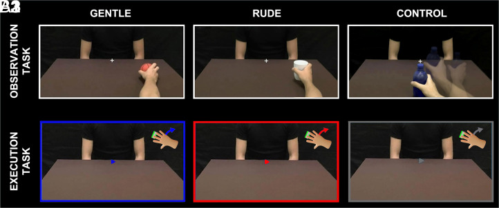
To ascertain the role of the cingulate cortex in vitality form processing, we carried out a new fMRI study. We required participants to perform two tasks: 1) observe an arm action (observation task [OBS]) and 2) execute the same action (execution task [EXE]). In the OBS task, participants observed video clips showing the right arm of an actor performing actions toward another actor (e.g., handing over a ball) with a gentle or rude vitality form (vitality form condition, Fig. 1, A1 and A2 and see also SI Appendix, SI Methods and Fig. S1) or with no vitality form (i.e., jerky actions; control condition, Fig. 1, A3). In the EXE task, participants moved a little box located on a plane while lying on their chest, as if offering it to the other person, with a gentle or rude vitality form (vitality form condition) or with no vitality form (i.e., using jerky movements as a control condition, Fig. 1, B3).
Fig. 1.
Design of the experimental task. First line: the OBS task. Participants observed the right hand of an actor moving an object rightward. Four objects were used. The observed action could be performed with a gentle (A1) or rude (A2) vitality form, and the task required participants to pay attention to the action’s vitality form. In the control condition, the participant observed the same action performed in a jerky way (A3). Second line: the EXE task. Participants held a little box and moved it with a gentle (B1, blue color) or rude (B2, red color) vitality form toward the actor in front of them, as shown in the screen. A cue positioned in the center of the screen indicated when to start the action. During action execution, they saw only the chest of the actor, and the actor’s hand was outside their field of vision. In the control condition, participants performed the same action in a neutral way (B3). All stimuli in the OBS and EXE tasks were viewed via digital visors (VisuaSTIM) with a 500,000 px × 0.25 square inch resolution and horizontal eye field of 30°.
The main finding of our study is that, in addition to the insula, the cingulate cortex is selectively involved in the observation and execution of actions performed with different vitality forms. Additionally, a voxel-based analysis carried out in these two cortical regions showed that a large proportion of the most active voxels are similarly activated during the observation and execution tasks. Very interestingly, we provided evidence that the blood-oxygen-level-dependent (BOLD) signal change extracted in these voxels exhibits a stronger modulation for rude vitality forms with respect to the gentle ones in both tasks. Finally, using a multifiber tractography investigation, we demonstrated that the two sites of DCI and MCC controlling vitality forms are anatomically connected.
Results
Cortical Activations during Observation and Execution of Vitality Forms.
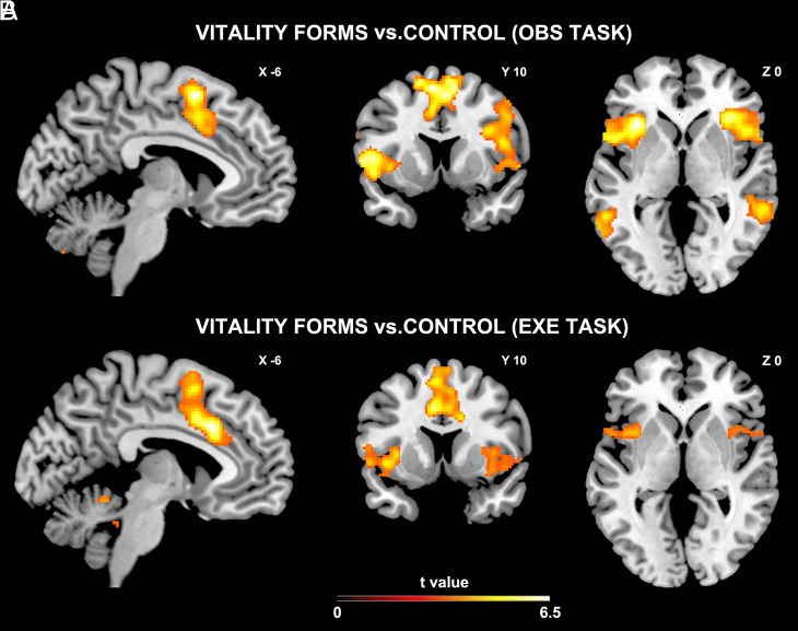
The main aim of the present study was to assess the activation of the cingulate cortex during the OBS and EXE tasks (see also SI Appendix, Fig. S1). As far as the OBS task is concerned, the contrast vitality forms vs. control (VF OBS vs. CT OBS) showed that the vitality form condition produced a consistent activation of the left MCC with an extension to the presupplementary motor area (pre-SMA) bilaterally (p value adjusted with family-wise error correction [pFWE – corr] 0.0001, Ke = 1,477 voxels), of the left DCI (pFWE – corr 0.0001, Ke = 1,486 voxels), of the middle temporal gyrus (left hemisphere: pFWE – corr 0.0001, Ke = 631 voxels; right hemisphere: pFWE – corr 0.0001, Ke = 993 voxels; Fig. 2A), and of the right premotor cortex extending to the inferior frontal gyrus (pFWE – corr 0.0001, Ke = 2,909 voxels; Fig. 2A). Similarly, for the EXE task, the contrast vitality forms vs. control (VF EXE vs. CT EXE) showed that the vitality form condition produced the activation of the same sectors of the left cingulate cortex extending to the right side (pFWE – corr 0.0001, Ke = 1,942 voxels) and of the insula bilaterally (left hemisphere: pFWE – corr 0.0001, Ke = 520 voxels; right hemisphere: pFWE – corr 0.002, Ke = 328 voxels; Fig. 2B; for coordinates and statistical values, see SI Appendix, Table S1).
Fig. 2.
Brain activations during vitality form processing (A1 and B1). Sagittal and coronal sections showing the activation of the cingulate and insular cortices in the two hemispheres during the direct contrasts VF OBS vs. CT OBS (A2) and VF EXE vs. CT EXE, respectively (B2). These activations are rendered using a standard Montreal Neurological Institute (MNI) brain template (PFWE < 0.05 at cluster level).
Conjunction Analysis of Observation and Execution Tasks.
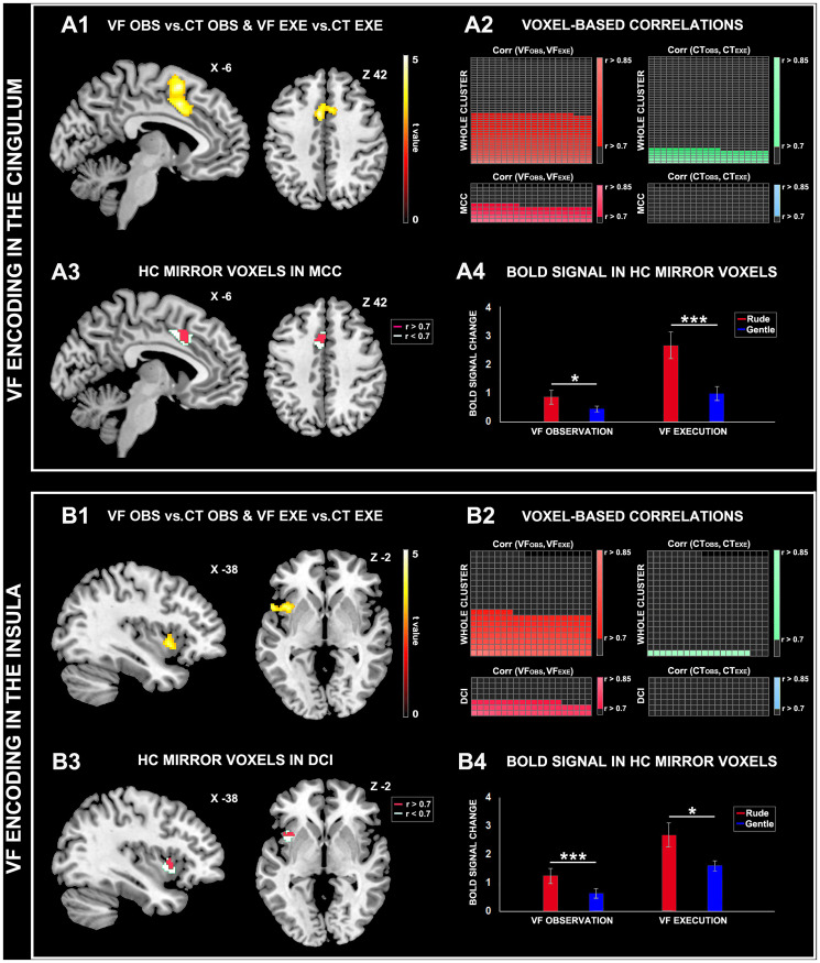
The results of the conjunction analysis of VF OBS vs. CT OBS and VF EXE vs. CT EXE contrasts revealed the activation of the MCC with an extension to the pre-SMA (pFWE – corr 0.0001, Ke = 826 voxels; Fig. 3, A1) and of the left DCI (pFWE – corr 0.001, Ke = 349 voxels; Fig. 3B; for coordinates and statistical values see SI Appendix, Table S1). These findings imply that the same voxels located in the MCC and DCI are selectively involved in the perception and execution of actions endowed with specific vitality forms (voxels endowed with mirror properties). Although this analysis showed voxels activated in both observation and execution tasks it is not informative about the trend of the BOLD signal between the two tasks. In order to identify voxels showing a similar trend of the BOLD signal during the two tasks (i.e., high signal for VF OBS and high signal for VF EXE), we carried out a voxel-based analysis. Specifically, for each voxel we correlated the BOLD signal obtained for each participant during the observation task (average value) with that obtained during the execution task. This analysis allowed us to highlight voxels showing a discreet/strong correlation effect between the two tasks i.e., mirror voxels characterized by a high correlated activity (HC mirror voxels). Most importantly, we considered only voxels that showed at least 50% of the explained variance between the two tasks. In this respect, we decided to use a cutoff correlation value of r > 0.7 (coefficient of determination R2 > = 0.49). Results of this analysis are shown in Fig. 3, A2. Specifically, this picture presents a grid representing the cingulum and adjacent cortical area (pre-SMA) subdivided into a series of small squares, each representing a single voxel. Orange squares indicate voxels selective for the encoding of vitality forms during both the OBS and EXE tasks. The results of the correlation analysis showed that 397 out of 826 voxels (48%) in the whole cluster of voxels (cingulum and pre-SMA) showed a significant correlation between vitality form tasks (VF OBS, VF EXE),112 out of 826 voxels (13.5%) showed a significant correlation between control tasks (CT OBS, CT EXE), whereas the remaining voxels (38.5%) showed a weak correlation between tasks (r < 0.7). A further analysis was restricted to voxels located in the left MCC. This analysis was carried out applying an inclusive mask of MCC obtained from a previous fMRI study (7), which showed that this brain sector was activated during the processing of action sounds conveying vitality forms. Results of this analysis revealed that 88 out of 181 voxels (48.6%) in the left MCC showed a strong significant correlation between vitality form observation and execution (HC mirror voxels; r > 0.7, P < 0.05; Fig. 3, A3). In this brain region, no voxels showed a significant correlation between control tasks.
Fig. 3.
Vitality form processing during the OBS and EXE tasks. Encoding of vitality forms in the cingulum (A) and insula (B). Activation maps of the left cingulum (A1) and insula (B1) resulting from the conjunction analysis of VF OBS vs. CT OBS and VF EXE vs. CT EXE contrasts. These activations are rendered on a standard MNI brain template (PFWE < 0.05 at cluster level). Maps of voxels showing a high correlated BOLD activity (r > 0.7) during the perception and execution of vitality form actions (hot color) or control actions (cold color) located in the cingulum (whole cluster, orange color, A2 Top), insula (whole cluster, orange color, B2 Top). Voxels located in the MCC (A3) and DCI (B3) showing a high correlated BOLD activity (r > 0.7) during vitality-forms OBS and EXE tasks (HC mirror voxels). Signal changes in the MCC (A4) and DCI (B4) during the processing of gentle and rude vitality forms. The horizontal line above the columns indicates the comparisons between vitality forms. Asterisks indicate significant differences at *P ≤ 0.05 and ***P ≤ 0.001.
Fig. 3, B2 presents voxels located in the insula selective for the encoding of vitality forms during both the OBS and EXE tasks. The results of the correlation analysis showed that 147 out of 349 voxels (42.1%) in the whole voxel cluster (insula and adjacent cortex) showed a significant correlation between vitality form task (VF OBS, VF EXE), 17 out of 349 voxels (4.8%) showed a significant correlation between control tasks (CT OBS, CT EXE), whereas the remaining voxels (53.1%) showed a weak correlation between tasks (r < 0.7). A further analysis was restricted to voxels located in the left DCI. This analysis was carried out applying an inclusive mask of DCI obtained from previous fMRI studies (3–6, 8) which showed that this brain sector was activated during the processing of action vitality forms. Results of this analysis revealed that 55 out of 140 voxels (39.2%) showed a strong significant correlation between vitality form observation and execution (HC mirror voxels; r > 0.7, P < 0.05; Fig. 3, B3). In this brain region, no voxels showed a significant correlation between control tasks.
Subsequently, from these HC mirror voxels, the BOLD signal change relative to gentle and rude vitality forms was extracted to assess their selectivity in vitality form processing. The comparison between gentle and rude conditions revealed a significant difference between the BOLD signal change during the OBS and EXE tasks in both the MCC (Fig. 3, A4) and DCI (Fig. 3, B4; paired sample t test, P ≤ 0.05).
White-Matter Tracts Connecting the Insula and Cingulate Cortices.
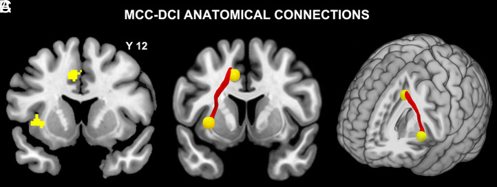
Connections between the DCI and MCC were found in the left hemisphere (Fig. 4). In particular, a three-dimensional (3D) reconstruction of the average tract, obtained using a single tract from each subject (with 10% threshold), is shown on a 3D brain template (Fig. 4 B and C).
Fig. 4.
Anatomical connectivity between the insula and cingulum. Activation of the left MCC and DCI resulting from the conjunction analysis of VF OBS vs. CT OBS and VF EXE vs. CT EXE contrasts (A). White-matter tract connecting the MCC and DCI (two-dimensional [2D] view in B; 3D view in C).
Discussion
Actions that have the same goal can be performed in different ways, which Stern termed vitality forms (13, 14). Vitality forms convey an agent’s internal states and thus play a crucial role in social interactions. In the last few years, several fMRI studies have been carried out to identify the neural substrate of vitality forms (3–12). These studies have shown that the dorso-central sector of the insula is the region that is selectively active in both the perception and expression of vitality forms. Recently, however, G.D.C. et al. (7) found that listening to action sounds performed gently or rudely produced activation not only of the DCI but also of the MCC.
To determine whether the MCC is also involved in the encoding of vitality forms, we carried out a new fMRI study with an experimental paradigm very similar to that used in the basic study we conducted previously (4), but with a major difference. This difference consisted of the type of control condition used. In our previous study, in the control condition participants were asked to observe or execute an action consisting of the accurate placement of a small ball in a little box. Therefore, the complexity of the task and the cognitive efforts associated with it (15) could explain the activation of MCC also in the control condition. In contrast, in the present study’s execution task, the control condition consisted of a simple hand action (moving a little box), whereas the observation task consisted of participants’ observation of hand actions identical to those in the experimental conditions but performed in a jerky way. These differences in the control condition may explain why our previous experiment did not show the activation of the cingulate cortex (see, e.g., Fig. 3). In fact, it is plausible that the previous experiment’s control condition masked the vitality form effect in the MCC.
In the present study, we also provided evidence that the MCC is endowed with the mirror mechanism. A voxel-based analysis showed that the BOLD signal strongly correlated during the observation and execution tasks in many voxels located in the MCC (48.6%) (i.e., HC mirror voxels). An identical analysis of the DCI showed similar results. The percentage of the HC mirror voxels located in the DCI was 39.2%. Additionally, we found that in the HC mirror voxels located in both the MCC and DCI, the BOLD signal change showed a stronger activity for rude vitality forms than the gentle ones.
Finally, to identify whether the MCC and DCI sites specifically related to action vitality forms are anatomically connected, we carried out a multifiber tractography investigation on the same group of participants. This analysis showed that these MCC and DCI sites are linked, forming a vitality form circuit for hand action. It is well known that the main tractrography limitation is the possible high false positive rate that has been observed in some studies (16). However, the robustness of our results is strongly confirmed by previous tract-tracing studies on macaque (17, 18). In fact, these connectional studies showed that in the monkey the sector homolog of the human middle cingulate cortex is tightly connected with the dorso-central part of the insula, a part of it known to be involved in modulating hand movements (19, 20). These data also indicate that the cingulo-insular circuit here described appears to be well conserved throughout the primate’s evolution.
Although our study indicates that DCI and MCC are both involved in encoding vitality form perception and expression, the fact that MCC is more strongly modulated during the execution of rude vitality form, relative to the gentle ones, than DCI suggests that the two areas can have a partial different functional role, in line with the existing literature.
In this respect it is noteworthy that: First, Craig described the whole insula as a sensory “interoceptive cortex” that integrates homeostatic, visceral, nociceptive, and somatosensory inputs (21), through which a representation of the internal state of the body is generated and, second, according to Kurth et al. (22), the insula can be subdivided in four main sectors: the sensorimotor, socioemotional, olfactory–gustatory, and cognitive ones. The DCI is located in the sensorimotor sector of the insula and is connected with the parieto-frontal circuit for reaching/grasping execution and observation (23) as well as with temporal territories encoding visual and acoustic biological stimuli (24). The DCI is also involved in processing the emotional aspects of visual stimuli; in fact a study on a blindsight patient demonstrated its selective activation for conscious, and not for unconscious, perception of fearful bodies (1).
Concerning MCC, in line with our results, previous studies showed that this cingulate sector is characterized by an evident motor scaffold. In fact, intracortical stimulation of MCC, carried out on epileptic drug-resistant patients, produced many typologies of motor acts, including arm, hand, body, and oral movements. Interestingly, the stimulation of this cingulate sector produced, before actual movements, the urge to move in relation to external contingencies (25).
Based on these considerations and on present results, we can hypothesize that DCI plays an essential role in encoding and integrating sensory and interoceptive information for generating the vitality form of the agent, during action execution, and for encoding those of the observer, during action observation. In contrast, MCC appears to be more involved in generating vitality form related to the external contingencies especially in the case of rude vitality form. However, further studies need to be carried out to test this hypothesis.
Methods
Sixteen healthy right-handed volunteers (nine females and seven males, mean age = 25.4, SD = 2) took part in the fMRI experiment. Due to the COVID-19 pandemic, 14 participants from the same group participated in the second scanning session, the purpose of which was to collect diffusion tensor imaging (DTI) images. All participants had normal or corrected-to-normal visual acuity. None of them reported a history of psychiatric or neurological disorders or current use of any psychoactive medications. They gave their written informed consent to be subjected to the experimental procedure, which was approved by the Local Ethics Committee of Parma (552/2020/SPER/UNIPR) in accordance with the Declaration of Helsinki.
Paradigm and Task.
The fMRI experiment consisted of two functional runs. In each run, we presented participants with video clips in two different tasks (OBS and EXE) and two different conditions (vitality forms, VF; control, CT). In total, four conditions were presented in independent miniblocks (VF OBS, VF EXE, CT OBS, CT EXE) in a randomized order (see also SI Appendix, Fig. S1). The OBS task started with the instruction “observe” and required the participants to pay attention to the action performed with vitality forms (VF OBS, Fig. 1, A1) or without vitality forms (CT OBS, Fig. 1, A3). The EXE task started with the instruction “execute” and required the participants to perform the action themselves (for details see also SI Appendix, Fig. S2). During the EXE tasks, we presented a static image of the actor seated opposite the observer and asked participants to move a little box toward the actor with vitality forms (gentle or rude, Fig. 1, B1 and B2) or without vitality forms (Fig. 1, B3) by simply rotating the wrist. A cue positioned in the center of the screen indicated when to start the action, and the color of the edge of the screen indicated the vitality form to use during the execution of the action (blue color: gentle, Fig. 1, B1; red color: rude, Fig. 1, B2; gray color: control, Fig. 1, B3). In each video, a fixation cross was introduced to control for restrained eye movements.
fMRI Data Acquisition and Analysis.
Anatomical T1-weighted and functional T2*-weighted MR images were acquired with a 3 Tesla General Electrics scanner (details in SI Appendix, SI Methods). After standard preprocessing steps, data were analyzed using a random-effects model, implemented in a two-level procedure. In the first level, the fMRI BOLD signal of each participant was modeled using two general linear models (GLMs), and analysis was carried out using SPM12 software (the Wellcome Department of Imaging Neuroscience). Subsequently, in the second level, the BOLD signal of all participants was modeled using two other GLMs, and the group analysis was carried out. Specifically, in the second-level analysis, the first group analysis was based on a GLM comprising four regressors (VF OBS, CT OBS, VF EXE, CT EXE) and enabled us to assess activations associated with each task versus implicit baseline and activations resulting from the direct contrast between conditions (VF OBS vs. CT OBS, VF EXE vs. CT EXE; Fig. 2, PFWE < 0.05 corrected at the cluster level).
In the second group analysis, the BOLD signal was modeled in a GLM comprising six regressors (GT OBS, RD OBS, CT OBS, GT EXE, RD EXE, CT EXE) and enabled us to examine and compare the BOLD signal change during the processing of gentle and rude vitality forms in the OBS and EXE tasks (details in SI Appendix, SI Statistical Analysis).
On the basis of the functional maps obtained from the first group analysis, we carried out a conjunction analysis of the brain activations resulting from the contrasts VF OBS vs. CT OBS and VF EXE vs. CT EXE (PFWE < 0.05 corrected at the cluster level) to highlight the brain areas active during both the perception and execution of vitality forms (Fig. 3 A and B). The results of this analysis highlighted two regions: the left DCI and the MCC. Subsequently, in these two regions, for each participant and for each single voxel, we extracted the BOLD signal change relative to each experimental condition (VF OBS, CT OBS, VF EXE, CT EXE) using the REX toolbox (https://www.nitrc.org/projects/rex/). Then, in order to identify and quantify voxels that showed a very similar BOLD signal change in the OBS and EXE tasks (HC mirror voxels), for each single voxel, the BOLD signal change obtained during the VF OBS condition was correlated with that obtained during the VF EXE condition (for details, see above), and a significant threshold was set at r > 0.7 (coefficient of determination R2 ≥ 0.49, P < 0.05). Finally, considering these voxels located in the DCI and MCC, in order to compare the BOLD signal during the processing of different vitality forms, we extracted from the functional maps obtained in the second group analysis the BOLD signal change relative to gentle and rude conditions for both tasks.
Diffusion Data Acquisition and Analysis.
In another scanning session, a diffusion spin-echo single shot echo planar imaging sequence with 64 diffusion directions (effective b-value of 1,000 s/mm2), eight images with no diffusion weight in the anterior–posterior phase encoding direction and eight images with no diffusion weight in the reverse phase encoding direction were collected from the same participants (details in SI Appendix, SI Methods). Diffusion data were processed using the FMRIB Software Library (FSL) tools (version 5.0.9). After standard preprocessing steps, a further probabilistic tractography analysis was performed with FSL’s PROBTRACKX tool, testing the connection of two regions of interest (diameter 12 mm) (DCI coordinates: −32 9 −2; MCC coordinates: −9 9 42).
Acknowledgments
G.D.C. and A.S. are supported by a starting grant from the European Research Council (ERC) to A.S. under the European Union’s Horizon 2020 research and innovation programme. Grant agreement No. 804388, wHiSPER. G.R. is supported by a grant Lombardia è Ricerca from the Lombardia region.
Footnotes
Reviewers: D.M.A.M., Universitatsklinikum Munster; M.T., Universita degli Studi di Torino; and C.W., University of Heidelberg.
The authors declare no competing interest.
This article contains supporting information online at https://www.pnas.org/lookup/suppl/doi:10.1073/pnas.2111358118/-/DCSupplemental.
Data Availability
All study data are included in the article and/or supporting information.
References
- 1.Tamietto M., et al., Once you feel it, you see it: Insula and sensory-motor contribution to visual awareness for fearful bodies in parietal neglect. Cortex 62, 56–72 (2015). [DOI] [PubMed] [Google Scholar]
- 2.de Gelder B., Towards the neurobiology of emotional body language. Nat. Rev. Neurosci. 7, 242–249 (2006). [DOI] [PubMed] [Google Scholar]
- 3.Di Cesare G., et al., The neural correlates of ‘vitality form’ recognition: An fMRI study: This work is dedicated to Daniel Stern, whose immeasurable contribution to science has inspired our research. Soc. Cogn. Affect. Neurosci. 9, 951–960 (2014). [DOI] [PMC free article] [PubMed] [Google Scholar]
- 4.Di Cesare G., Di Dio C., Marchi M., Rizzolatti G., Expressing our internal states and understanding those of others. Proc. Natl. Acad. Sci. U.S.A. 112, 10331–10335 (2015). [DOI] [PMC free article] [PubMed] [Google Scholar]
- 5.Di Cesare G., et al., Vitality forms processing in the insula during action observation: A multivoxel pattern analysis. Front. Hum. Neurosci. 10, 267 (2016). [DOI] [PMC free article] [PubMed] [Google Scholar]
- 6.Di Cesare G., Marchi M., Errante A., Fasano F., Rizzolatti G., Mirroring the social aspects of speech and actions: The role of the insula. Cereb. Cortex 28, 1348–1357 (2017). [DOI] [PubMed] [Google Scholar]
- 7.Di Cesare G., Marchi M., Pinardi C., Rizzolatti G., Understanding the attitude of others by hearing action sounds: The role of the insula. Sci. Rep. 9, 14430 (2019). [DOI] [PMC free article] [PubMed] [Google Scholar]
- 8.Di Cesare G., Gerbella M., Rizzolatti G., The neural bases of vitality forms. Natl. Sci. Rev. 7, 202–213 (2020). [DOI] [PMC free article] [PubMed] [Google Scholar]
- 9.Di Cesare G., Vannucci F., Rea F., Sciutti A., Sandini G., How attitudes generated by humanoid robots shape human brain activity. Sci. Rep. 10, 16928 (2020). [DOI] [PMC free article] [PubMed] [Google Scholar]
- 10.Di Cesare G., “The importance of the affective component of movement in action understanding” in Modelling Human Motion, Noceti N., Sciutti A., Rea F., Eds. (Springer, Cham, 2020), pp. 103–116. [Google Scholar]
- 11.Rizzolatti G., D’Alessio A., Marchi M., Di Cesare G., The neural bases of tactile vitality forms and their modulation by social context. Sci. Rep. 11, 9095 (2021). [DOI] [PMC free article] [PubMed] [Google Scholar]
- 12.Di Cesare G., Cuccio V., Marchi M., Sciutti A., Rizzolatti G., Communicative and affective components in processing auditory vitality forms: An fMRI study. Cereb. Cortex, 10.1093/cercor/bhab255 (2021). [DOI] [PMC free article] [PubMed] [Google Scholar]
- 13.Stern D. N., The Interpersonal World of the Infant (Basic Books, New York, 1985). [Google Scholar]
- 14.Stern D. N., Forms of Vitality Exploring Dynamic Experience in Psychology, Arts, Psychotherapy, and Development (Oxford University Press, United Kingdom, 2010). [Google Scholar]
- 15.Procyk E., et al., Midcingulate motor map and feedback detection: Converging data from humans and monkeys. Cereb. Cortex 26, 467–476 (2016). [DOI] [PMC free article] [PubMed] [Google Scholar]
- 16.Schilling K. G., et al., Limits to anatomical accuracy of diffusion tractography using modern approaches. Neuroimage 185, 1–11 (2019). [DOI] [PMC free article] [PubMed] [Google Scholar]
- 17.Vogt B. A., Pandya D. N., Cingulate cortex of the rhesus monkey: II. Cortical afferents. J. Comp. Neurol. 262, 271–289 (1987). [DOI] [PubMed] [Google Scholar]
- 18.Mesulam M. M., Mufson E. J., Insula of the old world monkey. III: Efferent cortical output and comments on function. J. Comp. Neurol. 212, 38–52 (1982). [DOI] [PubMed] [Google Scholar]
- 19.Jezzini A., Caruana F., Stoianov I., Gallese V., Rizzolatti G., Functional organization of the insula and inner perisylvian regions. Proc. Natl. Acad. Sci. U.S.A. 109, 10077–10082 (2012). [DOI] [PMC free article] [PubMed] [Google Scholar]
- 20.Jezzini A., et al., A shared neural network for emotional expression and perception: An anatomical study in the macaque monkey. Front. Behav. Neurosci. 9, 243 (2015). [DOI] [PMC free article] [PubMed] [Google Scholar]
- 21.Craig A. D., How do you feel? Interoception: The sense of the physiological condition of the body. Nat. Rev. Neurosci. 3, 655–666 (2002). [DOI] [PubMed] [Google Scholar]
- 22.Kurth F., Zilles K., Fox P. T., Laird A. R., Eickhoff S. B., A link between the systems: Functional differentiation and integration within the human insula revealed by meta-analysis. Brain Struct. Funct. 214, 519–534 (2010). [DOI] [PMC free article] [PubMed] [Google Scholar]
- 23.Di Cesare G., et al., Insula connections with the parieto-frontal circuit for generating arm actions in humans and macaque monkeys. Cereb. Cortex 29, 2140–2147 (2019). [DOI] [PubMed] [Google Scholar]
- 24.Almashaikhi T., et al., Functional connectivity of insular efferences. Hum. Brain Mapp. 35, 5279–5294 (2014). [DOI] [PMC free article] [PubMed] [Google Scholar]
- 25.Caruana F., et al., Motor and emotional behaviours elicited by electrical stimulation of the human cingulate cortex. Brain 141, 3035–3051 (2018). [DOI] [PubMed] [Google Scholar]
Associated Data
This section collects any data citations, data availability statements, or supplementary materials included in this article.
Data Availability Statement
All study data are included in the article and/or supporting information.


