In our paper (1), we draw two major conclusions.
-
1)
In cultured cells, we find strong evidence for a LINE1-mediated integration mechanism: target-site duplication, LINE1 endonuclease recognition sequence, and “poly-A tract” length ranging from 2 bp to 65 bp.
-
2)
Indirect evidence suggests that genomic integration of severe acute respiratory syndrome coronavirus 2 (SARS-CoV-2) sequences can also occur in patients, and integrated viral DNA can be expressed. This conclusion is based on the detection of large fractions of negative-strand viral RNA in lung tissues from some patients. SARS-CoV-2 is a positive-strand RNA virus, and the majority of viral RNAs detected in acutely infected cells are positive-strand genomic or subgenomic RNAs. Negative-strand viral RNAs are produced during virus replication and serve as templates for protein-coding positive-strand RNAs. We show that, in cells actively replicating virus, less than 1 in 1,000 viral RNAs are negative stranded. However, in some patient tissues, up to 50% of viral RNAs are negative stranded. Importantly, some of the negative-stranded viral RNAs are viral—cellular chimeras. This supports our inference that these sequences originate from SARS-CoV-2 sequences randomly integrated into genomic DNA, with ∼50% integrated viral DNA in the opposite orientation relative to host genes, rather than being sequencing artifacts.
Briggs et al. (2) performed a simple PCR assay for viral DNA and concluded that “all patients reported as positive were infected with SARS-CoV-2 virus, demonstrating no impact of possible viral integration on COVID-19 diagnosis.” We summarize our responses to Briggs et al.’s conclusions.
-
1)
Two out of 768 samples (0.26%) reproducibly amplified SARS-CoV-2 sequences in RT-absence qPCR.
Briggs et al. detected SARS-CoV-2 DNA signals from 2 out of 768 nasal swab samples in RT-absence qPCR. This result is consistent with our conclusion that SARS-CoV-2 sequences can be integrated into the genomic DNA. The detection of SARS-CoV-2 DNA in the two samples and the sensitivity of the signal to DNase treatment suggest these signals may not be due to contaminating DNA in the PCR. Briggs et al. provide no evidence to show that there was contamination.
-
2)
SARS-CoV-2 genome integration is a rare event.
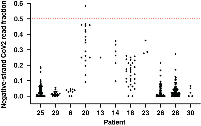
We agree that integration may be rare. However, in patients, a very large number of cells are infected. An important issue is whether there are nasal epithelial cells that survive an infection for a prolonged time and thus have a higher probability of integration. This is particularly relevant for acutely infected patients that have replicating virus in the nasal epithelium as studied by Briggs et al. However, if integration occurs in a highly expressed gene in longer-lived cells, even a small fraction of integrated sequences may cause a PCR-positive signal. Indeed, recently published nasal swab single-cell RNA sequencing (scRNA-seq) data (3) suggest that at least 1 out of 11 patients showed a consistently high fraction (up to ∼50%) of negative-strand viral RNA (Fig. 1), supporting our conclusion (1) and suggesting that there might be a higher level of viral DNA integration in nasal cells. We would point out that patients with more-severe symptoms showed higher fractions of negative-strand viral RNAs.
-
3)
SARS-CoV-2 integration has no practical impact on RT-qPCR diagnosis.
Fig. 1.
High fractions of RNA-seq reads derived from negative-strand viral RNAs in nasal swab cells support viral integrations. Fraction of viral reads that are derived from negative-strand viral RNAs in patient nasal swab single cells with more than 10 total viral reads per cell. Single cells are shown as black dots; red dashed line indicates the level at which 50% of viral reads are from negative-strand viral RNAs, a level expected if all the viral sequences are derived from integrated sequences. Data are from ref. 3.
We do not question the ability of RT-qPCR to detect actively replicating virus in patients during an acute infection. Rather, our data suggest that SARS-CoV-2 sequences integrated into the genome could produce a PCR-positive signal in patients that have cleared infectious virus. PCR assays are extremely sensitive. We suggest that prolonged viral RNA shedding in late-stage patients with no evidence for virus replication could be due to expression of integrated viral sequences, producing small amounts of viral RNA that are detectable by RT-qPCR. The study of Briggs et al. (2) does not differentiate patients who were in the initial stages of an infection from patients who had prolonged or recurrent low levels of viral RNA.
Footnotes
Author contributions: L.Z., A.R., M.I.B., S.H.H., R.A.Y., and R.J. analyzed data; and L.Z. and R.J. wrote the paper.
Competing interest statement: R.J. is an advisor/cofounder of Fate Therapeutics, Fulcrum Therapeutics, Omega Therapeutics, and Dewpoint Therapeutics. R.A.Y. is a founder and shareholder of Syros Pharmaceuticals, Camp4 Therapeutics, Omega Therapeutics, and Dewpoint Therapeutics. L.Z., A.R., M.I.B., and S.H.H. declare no competing interest.
References
- 1.Zhang L., et al., Reverse-transcribed SARS-CoV-2 RNA can integrate into the genome of cultured human cells and can be expressed in patient-derived tissues. Proc. Natl. Acad. Sci. U.S.A. 118, e2105968118 (2021). [DOI] [PMC free article] [PubMed] [Google Scholar]
- 2.Briggs E., et al., Assessment of potential SARS-CoV-2 virus integration into human genome reveals no significant impact on RT-qPCR COVID-19 testing. Proc. Natl. Acad. Sci. U.S.A., 10.1073/pnas.2113065118 (2021). [DOI] [PMC free article] [PubMed] [Google Scholar]
- 3.Ziegler C. G. K., et al., Impaired local intrinsic immunity to SARS-CoV-2 infection in severe COVID-19. Cell 184, 4713–4733.e22 (2021). [DOI] [PMC free article] [PubMed] [Google Scholar]


