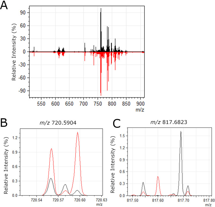
FIG 4.
Representative spectra of lipids detected in gastric tissue. (A) Average mass spectra over entire gastric tissue sections are shown for a representative uninfected stomach (black) and a representative H. pylori-infected stomach (red). The overall signal intensity and quality were very similar in infected versus uninfected tissue. (B) Zoomed-in image of the region surrounding m/z 720.5904, which was more abundant in infected tissues compared to uninfected, as shown in Fig. 5. (C) Zoomed-in image of the region surrounding m/z 817.6823, which was less abundant in infected tissues compared to uninfected, as shown in Fig. 6.

