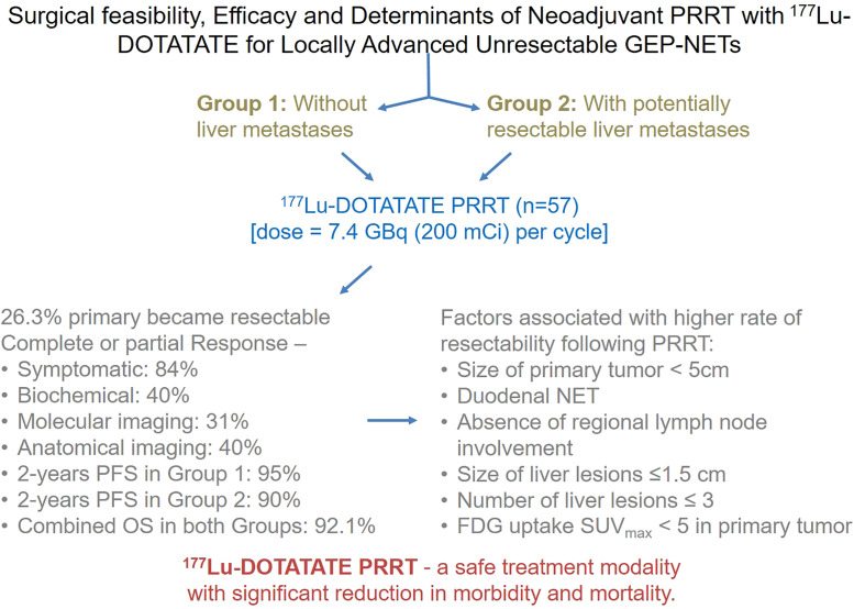
Visual Abstract
Keywords: neoadjuvant PRRT, gastroenteropancreatic neuroendocrine tumor, peptide receptor radionuclide therapy, 177Lu-DOTATATE therapy, 68Ga-DOTATATE PET/CT, triphasic CT
Abstract
We assessed 177Lu-DOTATATE peptide receptor radionuclide therapy (PRRT) in the neoadjuvant setting in patients with gastroenteropancreatic neuroendocrine tumors (GEP-NETs). We also evaluated the variables associated with resectability of the primary tumor after PRRT. Methods: This study included 57 GEP-NET patients who had a primary tumor that was unresectable (because of vascular involvement as defined using the pancreatic ductal adenocarcinoma criteria of the National Comprehensive Cancer Network) and who underwent 177Lu-DOTATATE therapy without any prior surgery. They were categorized into 2 groups: 23 patients without liver metastases (group 1) and 34 patients with potentially resectable liver metastases (group 2). 177Lu-DOTATATE was administered with mixed amino acid–based renal protection at a dose of 7.4 GBq (200 mCi) per cycle. Surgical resectability was evaluated using triphasic contrast-enhanced abdominal CT imaging at 3 different time points during the PRRT course. Four broad categories of overall PRRT response were evaluated. The Kaplan–Meier product-limit method was used to calculate progression-free survival (PFS) and overall survival (OS). Associations between variables and a resectable primary tumor after PRRT were analyzed using the χ2 test, with a P value of less than 0.05 considered statistically significant. Results: After 177Lu-DOTATATE therapy, the unresectable primary tumor became resectable in 15 of 57 (26.3%) patients (7 patients in group 1 and 8 patients in group 2). A complete or partial response to PRRT was seen in 48 patients (84%), 23 patients (40%), 18 patients (31%), and 23 patients (40%) using symptomatic, biochemical, molecular imaging, and anatomic imaging criteria, respectively. Estimated rates of PFS were 95% and 90% at 2 y in groups 1 and 2, respectively. The 2-y OS of the 2 groups combined was 92.1%. The rate at which the primary tumor was resectable after PRRT was significantly higher in patients who had duodenal neuroendocrine tumors, patients who had GEP-NETs with no regional lymph node involvement, patients for whom the primary tumor was smaller than 5 cm, patients for whom liver metastases were no larger than 1.5 cm, patients for whom there were no more than 3 liver metastases, and patients for whom 18F-FDG uptake in the primary tumor had an SUVmax of less than 5. Conclusion: In a moderate fraction of GEP-NET patients, with or without liver metastases, whose primary tumor was unresectable because of vascular involvement, the primary tumor converted from unresectable to resectable after 177Lu-DOTATATE therapy, signifying that neoadjuvant PRRT can be considered in such patients. The effective control of symptoms, favorable morphologic and functional imaging response, and durable PFS and OS that we observed after 177Lu-DOTATATE PRRT may lead to less morbidity and mortality in these patients.
Neuroendocrine tumors (NETs) represent a diverse group of neoplasms arising from neuroendocrine cells located at many different sites throughout the body, most commonly in the gastroenteropancreatic and respiratory systems. Because multiple therapeutic options are available (1,2), maximum therapeutic benefit to the patient is achieved with the involvement of a multidisciplinary team that includes medical oncologists, surgeons, gastroenterologists, radiologists, and nuclear medicine physicians.
In NETs, surgery is only definitive curative treatment option. The 5-y survival rate is more than 60% in patients with resectable gastroenteropancreatic NETs (GEP-NETs), whereas it drops to less than 30% in patients with unresectable tumors (3–9). Aggressive surgical resection of the primary tumor and liver metastases may improve symptoms and overall survival (OS) in GEP-NETs. However, the resectability of the primary tumor in GEP-NETs depends on the presence or absence of major abdominal vascular involvement and on the size and infiltration of the tumor into other adjacent tissues (6,8,10,11).
In somatostatin receptor–positive GEP-NETs, peptide receptor radionuclide therapy (PRRT) with targeted radiolabeled somatostatin analogs such as 90Y-DOTATOC and 177Lu-DOTATATE has the advantage of producing a selective treatment effect through a ligand that carries the radioisotope directly to the tumor cell population (12). PRRT has been reported to result in disease stabilization, partial remission, or even a reduction in tumor mass (more than 50%) in these patients (13). Thus, PRRT has been used as neoadjuvant therapy to decrease tumor size in a few case reports and studies on NET (14–16).
The aim of our study was to assess the performance of 177Lu-DOTATATE PRRT as neoadjuvant therapy in GEP-NET patients whose primary tumor is unresectable because of vascular involvement and who have either no liver metastases or potentially resectable liver metastases. In addition, we evaluated the overall efficacy of PRRT with the help of other response evaluation parameters and determined variables associated with the resectability of the primary tumor after PRRT.
MATERIALS AND METHODS
Patient Population
The study included patients with histopathologically proven GEP-NETs who had a primary tumor that was unresectable because of vascular involvement, who had no liver metastases or had potentially resectable liver metastases, and who had undergone 177Lu-DOTA-octreotate PRRT without any prior surgical intervention of the primary tumor. A primary tumor that was unresectable because of vascular involvement was defined using the National Comprehensive Cancer Network (NCCN) criteria for pancreatic ductal adenocarcinoma (17) and was classified as locally advanced when the tumor involved more than 180° of the circumference of the superior mesenteric artery, celiac trunk, aorta, inferior vena cava, portal vein, or superior mesenteric vein; when there was thrombosis of the portomesenteric venous system; or when there was unreconstructable occlusion of the superior mesenteric vein or portal vein. Patients were excluded if they had distant metastatic disease or extensive bilobar liver metastatic disease.
The patients were divided into 2 groups based on the presence or absence of metastatic liver disease: group 1 had no liver metastases, and group 2 had potentially resectable liver metastases (Table 1). In group 1, 10 patients had grade 1 tumors and 13 patients had grade 2. In group 2, 16 patients had grade 1 tumors, 17 patients had grade 2, and 1 patient had grade 3. All but one of the patients in this study had well-differentiated NETs.
TABLE 1.
Patient Characteristics
| Characteristic | Group 1 | Group 2 |
|---|---|---|
| Total patients (n) | 23 | 34 |
| Sex (n) | ||
| Male | 15 | 18 |
| Female | 8 | 16 |
| Age (y) | ||
| Range | 30–76 | 30–78 |
| Average | 52 | 51 |
| Symptomatic patients before PRRT (n) | 23 | 34 |
| Prior therapy (n) | ||
| Chemotherapy | 9 | 6 |
| Octreotide analog | 5 | 7 |
| Primary site (n) | ||
| Pancreatic | 12 | 20 |
| Duodenal | 4 | 8 |
| Jejunal | 1 | 4 |
| Ileal | 6 | 2 |
| MIB-1 index | ||
| Range | 1%–15% | 1%–25% |
| Median | 3% | 4% |
| Primary tumor size before PRRT (cm) | ||
| Range | 3.5–11 | 4–12 |
| Average | 5.8 | 6 |
| Liver metastasis size before PRRT (cm) | ||
| Range | — | 0.8–5.6 |
| Average | — | 3 |
| Cumulative 177Lu-DOTATATE dose | ||
| Range | 14.8–40.7 GBq (400–1,100 mCi) | 14.8–40.7 GBq (400–1,100 mCi) |
| Average | 22.2 GBq (600 mCi) | 27.45 GBq (742 mCi) |
| PRRT cycles (n) | ||
| Range | 2–5 | 2–5 |
| Average | 4 | 4 |
The study was approved by the Institutional Scientific Committee and the Institutional Ethics Committee. The need to obtain informed consent was waived because the study was retrospective.
PRRT Regimen
The patients had undergone triphasic contrast-enhanced abdominal CT and dual-tracer PET/CT (68Ga-DOTATATE and 18F-FDG PET/CT) before the start of the PRRT. According to our institutional protocol, neoadjuvant 177Lu-DOTATATE PRRT is given to patients who have somatostatin receptor–positive GEP-NETs with a Krenning score of at least 3 (as compared on maximum-intensity projection, coronal, and transaxial 68Ga-DOTATATE PET/CT images), whose primary tumor is unresectable because of vascular involvement, and who have either no liver metastases or potentially resectable liver metastases. Mixed amino acid–based renal protection is followed along with PRRT and 177Lu-DOTATATE is administered at a dose of 7.4GBq (200 mCi) per cycle. The PRRT cycles are repeated at intervals of 8–10 wk (4–5 cycles in total).
Surgical Resectability Evaluation
Surgical resectability after PRRT was evaluated using abdominal CT at 3 phases: triphasic contrast-enhanced abdominal CT imaging was acquired 4 mo after the second cycle (i.e., 2 mo after the first cycle of PRRT), and next, 3 mo after completion of the last cycle. The surgical resectability criteria given by the NCCN for pancreatic ductal adenocarcinoma were used in this study. These criteria define a post-PRRT resectable primary tumor as one that shows a decrease in size on contrast-enhanced CT and clear fat planes around major abdominal vessels, or as one that involves less than 180° of the circumference of the superior mesenteric vein or portal vein, celiac trunk, or superior mesenteric or hepatic artery and that encases or occludes a short segment of the superior mesenteric vein or portal vein.
Response Evaluation
After PRRT, all patients were followed up with symptomatic and biochemical (serum chromogranin-A level) response evaluations and with molecular (68Ga-DOTATATE PET/CT) and anatomic (contrast-enhanced CT) imaging.
Symptomatic Response
For the symptomatic response evaluation, the patients were asked to evaluate—on a scale of 0%–100% compared with baseline—whether their tumor-related symptoms had disappeared (90%–100% improvement; complete response [CR]), had improved (30%–89% improvement; partial response [PR]), were stable (<30% improvement or <30% deterioration; stable disease [SD]), or had worsened (≥30% increase in symptoms or new symptoms; progressive disease [PD]).
Biochemical Response
Biochemical response was assessed using serum chromogranin-A levels. The baseline level before the start of PRRT was measured, and the percentage change at the time of analysis was calculated. More than a 75% reduction or normalization of the level was considered CR, a 30%–75% reduction was PR, a less than 30% reduction to a less than 30% increase was SD, and an increase by 30% or more was PD.
Molecular Imaging Response
The 68Ga-DOTATATE PET/CT response evaluation was done using PERCIST.
Anatomic Imaging Response
The contrast-enhanced CT response evaluation was done using RECIST, version 1.1.
Progression-Free Survival (PFS) and OS
PFS and OS were also assessed. PFS was defined as the time from the first cycle of PRRT to documented disease progression on an imaging study, and OS was defined as the time from the first cycle of PRRT to death of the patient. If death did not occur during the observation period, the survival time was censored on the last date at which the subject was known to be alive.
Statistics
Patient characteristics were summarized as count and percentage, and the number of patients with a resectable primary tumor after PRRT was determined. CR, PR, SD and PD were determined for each of the 4 types of response evaluation. Median and 95% CI for PFS and OS were calculated by the Kaplan–Meier method. PFS curves for groups 1 and 2 were determined using the Kaplan–Meier product-limit method.
The χ2 test was used to test the association between the following categoric variables and a resectable primary tumor after PRRT, with a P value of less than 0.05 considered statistically significant: patient age at start of PRRT (20–45 y, 46–60 y, or ≥61 y), site of primary tumor (pancreatic, duodenal, jejunal, or ileal), total cumulative radionuclide dose (14,800–22,200 MBq [400–600 mCi], 22,237–29,600 MBq [601–800 mCi], or 29,637–40,700 MBq [801–1,100 mCi]), number of PRRT cycles (2, 3–4, or 5), MIB-1 index (<3%, 3%–20%, or >20%), previous chemotherapy and previous octreotide analog therapy (received vs. not received), regional lymph node involvement (involved vs. not involved), baseline size of primary tumor (<5 cm, 5–7 cm, or >7 cm), and baseline 68Ga-DOTATATE uptake (SUVmax < 20, SUVmax = 20–50, or SUVmax > 50) and 18F-FDG uptake in liver metastases (SUVmax < 5, SUVmax = 5–7, or SUVmax > 7) in primary tumor. Additionally, the following variables were evaluated in group 2 patients: baseline 68Ga-DOTATATE uptake and 18F-FDG uptake, size of liver metastases (≤1.5 cm, 1.6–3.5 cm, or >3.5 cm), and number of liver metastases (≤3, 4–6, or ≥7).
RESULTS
The study included and analyzed 57 patients with GEP-NETs: 23 in group 1 and 34 in group 2 (Table 1).
The pancreas was the most common site for the primary NET (32 patients [56%]), with the head and body of the pancreas being most commonly involved (27 patients [47%]). The superior mesenteric vein or portal vein was the commonly involved blood vessel (35 patients [61%]), followed by the superior mesenteric artery (27 patients [47%]). The baseline size of primary GEP-NETs was 3.5–11 cm (average, 5.8 cm) in group 1 and 4–12 cm (average, 6 cm) in group 2. The baseline size of liver metastases was 0.8–5.6 cm (average, 3 cm) in group 2. The MIB-1 labeling index was 1–15 (median, 3) in group 1 and 1–25 (median, 4) in group 2. All 57 patients were symptomatic before the start of PRRT, with abdominal pain, vomiting, weakness, and weight loss being the most common complaints. Before PRRT, systemic chemotherapy and somatostatin analog therapy were administered to 9 and 5 patients, respectively, in group 1 and 6 and 7 patients, respectively, in group 2 and produced either no response or PD.
The total cumulative dose of 177Lu-DOTATATE per patient in groups 1 and 2 was, respectively, 14.8–40.7 GBq (400–1,100 mCi; average, 22.2 GBq [600 mCi]) and 14.8–40.7 GBq (400–1,100 mCi; average, 27.45 GBq [742 mCi]). The number of cycles per patient ranged from 2 to 5 and averaged 4.
After PRRT, the size of the primary GEP-NETs was 2.0–10 cm (average, 4.8 cm) in group 1 and 2.0–9.5 cm (average, 4.6 cm) in group 2, and the size of the liver metastases in group 2 was 0.5–7 cm (average, 2.4 cm).
PRRT was well tolerated in all 57 GEP-NET patients, none of whom showed any major hematologic or renal toxicity. Two patients in group 1 and one patient in group 2 showed mild (grade I) hematologic toxicity and renal toxicity, respectively, during the initial PRRT cycles but were found to have recovered during the subsequent follow-up.
Surgically Resectable Primary Tumor After PRRT
According to the NCCN criteria, an unresectable primary GEP-NET became resectable after PRRT in 7 of 23 patients in group 1 (2 pancreatic, 3 duodenal, and 2 ileal) and 8 of 34 patients in group 2 (4 pancreatic, 3 duodenal, and 1 jejunal). Thus, the overall rate at which the primary tumors became resectable in the 2 groups was 26.3% (15/57 patients). Imaging was repeated after 2 cycles in all 57 patients and after 4–5 cycles in 56 patients; of the 15 patients who became operable after PRRT, 1 became operable after 2 cycles and 14 after 4–5 cycles.
Response Evaluation
Symptomatic Response
All GEP-NET patients had symptomatic disease before PRRT. After PRRT, 19 of the 23 patients in group 1 had CR (82.8%), 1 had PR (4.3%), 2 had SD (8.6%), and 1 had PD (4.3%), whereas 24 of the 34 patients in group 2 had CR (70.5%), 4 had PR (11.9%), 3 had SD (8.8%), and 3 had PD (8.8%)
Biochemical Response
Regarding biochemical response, no patients among the 23 in group 1 had CR, whereas 10 had PR (43.5%), 10 had SD (43.5%), and 3 had PD (13%). No patients among the 34 in group 2 had CR, whereas 13 had PR (38.2%), 18 had SD (53%), and 3 had PD (8.8%).
Molecular Imaging Response
Regarding the response on 68Ga-DOTATATE PET/CT, 1 of the 23 patients in group 1 had CR (4.3%), 8 had PR (34.8%), 12 had SD (52.3%), and 2 had PD (8.6%). None of the 34 patients in group 2 had CR, whereas 9 had PR (26.5%), 24 had SD (70.5%), and 1 had PD (3%).
Anatomic Imaging Response
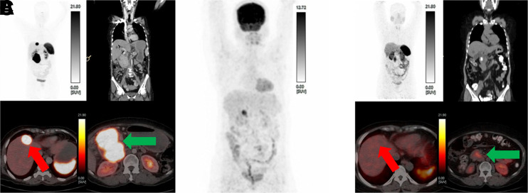
Regarding the response on contrast-enhanced CT, none of the 23 patients in group 1 had CR, whereas 7 had PR (30.4%), 15 had SD (65.3%), and 1 had PD (4.3%). In group 2, none of the 34 patients had CR, whereas 16 had PR (47%; Fig. 1), 15 had SD (44.1%), and 3 had PD (8.9%) (Table 2).
FIGURE 1.
A 56-y-old women with unresectable pancreatic NET. (A) Baseline 68Ga-DOTATATE maximum-intensity-projection (MIP) PET image (left upper panel) and transaxial fused PET/CT images (left panel, both lower images) showed intensely somatostatin receptor–avid unresectable primary pancreatic lesion (8.0 × 8.8 cm, green arrow), and left lower panel showed intensely somatostatin receptor–avid single metastasis (2.5 × 2.8 cm, red arrow) in segment IV of the liver. Contrast-enhanced CT image (right upper panel) is coronal view showing pancreatic lesion involving portal vein and superior mesenteric vein (>180°). (B) Baseline 18F-FDG PET (MIP) image demonstrated no uptake in primary tumor or metastasis. (C) Post-PRRT 68Ga-DOTATATE PET/CT: PET (MIP) image (left upper panel), transaxial fused PET/CT images (both lower panels), and coronal view of CT image (right upper panel). After 5 cycles of PRRT (total cumulative dose, 33.3 GBq), image in left lower panel showed complete morphologic disappearance of liver metastasis (red arrow), significant reduction in size and 68Ga-DOTATATE uptake of pancreatic lesion (green arrow), and no major abdominal vessel involvement. Patient underwent Whipple procedure to resect primary tumor without any major complications in perioperative period or in subsequent follow-up.
TABLE 2.
PRRT Response Evaluation Results
| Symptomatic | Biochemical | Molecular imaging | Anatomic imaging | |||||
|---|---|---|---|---|---|---|---|---|
| Response | Group 1 | Group 2 | Group 1 | Group 2 | Group 1 | Group 2 | Group 1 | Group 2 |
| CR | 19 | 24 | 0 | 0 | 1 | 0 | 0 | 0 |
| PR | 1 | 4 | 10 | 13 | 8 | 9 | 7 | 16 |
| SD | 2 | 3 | 10 | 18 | 12 | 24 | 15 | 15 |
| PD | 1 | 3 | 3 | 3 | 2 | 1 | 1 | 3 |
PFS and OS
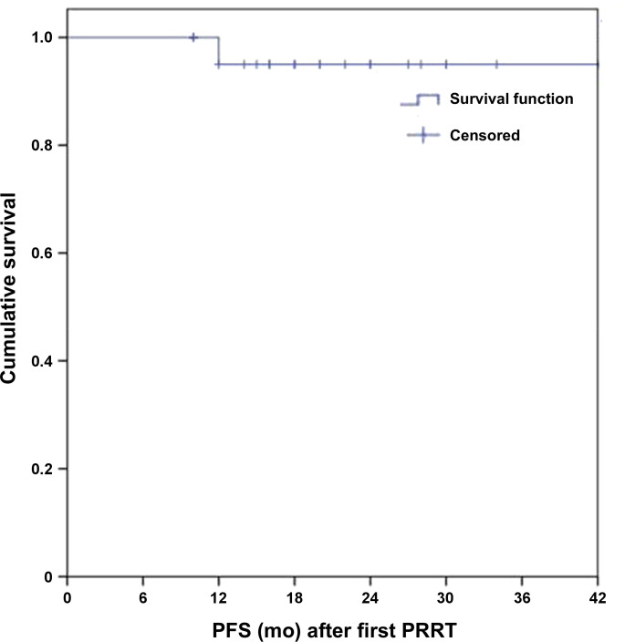
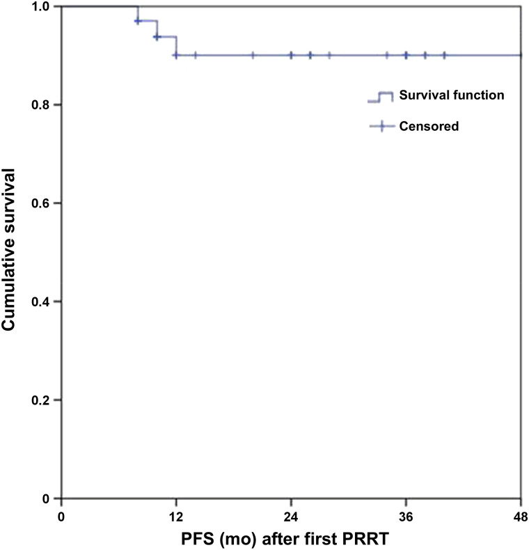
In this study, with a median follow-up period of 24 mo, the median PFS and OS were not reached. The estimated rates of PFS were 95% and 90% at 2 y in groups 1 and 2, respectively (Figs. 2 and 3). No deaths occurred in group 1, whereas 1 patient in group 2 died. The 2-y OS of both groups combined was 92.1%.
FIGURE 2.
Kaplan–Meier curve of PFS in group 1.
FIGURE 3.
Kaplan–Meier curve of PFS in group 2.
Association of Tumor Resectability After PRRT
A resectable primary tumor after PRRT was found to be significantly associated with site of primary tumor (duodenal NET), regional lymph node involvement (no involvement), size of primary tumor (<5 cm), and baseline 18F-FDG uptake in primary tumor (SUVmax < 5) for groups 1 and 2 combined. For group 2, a significant association was found for size of liver metastases (≤1.5 cm) and number of liver metastases (≤3).
DISCUSSION
In GEP-NETs, surgery offers the only chance for cure, and aggressive surgical resection of the tumor has been reported to result in long-term survival with acceptable morbidity and mortality. Neoadjuvant therapy—mainly chemotherapy, radiation therapy, or hormonal therapy—is intended to reduce the tumor size, enabling surgical resection of many gastrointestinal cancers. Treatment options are limited in patients with unresectable and locally advanced GEP-NETs. For these tumors, chemotherapy has limited efficacy and a high incidence of significant side effects (18). Biologic therapy with somatostatin analogs and interferon-α can reduce symptoms but fails to produce an objective response in terms of tumor shrinkage in the neoadjuvant setting (19).
Somatostatin analogs labeled with radionuclides have been used in diagnosis and therapy of GEP-NETs, as these tumors express somatostatin receptors on their surface. PRRT has been used in disseminated metastatic and unresectable GEP-NETs, with positive somatostatin receptor expression confirmed by molecular imaging. 177Lu-DOTATATE PRRT has shown promising response and survival rates, with minimal associated toxicity (13,20,21). A few reports demonstrated the use of preoperative PRRT in unresectable pancreatic NETs for reducing tumor size and enabling surgical intervention (14–16).
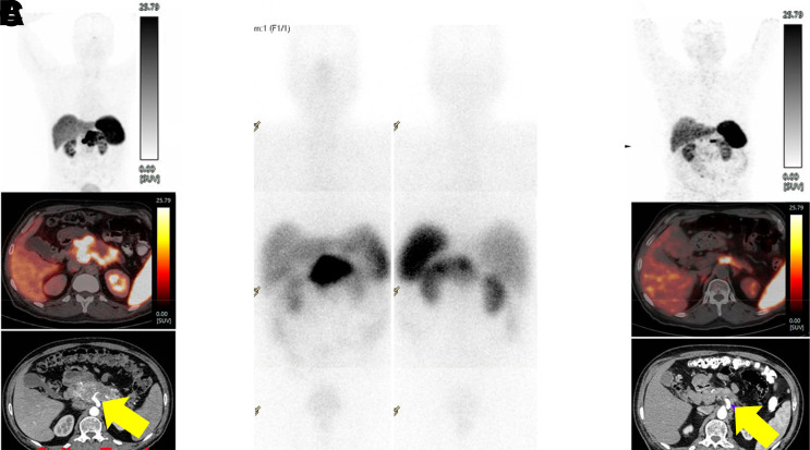
One particularly challenging aspect of GEP-NETs is defining resectable and unresectable primary tumors, a task that often is subjective and surgeon-dependent. Hence, in our study, we used objective resectability criteria (from the NCCN) for determining the resectability of the primary tumor (Figs. 2 and 4). All GEP-NET patients in our study were deemed by an expert gastrointestinal and hepatopancreatobiliary surgeon to have an unresectable primary tumor before PRRT (22). PRRT is generally better tolerated than chemotherapy and other treatment modalities in GEP-NETs. In our study, 177Lu-DOTATATE PRRT was well tolerated and produced no major hematologic or renal toxicity in any patient, and using imaging criteria, we found that an unresectable primary tumor became resectable in 7 patients (30.43%) in group 1 and 8 patients (23.5%) in group 2. The results of our study are similar to those of other NET series reported in the literature (23).
FIGURE 4.
A 67-y-old man with unresectable pancreatic NET. (A) Baseline 68Ga-DOTATATE PET (maximum-intensity projection [MIP]) image (upper panel); transaxial fused PET/CT image (middle panel); and axial contrast-enhanced CT image showed complete encasement (>180°) of celiac trunk (yellow arrow) by intensely somatostatin receptor–avid pancreatic lesion (7.0 × 6.6 cm; SUVmax 80) after PRRT (total cumulative dose, 31.45 GBq). (B) Post-177Lu-DOTATATE therapy planar gamma camera–based scan showed good tracer concentration in primary tumor. (C) Post-PRRT follow-up 68Ga-DOTATATE PET (MIP) image (upper panel); transaxial fused PET/CT image (middle panel); and axial contrast-enhanced CT (lower panel) showed significant reduction in size (2.0 × 1.5 cm) and uptake (SUVmax, 30) of primary tumor, with less than 180° encasement of celiac trunk (yellow arrow). Unresectable primary tumor became resectable.
Barber et al. (15) used 177Lu-octreotate as neoadjuvant PRRT in 5 patients, 4 of whom had pancreatic NET confined to local or locoregional sites and 1 of whom had a duodenal NET with a solitary liver metastasis. In their study, PRRT was administered with a concurrent radiosensitizing dose of fluorouracil chemotherapy (200 mg/m2/24 h) commencing 4 d beforehand and continuing for a total of 3 wk in 4 patients and accompanied by external-beam radiotherapy (45 Gy in 25 fractions) in the remaining patient to maximize delivery of the radiation dose to the tumor. All 5 patients responded well to PRRT, and 1 patient underwent curative surgery after neoadjuvant PRRT.
van Vliet et al. (16) demonstrated the use of 177Lu-octreotate PRRT as neoadjuvant therapy in 29 pancreatic NET patients who had a borderline or unresectable primary tumor with or without oligometastatic liver lesions. The investigators found extensive vascular involvement of the primary tumor and thrombosis of the portal and mesenteric veins before the start of PRRT. They suggested that sufficient venous collaterals may form during the course of PRRT cycles, leading to surgical resection of the primary tumor along with safe and easy reconstruction of the portal and mesenteric veins because of intact collateral circulation. In their study, surgery was performed on 9 (31%) of 29 patients after neoadjuvant PRRT.
Stoeltzing et al. (24) and Sowa-Staszczaket et al. (14) studied the use of neoadjuvant PRRT with the help of 90Y-DOTATOC and 90Y-DOTATATE, respectively, in pancreatic NETs with liver metastases. Liver metastases regressed significantly after neoadjuvant PRRT, facilitating surgical removal of liver metastases. Similarly, in our study the average size of liver metastases changed from 3 to 2.4 cm after 177Lu-DOTATATE PRRT, and there was PR on anatomic imaging in 16 (47%) of 34 patients. This reduction in liver metastasis size will be helpful for surgical intervention in these patients with liver metastases, as shown by Stoeltzing et al. (24) and Sowa-Staszczaket et al. (14) in their studies.
Partelli et al. (25) adopted neoadjuvant PRRT in 23 pancreatic NET patients with features of high disease recurrence. They found that the size of the primary pancreatic tumor decreased after neoadjuvant PRRT and that there was a low risk of pancreatic fistula development (after surgery) and a low incidence of nodal metastases (at the time of surgery) in the neoadjuvant PRRT group as compared with the group treated up front with surgery. Similarly, in our study the average size of the primary tumor changed from 5.8 to 4.8 cm in group 1 and from 6.0 to 4.6 cm in group 2. This shrinkage could also facilitate surgery, as there would be a low incidence of nodal metastases and a low risk of pancreatic fistula formation at the time of surgery or thereafter, respectively, as mentioned by Partelli et al. (25), reducing the risk of morbidity and mortality associated with surgery.
Our observation of the significant association we found with site of primary tumor (duodenal NET), regional lymph node involvement (absent), baseline size of primary tumor (<5 cm), baseline size (≤1.5 cm) and number (≤3) of liver metastases, and baseline 18F-FDG uptake (SUVmax < 5) in the primary tumor indicates that GEP-NET patients with these variables have high rate of converting to a resectable primary tumor after PRRT.
Our finding of an 82% and 70% CR rate for groups 1 and 2, respectively, in the symptomatic response evaluation indirectly shows an improvement in global health status and quality of life in these patients after PRRT, and our observed longer PFS and OS after PRRT may have additional importance in patient care management.
The limitations of our study were its retrospective nature, its nonfixed total cumulative dose of 177Lu-DOTATATE, and its variable number of PRRT cycles. However, the average total cumulative doses of 177Lu-DOTATATE and average number of PRRT cycles were in the usual range for PRRT and similar to those reported for other neoadjuvant PRRT studies.
CONCLUSION
In a moderate fraction of GEP-NET patients whose primary tumor was unresectable because of vascular involvement—either without liver metastases or with potentially resectable liver metastases—the unresectable primary tumor became resectable after 177Lu-DOTATATE PRRT. We therefore conclude that this neoadjuvant therapy can be useful in such patients. 177Lu-DOTATATE PRRT can be considered safe; it does not have a high incidence of major hematologic or renal toxicity and would likely be helpful in reducing the overall morbidity and mortality associated with surgery or other treatment modalities. Our study showed a favorable imaging response in most patients, who became symptom-free after 177Lu-DOTATATE PRRT. The success rate of tumor resectability after PRRT depends on the site of the primary tumor, the presence or absence of regional lymph node involvement, the size of the primary tumor, the size and number of liver metastases in those patients who have them, and the intensity of 18F-FDG uptake in the primary tumor.
DISCLOSURE
No potential conflict of interest relevant to this article was reported.
KEY POINTS
QUESTION: In a real-life clinical scenario at a large-volume tertiary-care cancer center, how well does neoadjuvant 177Lu-DOTATATE PRRT perform in patients with locally advanced, unresectable GEP-NETs whose primary tumor is unresectable because of vascular involvement and who either have no liver metastases or have potentially resectable liver metastases?
PERTINENT FINDINGS: An unresectable primary tumor became resectable in a moderate fraction of GEP-NET patients (26.3%) after 177Lu-DOTATATE PRRT. The success rate of tumor resectability after PRRT depended on the site of the primary tumor, the presence or absence of regional lymph node involvement, the size of the primary tumor, the size and number of liver metastases in those patients who had them, and the intensity of 18F-FDG uptake in the primary tumor.
IMPLICATIONS FOR PATIENT CARE: The role of neoadjuvant PRRT as a potentially useful option in GEP-NET patients is of significant clinical interest from the perspective of the gastrointestinal surgeons who must select patients for such therapy.
REFERENCES
- 1. Cives M, Soares HP, Strosberg J. Will clinical heterogeneity of neuroendocrine tumors impact their management in the future? Lessons from recent trials. Curr Opin Oncol. 2016;28:359–366. [DOI] [PubMed] [Google Scholar]
- 2. Oberg KE. The management of neuroendocrine tumours: current and future medical therapy options. Clin Oncol (R Coll Radiol). 2012;24:282–293. [DOI] [PubMed] [Google Scholar]
- 3. Frilling A, Li J, Malamutmann E, et al. Treatment of liver metastases from neuroendocrine tumours in relation to the extent of hepatic disease. Br J Surg. 2009;96:175–184. [DOI] [PubMed] [Google Scholar]
- 4. Schurr PG, Strate T, Rese K, et al. Aggressive surgery improves long-term survival in neuroendocrine pancreatic tumors: an institutional experience. Ann Surg. 2007;245:273–281. [DOI] [PMC free article] [PubMed] [Google Scholar]
- 5. Sarmiento JM, Heywood G, Rubin J, et al. Surgical treatment of neuroendocrine metastases to the liver: a plea for resection to increase survival. J Am Coll Surg. 2003;197:29–37. [DOI] [PubMed] [Google Scholar]
- 6. Chen H, Hardacre JM, Uzar A, et al. Isolated liver metastases from neuroendocrine tumors: does resection prolong survival? J Am Coll Surg. 1998;187:88–92. [DOI] [PubMed] [Google Scholar]
- 7. Thompson GB, van Heerden JA, Grant CS, et al. Islet cell carcinomas of the pancreas: a twenty-year experience. Surgery. 1988;104:1011–1017. [PubMed] [Google Scholar]
- 8. Chamberlain RS, Canes D, Brown KT, et al. Hepatic neuroendocrine metastases: does intervention alter outcomes? J Am Coll Surg. 2000;190:432–445. [DOI] [PubMed] [Google Scholar]
- 9. Kulke MH, Lenz HJ, Meropol NJ, et al. Activity of sunitinib in patients with advanced neuroendocrine tumors. J Clin Oncol. 2008;26:3403–3410. [DOI] [PubMed] [Google Scholar]
- 10. Malafa MP. Defining borderline resectable pancreatic cancer: emerging consensus for an old challenge. J Natl Compr Canc Netw. 2015;13:501–504. [DOI] [PubMed] [Google Scholar]
- 11. Sarmiento JM, Que FG. Hepatic surgery for metastases from neuroendocrine tumors. Surg Oncol Clin N Am. 2003;12:231–242. [DOI] [PubMed] [Google Scholar]
- 12. Hicks RJ, Kwekkeboom DJ, Krenning E, et al.; Antibes consensus conference participants. ENETS consensus guidelines for the standards of care in neuroendocrine neoplasia: peptide receptor radionuclide therapy with radiolabeled somatostatin analogues. Neuroendocrinology. 2017;105:295–309. [DOI] [PubMed] [Google Scholar]
- 13. van der Zwan WA, Bodei L, Mueller-Brand J, et al. GEPNETs update: radionuclide therapy in neuroendocrine tumors. Eur J Endocrinol. 2015;172:R1–R8. [DOI] [PubMed] [Google Scholar]
- 14. Sowa-Staszczak A, Pach D, Chrzan R, et al. Peptide receptor radionuclide therapy as a potential tool for neoadjuvant therapy in patients with inoperable neuroendocrine tumors (NETs). Eur J Nucl Med Mol Imaging. 2011;38:1669–1674. [DOI] [PMC free article] [PubMed] [Google Scholar]
- 15. Barber TW, Hofman MS, Thomson BN, et al. The potential for induction peptide receptor chemoradionuclide therapy to render inoperable pancreatic and duodenal neuroendocrine tumours resectable. Eur J Surg Oncol. 2012;38:64–71. [DOI] [PubMed] [Google Scholar]
- 16. van Vliet EI, van Eijck CH, de Krijger RR, et al. Neoadjuvant treatment of nonfunctioning pancreatic neuroendocrine tumors with [177Lu-DOTA0,Tyr3]octreotate. J Nucl Med. 2015;56:1647–1653. [DOI] [PubMed] [Google Scholar]
- 17. NCCN clinical practice guidelines in oncology (NCCN guidelines®): pancreatic adenocarcinoma—version 2.2021. NCCN website. https://www.nccn.org/professionals/physician_gls/pdf/pancreatic.pdf. Published February 25, 2021. Accessed May 3, 2021.
- 18. O’Toole D, Hentic O, Corcos O, et al. Chemotherapy for gastro-enteropancreatic endocrine tumours. Neuroendocrinology. 2004;80:79–84. [DOI] [PubMed] [Google Scholar]
- 19. Appetecchia M, Baldelli R. Somatostatin analogues in the treatment of gastroenteropancreatic neuroendocrine tumours, current aspects and new perspectives. J Exp Clin Cancer Res. 2010;29:19. [DOI] [PMC free article] [PubMed] [Google Scholar]
- 20. Kwekkeboom DJ, de Herder WW, Kam BL, et al. Treatment with the radiolabeled somatostatin analog [177Lu-DOTA0, Tyr3] octreotate: toxicity, efficacy, and survival. J Clin Oncol. 2008;26:2124–2130. [DOI] [PubMed] [Google Scholar]
- 21. Kwekkeboom DJ, Teunissen JJ, Bakker WH, et al. Radiolabeled somatostatin analog [177Lu-DOTA0, Tyr3]octreotate in patients with endocrine gastroenteropancreatic tumors. J Clin Oncol. 2005;23:2754–2762. [DOI] [PubMed] [Google Scholar]
- 22. Shrikhande SV, Shinde RS, Chaudhari VA, et al. Twelve hundred consecutive pancreato-duodenectomies from single centre: impact of centre of excellence on pancreatic cancer surgery across India. World J Surg. 2020;44:2784–2793. [DOI] [PubMed] [Google Scholar]
- 23. Sowa-Staszczak A, Hubalewska-Dydejczyk A, Tomaszuk M. PRRT as neoadjuvant treatment in NET. Recent Results Cancer Res. 2013;194:479–485. [DOI] [PubMed] [Google Scholar]
- 24. Stoeltzing O, Loss M, Huber E, et al. Staged surgery with neoadjuvant 90Y-DOTATOC therapy for down-sizing synchronous bilobular hepatic metastases from a neuroendocrine pancreatic tumor. Langenbecks Arch Surg. 2010;395:185–192. [DOI] [PubMed] [Google Scholar]
- 25. Partelli S, Bertani E, Bartolomei M, et al. Peptide receptor radionuclide therapy as neoadjuvant therapy for resectable or potentially resectable pancreatic neuroendocrine neoplasms. Surgery. 2018;163:761–767. [DOI] [PubMed] [Google Scholar]


