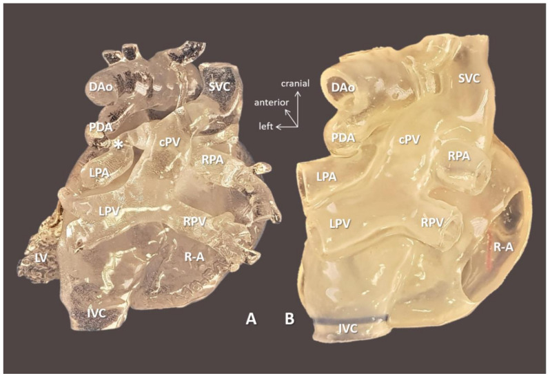
Figure 5.
3D-printed blood volume (A) and hollow (B) models of right atrial isomerism, visceral heterotaxy, and dextrocardia (Case 10). Posterior view: right-sided atrium is opened on the hollow model. Complex anomalies are illustrated on the models left-sided IVC; right-sided SVC receives inflow from common pulmonary vein (cPV), i.e., supracardiac total anomalous pulmonary venous return. Tortuous patent arterial duct (PDA) reaches the left pulmonary artery (LPA); there is pulmonary coarctation (*) at the entry point. The models were instrumental in planning for complete biventricular repair the patient successfully underwent subsequently. Abbreviations: cPV: common vertical pulmonary vein, DAo: descending aorta, IVC: left-sided inferior vena cava, LPA: left pulmonary artery, LPV: left pulmonary vein, LV: left ventricle, PDA: patent arterial duct, R-A: right-sided atrium, RPA: right pulmonary artery, RPV: right pulmonary vein, SVC: right-sided superior vena cava.

